


1. Background
One determining factor in the prognosis of root canal therapy is the appropriate chemical and mechanical preparation that eliminates pathogenic microorganisms and debris from the canal (1-5). Stainless steel hand files were the first instruments used in root canal cleaning and shaping. However, due to the stiffness of these files, the incidence of endodontic procedure accidents has increased (1, 6, 7). Additionally, hand instrumentation is highly time-consuming and can lead to reduced pinch grip strength in dentists (1, 6, 7). To address the difficulties associated with hand files, engine-driven instruments were introduced to the dentistry market. Nickel-titanium (NiTi) rotary files are more flexible, faster, and safer, reducing the risk of accidents during cleaning and shaping (8-10). However, it is estimated that they can lead to dentinal microcracks with a prevalence of 12 - 60% (2, 11-13). Canal preparation can cause microcracks and dentinal damage due to the interaction between files and the walls of the root canal. Lateral forces produced by either hand or rotary files and spreaders during preparation and obturation can stress the canal walls. This stress can be more significant when using large-tapered instruments (14, 15). Failure in root canal therapy due to vertical root fracture (VRF), which results in tooth extraction, can be one of the consequences of microcracks (16, 17).
Nowadays, different rotary systems are available. The Mtwo rotary system has two cutting blades, an S-tip shape, and four instruments. Unlike other rotary systems, Mtwo is used through a single-length approach (12, 18, 19). The M3 rotary system includes three NiTi files with triangular tip shapes, one opener file, and one glide path file. These files are produced from advanced-mechanism shape memory alloys, which can enhance cyclic fatigue. M3 Pro Gold is an advanced version of the M3 rotary system, offering more flexibility due to various heat treatment procedures (20).
2. Objectives
This study aimed to evaluate surface defects in the root canal dentine after preparation with two NiTi rotary files, Mtwo and M3 Pro Gold, and a hand K-file, which remain essential tools in endodontics due to their historical significance, simplicity, and utility in difficult cases, such as curved canals where precision is required. They serve as a baseline for comparisons when evaluating newer instruments.
3. Methods
This study was conducted in the Endodontic Laboratory of the North Khorasan University of Medical Sciences, Bojnurd, Iran, between January 2020 and June 2021.
3.1. Sample Size Calculation
The sample size for this study was determined using Gehan’s procedure (21). Based on the table provided by Gehan (21), assuming a type I error (α) of 0.05, a power of 0.84, and a minimum acceptable success rate (π) of 0.60, 25 samples were required in each group.
3.2. Sample Selection
One hundred mandibular premolars, extracted due to orthodontic or periodontal problems from January 2020 to June 2021, were selected for this study. Teeth with a single canal, closed apex, and less than 10º curvature were included to meet the study's inclusion criteria. The samples were chosen using the convenience sampling method due to the practical limitations of obtaining extracted teeth. Teeth with abnormalities, previous root canal treatment, extensive restoration, or existing cracks were excluded to ensure homogeneity. The degree of root canal curvature was evaluated using the Schneider method. While this sampling method was practical, it may limit the generalizability of the findings. The presence of cracks or fractures was analyzed before instrumentation by two dentists, one of whom was an endodontist. This detection was performed using a stereomicroscope (Austria-MZ1000) at 12x magnification. The specimens were randomly divided into four groups: (1) Unprepared, (2) prepared with hand K-files, (3) prepared with Mtwo, and (4) prepared with M3 Pro Gold. None of the teeth were extracted for this study.
3.3. Tooth Preparation
In the first stage, all selected teeth were immersed in 5.25% sodium hypochlorite (NaOCl) (Shamin Company, Tehran, Iran) for 2 hours (22, 23). Any remaining periodontal tissue was then removed using a periodontal curette. The coronal part of each tooth was sectioned using a diamond fissure bur (Tizkavan: Jota, 837L, Iran) with water cooling. Canal patency was established with a #10 K-file (Mani hand file, China), and radiographs along with a digital linear ruler were used to determine the working length. The final working length was set at 1 mm less than the measured length. In the first group (control), no preparation was done. In the remaining three groups, 2.5% NaOCl was used to irrigate the canal during preparation with a 10 mL irrigating syringe (Pars Syringe company, Iran), and PC-Prep (Pulpdent, USA) was used for canal lubrication.
- Group 2: Hand K-files (Mani hand file, China) were used for canal preparation using the step-back technique, incorporating watch-winding, push-pull, and circumferential motions. The initial file size was #10, and preparation continued up to at least size #25 and at most size #30.
- Group 3: After assessing canal morphology with #10 and #15 K-files, the Mtwo rotary system (VDW, Munich, Germany) was used according to the manufacturer’s instructions (initial file size #10/.04, file size #15/.05, file size #20/.06, file size #25/.06). Each file was used for only 5 teeth. The rotary files were operated with an NSK endodontic micromotor (Endo-Mate AT, Japan) at a torque level of 2 N/cm² and a speed of 300 rpm. The single-length technique with a brushing motion was employed without applying additional force.
- Group 4: Similarly, after initial assessment with #10 and #15 K-files, the M3 ProGold rotary system (United Dental, China) was used as per the manufacturer’s guidelines (#12/.02vt, #16/.02vt, #18/.05, #25/.06). The rotary files were used with the same micromotor settings (2 N/cm² torque, 300 rpm). Gentle pecking motions were applied with 1 to 2 mm forward movement and 4 mm backward movement.
After preparation, each root canal was irrigated with 2 mL of normal saline. All specimens were then stored in distilled water until they were sectioned for defect evaluation.
3.4. Dentinal Defect Evaluation
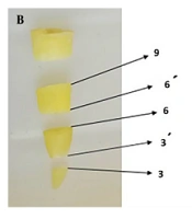
The specimens were sectioned perpendicular to their long axis at 3, 6, and 9 mm distances from the apex using a diamond disk (Resista Company, Iran) with water cooling (Figure 1). Each section was 0.2 mm thick. Each diamond disk was used for only 10 teeth before being discarded. For each tooth, 5 sections were evaluated (n = 125). The specimens were analyzed under 12x magnification with a stereomicroscope (Austria-MZ1000, Austria) by two dentists, one of whom was an endodontist. In cases of disagreement between the two examiners, a third expert evaluated the sections. Blinding of observers during the stereomicroscopic evaluation was ensured by masking group identities.

A, sectioned teeth using diamond disk; B, the location of each section
The defects were categorized into three groups: (1) No defects: The internal or external surface of root dentin is free of any lines or cracks; (2) fractured: A complete crack extends from the canal wall to the external tooth surface; (3) defective: An incomplete crack extends from the canal wall into the dentin but does not reach the tooth's outer surface, or vice versa.
Specimens categorized in groups 2 or 3 exhibited at least one incomplete crack or fracture in one of the five sections evaluated under the stereomicroscope. Finally, the frequency of cracks was compared across all groups (24).
3.5. Statistical Analysis
The statistical analysis aimed to compare the incidence of dentinal microcracks across the four experimental groups and assess the interaction between preparation methods and section levels. Descriptive statistics were presented as frequencies and percentages for categorical data. The normality of data was assessed using the Shapiro-Wilk test, which is considered more powerful than alternatives like the Kolmogorov-Smirnov test, especially for detecting deviations from normality in smaller datasets (less than 50 samples per group). The incidence of dentinal microcracks was compared across the groups using the chi-squared test. For variables where the assumptions of the chi-squared test were violated (expected frequency < 5), Fisher’s exact test was used. A two-way ANOVA was performed to evaluate the interaction between preparation methods and section levels. When significant differences were found in the ANOVA, Tukey’s post hoc test was conducted to determine which groups differed significantly. All statistical decisions were made at a significance level of α = 0.05 and a confidence level of 95%. All statistical analyses were performed using SPSS (version 25.0, IBM Corp., Armonk, NY).
4. Results
In this study, there was no disagreement between the two examiners. For each specimen, 5 sections were evaluated, resulting in a total of 500 sections analyzed. The Shapiro-Wilk test confirmed that the data were normally distributed (P > 0.05). Table 1 shows the distribution of defects in the 4 examined groups.
| Groups | Types of Cracks | ||
|---|---|---|---|
| Non-defected | Incomplete | Complete | |
| Control | 125 (100) | 0 (0) | 0 (0) |
| K-files | 119 (95.2) | 5 (4) | 1 (0.8) |
| Mtwo rotary files | 114 (91.2) | 10 (8) | 1 (0.8) |
| M3 Pro Gold rotary files | 122 (97.6) | 3 (2.4) | 0 (0) |
- Unprepared group (control group): No defects were observed.
- Group 2 (prepared with hand K-files): Four specimens (16%) exhibited dentinal microcracks, with 5 sections showing incomplete cracks and 1 section showing complete cracks.
- Group 3 (prepared with Mtwo rotary system): Eight specimens (32%) exhibited dentinal microcracks, with 10 sections showing incomplete microcracks and 1 section showing a complete microcrack.
- Group 4 (M3 Pro Gold group): Three specimens (12%) showed incomplete microcracks, and none demonstrated complete microcracks.
Table 2 shows the status of the microcracks according to the examined files. There was a significant difference between the three preparation instruments regarding crack incidence (Pearson chi-square = 75.13, P-value = 0.003). Mtwo rotary files caused the most significant number of microcracks (8.8%), followed by hand K-files (4.8%). The fewest number of microcracks was seen in M3 Pro Gold (2.4%).
A two-way ANOVA showed significant interactions between preparation methods and section levels (P-value = 0.012). Tukey’s post hoc test identified group 3 as having significantly more defects than groups 2 and 4 (P < 0.05). Figure 2 demonstrates the location of each section, and Table 3 shows the status of microcracks in the different section areas.

Evaluation of cracks in 3 examined groups using stereomicroscope: A, a section of the control group specimens with no crack; B, an incomplete and; C, a complete crack in sections prepared with K-files; D and E, incomplete cracks and; F, complete crack in sections prepared with Mtwo rotary systems; G, incomplete cracks in sections prepared using M3 Pro Gold rotary system
The most significant number of incomplete microcracks was seen at a 3 mm distance from the apex (33.3% at 3 and 27.8% at 3′). The amount of these types of cracks ranked second at 6 mm-distance sections (22.2% at 6 and 16.7% at 6′). No cracks were observed at a 9 mm distance from the apex (9 and 9′).
| Types of Microcracks | Section Levels | Total | ||||
|---|---|---|---|---|---|---|
| 3 | 3′ | 6 | 6′ | 9 | ||
| Non-defected | 93 (19.4) | 94 (19.6) | 96 (20) | 97 (20.2) | 100 (20.8) | 480 (100) |
| Incomplete | 6 (33.3) | 5 (27.8) | 4 (22.2) | 3 (16.7) | 0 (0) | 18 (100) |
| Complete | 1 (50) | 1 (50) | 0 (0) | 0 (0) | 0 (0) | 2 (100) |
| Total | 100 (20) | 100 (20) | 100 (20) | 100 (20) | 100 (20) | 500 (100) |
5. Discussion
In this in vitro study, both hand and rotary system instruments resulted in dentinal microcracks in root canal walls. The most significant number of microcracks was observed in roots prepared with Mtwo rotary files, followed by hand K-files, with the fewest microcracks seen in the M3 Pro Gold group. No microcracks were observed in the control group, suggesting that using diamond disks for root sectioning does not produce microcracks. While this study found differences in the incidence of microcracks among the three groups, some previous studies reported no difference between hand instruments and rotary systems regarding microcrack incidence (24-27). This disparity might be due to differences in the types of evaluated cracks. In most of the mentioned studies, only complete dentinal cracks and craze lines were analyzed, whereas in our study, both complete and incomplete cracks were evaluated. Additionally, although all specimens in both study groups were extracted for periodontal and orthodontic reasons, there is no further information about the forces that teeth experienced in the oral environment. This lack of information was a limitation of our study and may explain the different results.
In this study, a significant difference was observed between the use of the three types of instruments regarding the location of microcracks. The 3 - 6 mm distance from the apex is at the highest risk of microcrack incidence, possibly due to more interaction between files and the root walls in these areas. In the study conducted by Mavani et al., the most significant number of microcracks was observed at 2 to 4 mm distances from the apex (28). Therefore, conservative preparation seems necessary in apical areas to reduce the number of microcracks (29). By comparing different filling instruments, the results of various studies confirm our findings that Mtwo rotary files created the highest number of microcracks (12, 28, 30).
M3 Pro Gold caused fewer microcracks than hand K-files and Mtwo rotary files. This may be attributed to differences in flexibility, blade shape, file flute design and construction, and the cross-section of each file. Stainless steel hand files have an inherent stiffness that can be more pronounced in larger files. This inflexibility can produce more microcracks due to increased strain on root walls and greater dentinal removal (31). In the control group, no cracks were found; however, some studies have detected microcracks in this group (32-34). It has been suggested that these cracks be classified as “experimental dentinal microcracks”, referring to defects in the dentin of stored or extracted teeth caused by dehydration. This terminology is supported by multiple studies, which attribute the formation of these cracks to environmental pressures and conditions both before and during experimentation, leading to the natural dehydration of extracted teeth (13, 25, 32-34).
The incidence of dentinal microcracks can vary based on the instruments and techniques used in the cleaning and shaping process. It is reported that the incidence of microcracks can range from 0 to 50% when hand instruments are used, while for rotary and reciprocating instruments, it is higher and can range from 0 to 80% (35). There is a positive relationship between excessive dentin removal and the incidence of dentinal microcracks (2, 30, 36). Highly tapered instruments can lead to a high tendency for root fractures. Bier showed that using finishing files in the Protaper rotary system, commonly used to prepare the apical part of roots, can cause severe microcracks in this area. These files can cause a taper preparation up to 9% and result in extreme stress on root walls (37).
The importance of dentinal microcracks was first elaborated by Wilcox, who announced that VRF does not occur unless the teeth were previously crazed in a certain way (38, 39). Vertical root fracture is not a common phenomenon after root canal therapy, but it is a major concern for dentists due to its devastating potential. Endodontists should pay close attention to crazes and microcracks, as each craze has the potential to extend and result in a fracture (38). Bier reported that although VRF might not occur during root canal therapy, dentinal microcracks can be produced by 16%, leading to VRF due to continuous occlusal forces (37).
While this study has several strengths, it is important to acknowledge its limitations. First, as the findings are based on an in vitro study, their applicability to clinical scenarios should be approached with caution. Future in vivo studies are necessary to validate these results. Second, hand K-files were used as a baseline for comparison, which is common practice. However, we recommend that future studies include comparisons of the M3 Pro Gold and Mtwo systems with state-of-the-art rotary instruments to provide more robust clinical guidance. Third, the absence of blinding during the preparation phase may have introduced observer bias. Addressing this in future studies would strengthen the reliability of the results.
5.1. Conclusions
As a result of this in vitro study, using hand or reciprocating instruments during root canal preparation can induce dentinal defects with varying frequencies. However, M3 Pro Gold rotary files showed fewer microfractures compared to the Mtwo rotary system and hand K-files.
