1. Background
The meniscus, an integral component of the knee joint capsule, is highly susceptible to injury (1) and is crucial for load distribution, shock absorption, lubrication, nutrition of articular cartilage, and joint stability (2). Meniscal injuries are among the most common knee pathologies, significantly affecting muscle strength, proprioception, and joint stability, leading to functional limitations and a reduced quality of life (QOL) (3).
Longitudinal meniscal tears are among the most common meniscal injuries, frequently occurring in the vascular zone where surgical intervention is typically necessary (4), and meniscal repair and meniscectomy are the most frequently performed surgeries to treat them (5, 6). While meniscal repair aims to simplify surgical procedures, accelerate recovery periods, and reduce the risk of neuromuscular complications, it carries risks such as potential meniscal damage caused by surgical instruments, implant displacement, the body's immune response to foreign substances, and increased financial expenditures (7). Conversely, meniscectomy, whether total or partial, offers benefits such as preservation of the peripheral rim responsible for the biomechanical function of the knee and minimized meniscal trauma, but also carries drawbacks like predisposition to early-onset osteoarthritis (8), radiographic alterations (9), and diminished long-term functional outcomes (10).
Meniscal injuries involve both biomechanical and neurophysiological factors that are critical for joint stability and proprioception (11). While proprioception plays a significant role in maintaining functional balance and overall function (12), it is often overlooked in traditional rehabilitation approaches after surgeries. Furthermore, to the authors’ knowledge, many studies compare the outcomes of meniscectomy and meniscal repair (6, 10, 13).
Despite this, there remains a significant gap and controversy regarding the optimal surgical approach for managing meniscal tears. To date, the biomechanical implications and functional outcomes of longitudinal tears have been underexplored in comparison to other tear types.
2. Objectives
To address these gaps, this study aimed to compare the biomechanical indices, function, functional balance, and knee proprioception after meniscectomy and meniscal repair in patients with longitudinal meniscal tears using comprehensive, validated assessment tools. Understanding these outcomes can provide valuable insights into surgical decision-making and rehabilitation strategies to help patients experience a faster return to work and also achieve higher post-surgical QOL, function, and biomechanical indices.
3. Methods
3.1. Participants
In this cross-sectional study, patients with longitudinal meniscal tears were selected using convenience sampling due to practical constraints. Patients were categorized into two groups based on the procedure received: Meniscectomy or meniscal repair at the vascular zone. All surgeries were conducted at Akhtar Hospital. During arthroscopy, the surgical team assessed the tear's pattern, location, and vascularity. Meniscal repair used all-inside techniques for posterior, body, or anterior tears, while non-repairable tears were trimmed to achieve a stable peripheral rim. The postoperative outcomes of these patients from Shahid Beheshti University of Medical Sciences were evaluated one year after surgery at the Orthopedic and Biomechanical Laboratory. The study was conducted between January 2021 and December 2022.
The sample size was calculated based on a type I error rate (α) of 0.05, a type II error rate (β) of 0.2 (80% power), and mean values of 68 and 91 with standard deviations of 35 and 15, derived from prior studies. Using these parameters, the required sample size was determined to be 24 participants per group, totaling 48 participants. This aligns with the recommendations of Seltman (14) and ensures sufficient power to detect statistically significant differences. Participants were recruited based on predefined inclusion and exclusion criteria to minimize potential biases.
Patients who met the following inclusion criteria were invited to participate: One year (9 - 12 months) had passed since the surgery (15), aged between 20 - 40 years, had completed at least 16 physiotherapy sessions, were able to walk without assistance, had achieved full recovery of function at the one-year assessment, and had a dominant right leg (16). Exclusion criteria encompassed conditions such as knee pain or inflammation (redness, swelling, warmth), history of musculoskeletal diseases (e.g., rheumatoid arthritis, osteoarthritis) (17), previous surgical interventions for inflammatory conditions in the hip or knee, history of ankle trauma or surgery, limb length discrepancy, use of corticosteroids or medications affecting balance, substance addiction, peripheral or central nervous system complications, neurological disorders, obesity [Body Mass Index (BMI) ≥ 30] (19), diabetes (18), cognitive impairments (19), vision or hearing impairments, fractures or balance-affecting complications post-study commencement, and patient unwillingness or inability to continue research participation. The Ethics Committee of Shahid Beheshti University of Medical Sciences (IR.SBMU.RETECH.REC.1399.1313) and the affiliated institutions of the authors approved this study.
Participants provided demographic data and completed a questionnaire. The questionnaires were administered by an occupational therapist (first author). The therapist was trained in the standardized administration of the Knee Injury and Osteoarthritis Outcome Score (KOOS) and other assessment tools used in this study to ensure consistency and reliability in data collection. Tests were conducted in one session by a single therapist on both surgical and non-surgical knees, except for the KOOS Questionnaire, which was only for the surgical knee. All participants followed standard post-surgical rehabilitation guidelines as recommended by their healthcare providers. This program included mobility exercises and functional training to restore movement and balance. Patients' physical conditions were monitored, and tests were waived if conditions were inappropriate. A five-minute break was given between tests to prevent fatigue.
3.2. Measurement Tools
3.2.1. Demographic Information Questionnaire
A study form was used at baseline to gather demographic details, incorporating gender, age, weight, height, BMI, dominant leg, operated side, and time interval between surgery and evaluation.
3.2.2. Knee Injury and Osteoarthritis Outcome Score
The KOOS tool is utilized to assess both the short-and-long-term effects of knee injuries, including those recovering from surgery. It has demonstrated excellent test-retest reliability and validity in populations with meniscal injuries, osteoarthritis, and other knee pathologies (20). It includes 42 items across five subscales: Pain, symptoms, daily living function, sport and recreation function, and QOL. Each subscale is rated on a 5-point Likert scale (0 = no problems, 4 = extreme problems), with scores converted to a 0 - 100 scale, where higher scores indicate better knee function. Completing the KOOS typically takes about 10 minutes, and it does not provide an overall score, focusing instead on individual subscale analysis. The Persian interpretation of the KOOS has been validated and shown to be reliable for use with Persian patients (21). The questionnaire was administered for each surgical method, providing insights into the recovery trajectory after meniscectomy and meniscal repair. The KOOS was chosen for its high sensitivity in detecting functional deficits and patient-reported outcomes in post-surgical rehabilitation.
3.2.3. Biomechanical Indicators
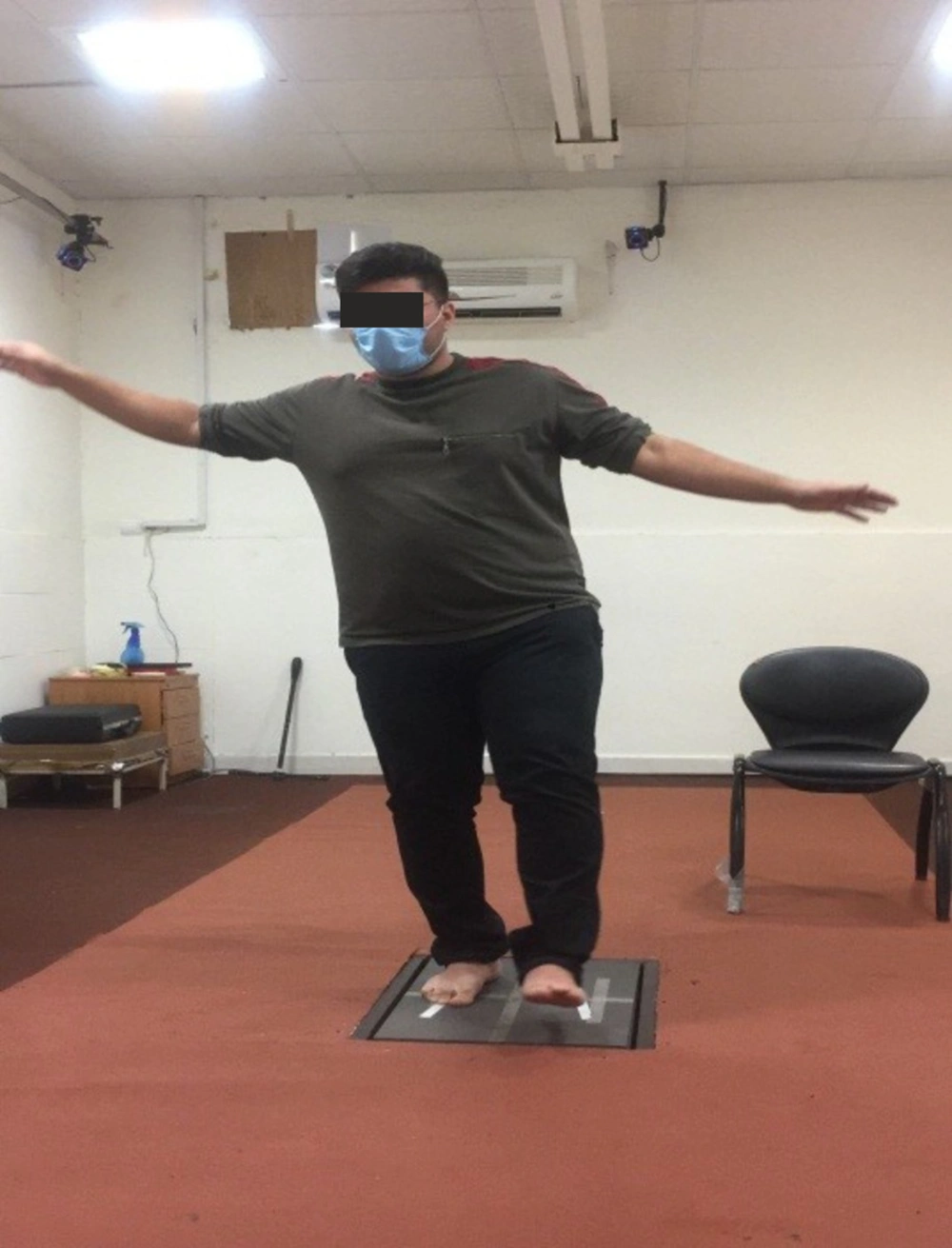
Biomechanical indicators, including static and dynamic balance, were assessed using the Bertec 9090 force plate (15.2 cm height, sensitivity Div./10, 100 Hz frequency). Static balance tests included six positions (open/closed eyes, single/double-leg stance) on a firm surface (Figure 1), which provides reliable measures of postural sway, including velocity, displacement, and confidence ellipse (22). The center of pressure changes was calculated by measuring the displacement of the center of pressure in the anteroposterior and medial-lateral directions. Additionally, the center of pressure path length was considered as the amount of postural sway. The difference between maximum and minimum displacement in the anterior-posterior and internal-external directions was also calculated. Other parameters included average displacement speed and the amount of swings. Three attempts were made for each test.
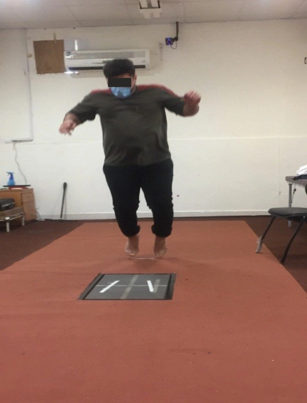
Dynamic balance was evaluated by having participants jump forward from half their maximum jump distance, measuring force in the vertical (Fz), anterior, and total directions (Figure 2). Each test was performed three times, with six-second breaks between jumps.
3.2.4. Star Excursion Balance Test
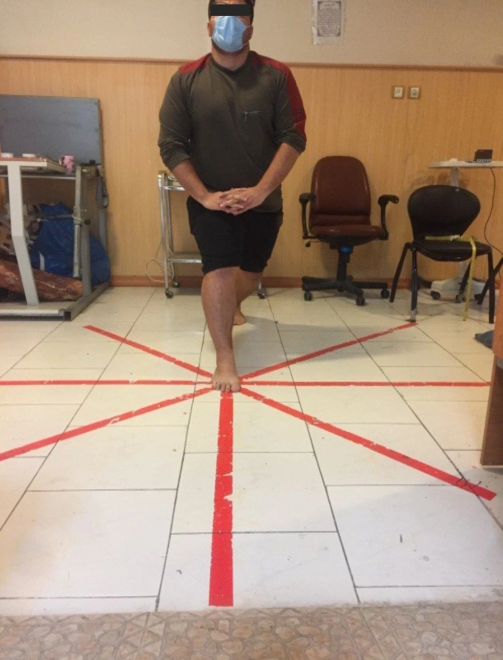
The functional balance of participants was assessed using the Star Excursion Balance Test (SEBT). The length of the lower limb was measured from the anterior superior iliac spine to the medial malleolus. To ensure standardized testing, two main lines were drawn along the posterior-anterior and medial-lateral directions, using a goniometer to ensure these lines were perpendicular. The secondary directions were placed at a 45-degree angle to the main directions. The SEBT requires participants to hold a single-leg stance while reaching as far as possible with the opposite leg in eight different directions with the same angles (Figure 3). The test was conducted on both surgical and non-surgical knees, with each excursion being measured using a standard tape measure (23). Each participant performed each direction three times. The average reach distance was calculated, divided by the length of the lower limb in centimeters, and then multiplied by 100 to obtain the reach distance as a percentage of the length of the lower limb (24). The SEBT is a reliable and valid tool for identifying functional balance deficits in patients with lower extremity conditions (25).
3.2.5. Proprioception
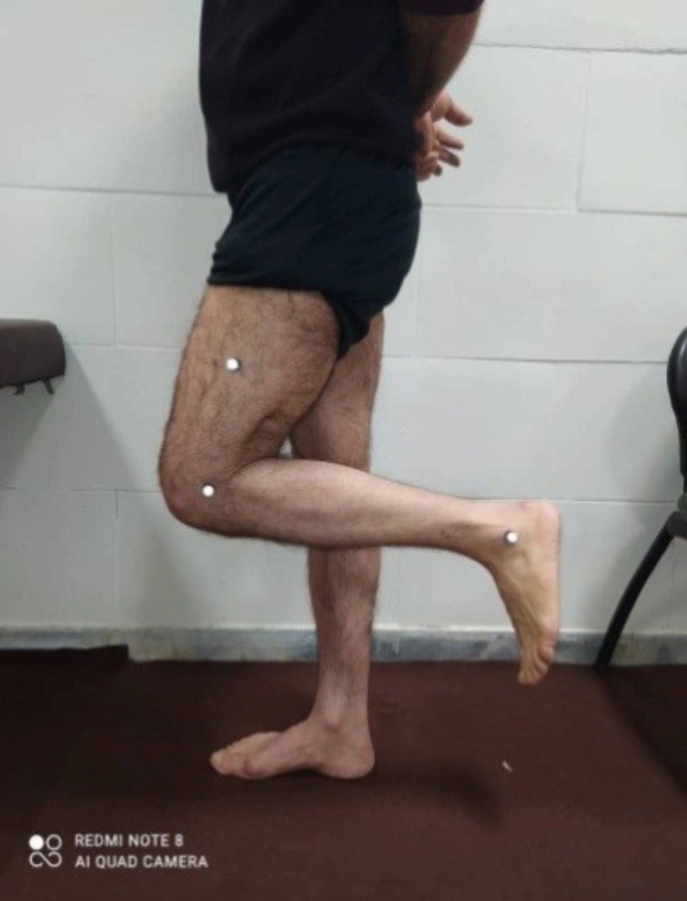
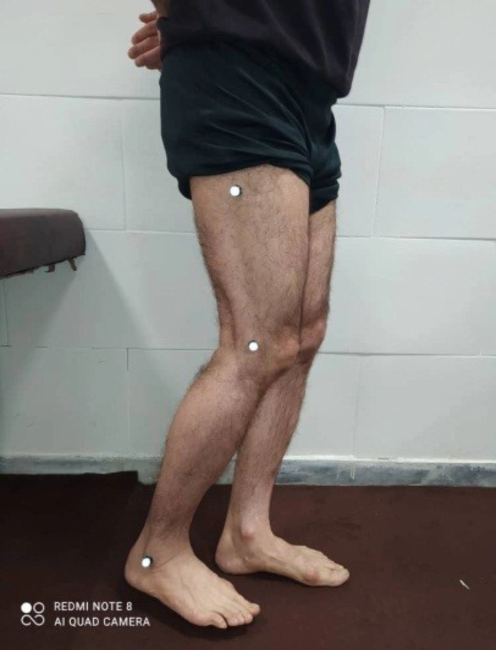
Digital photography evaluated proprioception as participants stood on one leg with knee markers at specific points of the knee (Figures 4 and 5). A camera positioned 185 cm away captured images at 30°, 45°, and 90° flexion, held for 5 seconds. After a 6-second rest, participants reconstructed the angles, and photos were taken. Each angle was measured three times, and the average was analyzed using Digimizer 5.3.4 software. This method has demonstrated good validity and reliability for assessing proprioceptive errors by calculating the absolute difference between target and reconstructed angles (20). Both surgical and non-surgical knees were tested (26).
3.3. Statistical Analysis
Considering that the number of our participants was 48, we applied the Shapiro-Wilk test to assess the normality of the data (27). Given that the value of this statistic in all the indicators studied had a significance level greater than 0.05 (P > 0.05), it can be concluded that the distribution of the variables is normal. As all variables were distributed normally, parametric tests were used for the analyses. The independent samples t-test was used for comparisons between groups, and the paired t-test was applied for within-group comparisons. To determine if there were any differences in multiple dependent variables over time or between treatments, we used multivariate analysis of variance. Additionally, covariance analysis was conducted to control for any potential confounding variables. The data was analyzed and modeled with the SPSS statistical software (version 27.0).
4. Results
Fifty patients met the inclusion criteria, and two patients (4%) declined participation, leading to a final sample size of 48 patients, including 24 patients who underwent meniscectomy (with a mean age of 30.91 ± 6.04 years) and 24 patients who underwent meniscal repair (with a mean age of 33.04 ± 6.42 years). Table 1 provides a summary of participant characteristics. Chi-square analysis revealed a statistically significant difference between the surgical knee and the meniscectomy and meniscal repair surgery groups (P < 0.05).
| Variables | Participants | P-Value | ||
|---|---|---|---|---|
| Range | Meniscectomy | Meniscal Repair | ||
| Age | 20 - 40 (y) | 30.91 ± 6.04 | 33.04 ± 6.42 | 0.117 |
| Sex | - | 12 M, 12 F | 8 M, 16 F | 0.190 |
| Weight | - | 79.33 ± 15.49 | 83.21 ± 13.59 | 0.627 |
| Height | - | 174.95 ± 8.96 | 177.50 ± 6.90 | 0.030 c |
| BMI | - | 25.75 ± 3.57 | 26.60 ± 4.02 | 0.663 |
| Dominant leg | - | 24 R | 24 R | |
| Operated side | - | 17R, 7 L | 10R, 14 L | 0.040 c |
| Time interval between surgery and evaluation | 9 - 12 (mo) | 11.03 ± 1.04 | 11.71 ± 1.12 | 0.102 |
4.1. Function
The KOOS scores for symptoms, pain, activity of daily living, sport and recreation, as well as QOL, were calculated for patients who underwent meniscectomy or meniscus repair. In the meniscectomy group, the mean and standard deviation for symptoms, pain, daily living activities (ADL), sport and recreation, and QOL were 12.25 ± 4.34, 13.41 ± 7.07, 20.37 ± 8.02, 11.58 ± 3.32, and 9.41 ± 2.61, respectively. In the meniscal repair group, the mean and standard deviation for the same categories were 13.12 ± 4.50, 15.12 ± 6.40, 23.00 ± 11.66, 12.16 ± 3.70, and 9.70 ± 2.33. However, no significant differences were found in any of the sub-scores (P > 0.05, Table 2). Therefore, the total KOOS score (P = 0.71) and sub-scores were not significantly different between the two groups.
| Variables | Meniscal Repair | Meniscectomy | P-Value (Between Groups) |
|---|---|---|---|
| Symptoms | 4.50 ± 13.12 | 4.34 ± 12.25 | 0.72 |
| Pain | 6.40 ± 15.12 | 7.07 ± 13.41 | 0.56 |
| ADL | 11.66 ± 23.00 | 8.02 ± 20.37 | 0.20 |
| Sport | 3.70 ± 12.16 | 3.32 ± 11.58 | 0.72 |
| QOL | 2.33 ± 9.70 | 2.61 ± 9.41 | 0.16 |
| Total function | 25.64 ± 73.12 | 21.21 ± 67.04 | 0.71 |
Function Results in Meniscectomy and Meniscal Repair Groups a
Given that the t-test showed that the height of the participants in the two types of treatment was significantly different, the analysis of covariance (ANCOVA) test was used to eliminate this variable. The results of this test are reported in Appendix 1 in Supplementary File in terms of the components and total score of the KOOS test. According to the table, the participants' height (as a covariate variable) did not have a significant effect on any of the KOOS test components (P > 0.05). It should also be noted that the total KOOS test score was not affected by this variable.
4.2. Proprioception
According to the proprioceptive results, there was a significant difference in digital photography scores for knee motion at 30°, 45°, and 90° between the surgical and non-surgical knees in both groups (P < 0.05; Table 3). The average digital photography analysis showed that patients with meniscectomy had scores of 26.16 ± 2.16, 41.25 ± 2.99, and 83.33 ± 3.40 for three ranges of knee motion (30°, 45°, and 90°), respectively. Meanwhile, the average digital photography scores in patients with meniscal repair were 26.91 ± 1.52, 41.00 ± 2.99, and 84.70 ± 3.82. The results indicated no significant difference between the meniscal repair group's surgical knee and the meniscectomy group's surgical knee (30° P = 0.17, 45° P = 0.77, and 90° P = 0.19, Table 3). The ANCOVA test (Appendix 2 in Supplementary File) showed that the participants' height had no effect on proprioception at 30°, 45°, and 90° (P > 0.05).
| Variables | Meniscal Repair | Meniscectomy | P-Value (Between Groups) | ||
|---|---|---|---|---|---|
| P-Value b | Mean ± SD | P-Value b | Mean ± SD | ||
| 30° | 0.001 | 0.001 | 0.172 | ||
| Surgical knee | 26.91 ± 1.52 | 26.16 ± 2.16 | 0.670 | ||
| Non-surgical knee | 28.62 ± 1.17 | 28.79 ± 1.50 | 0.774 | ||
| 45° | 0.001 | 0.001 | |||
| Surgical knee | 41.00 ± 2.99 | 41.25 ± 2.99 | 0.439 | ||
| Non-surgical knee | 43.04 ± 2.44 | 43.54 ± 1.97 | 0.195 | ||
| 90° | 0.001 | 0.001 | |||
| Surgical knee | 84.70 ± 3.82 | 83.33 ± 3.40 | 0.706 | ||
| Non-surgical knee | 87.83 ± 2.56 | 88.08 ± 1.95 | |||
Proprioception Results in Meniscectomy and Meniscal Repair Groups a
4.3. Functional Balance Results
According to the functional balance data, there was a significant within-group difference in the reach distances between the surgical and non-surgical knee in meniscectomy and meniscal repair for each of the 8-reach distances (P < 0.05; Table 4). However, the two surgical groups did not exhibit a significant difference in reach distances in the surgical knee across the anterior, anteromedial, medial, posteromedial, posterolateral, and anterolateral directions (anterior P = 0.99, anterolateral P = 0.80, anteromedial P = 0.58, medial P = 0.09, lateral P = 0.53, posterior P = 0.28, posterolateral P = 0.37, posteromedial P = 0.21; Table 4).
| Variables | Meniscectomy | Meniscal Repair | P-Value (Between Groups) | ||
|---|---|---|---|---|---|
| Mean ± SD | P-Value b | Mean ± SD | P-Value b | ||
| Anterior | 0.001 | 0.001 | |||
| Surgical knee | 92.45 ± 13.95 | 92.42 ± 11.37 | 0.995 | ||
| Non-surgical knee | 99.82 ± 13.68 | 100.91 ± 13.65 | 0.784 | ||
| Anterolateral | 0.003 | 0.001 | |||
| Surgical knee | 87.15 ± 18.62 | 88.52 ± 19.81 | 0.806 | ||
| Non-surgical knee | 87.93 ± 18.88 | 90.58 ± 21.69 | 0.654 | ||
| Anteromedial | 0.001 | 0.001 | |||
| Surgical knee | 91.98 ± 20.22 | 95.11 ± 18.59 | 0.581 | ||
| Non-surgical knee | 97.63 ± 22.58 | 100.16 ± 22.36 | 0.718 | ||
| Medial | 0.001 | 0.001 | |||
| Surgical knee | 97.86 ± 28.72 | 102.77 ± 25.58 | 0.091 | ||
| Non-surgical knee | 99.42 ± 30.23 | 140.77 ± 17.11 | 0.626 | ||
| Lateral | 0.011 | 0.023 | |||
| Surgical knee | 68.73 ± 18.40 | 79.55 ± 24.56 | 0.535 | ||
| Non-surgical knee | 78.70 ± 21.05 | 72.09 ± 26.53 | 0.247 | ||
| Posterior | 0.001 | 0.010 | |||
| Surgical knee | 85.15 ± 25.53 | 93.13 ± 26.10 | 0.289 | ||
| Non-surgical knee | 89.82 ± 25.35 | 100.62 ± 29.49 | 0.180 | ||
| Posterolateral | 0.001 | 0.001 | |||
| Surgical knee | 62.58 ± 10.99 | 65.53 ± 11.87 | 0.376 | ||
| Non-surgical knee | 74.09 ± 17.99 | 74.94 ± 22.69 | 0.887 | ||
| Posteromedial | 0.003 | 0.005 | |||
| Surgical knee | 90.43 ± 20.74 | 97.94 ± 20.50 | 0.214 | ||
| Non-surgical knee | 92.57 ± 20.24 | 101.29 ± 20.81 | 0.148 | ||
Functional Balance Results in Meniscectomy and Meniscal Repair Groups a
As can be seen in Appendix 3 in Supplementary File, in the two types of treatment, the height of the participants had no effect on any of the components (anterolateral, anteromedial, lateral, anterior, medial, posterior, posterolateral, and posteromedial) (P > 0.05).
4.4. Static and Dynamic Balance
Based on the statistical distribution analysis of biomechanical indicators for both one-leg and two-leg postures, it was found that the average value of all variables was higher (P < 0.05) when the participants performed the test with their eyes closed compared to when their eyes were opened. The study found that regardless of the group, the balance characteristics in one leg versus two legs and balance characteristics with eyes closed versus eyes opened had significant effects on all dependent variables analyzed. The P-value for the main effects of foot and eyesight in two groups for each dependent variable was less than 0.05 (P < 0.05). The intra-group scores of the balance test for the surgical knee were worse than those of the non-surgical knee in both groups. However, the two knees had no significant difference (P > 0.05).
For anterior-posterior velocity, the difference in means and P-value between meniscectomy and meniscal repair were 91.79 ± 91.48, 50.93 ± 40.23 (P = 0.05; Appendix 4 in Supplementary File) for eyes-opened knee surgery, and 93.98 ± 24.04, 93.81 ± 36.37 (P=0.98; Appendix 5 in Supplementary File) for eyes-closed knee surgery. Mediolateral velocity was significantly higher (P < 0.05; Appendix 4 in Supplementary File) in meniscectomy (61.82 ± 31.59) compared to meniscal repair (45.80 ± 13.83) for eyes-opened knee surgery. There was no significant difference between groups (meniscectomy 78.97 ± 13.33, meniscal repair 79.92 ± 15.13) regarding mediolateral velocity for eyes-closed knee surgery (P = 0.82; Appendix 5 in Supplementary File).
Additionally, anterior-posterior displacements were significantly higher (P < 0.05; Appendix 6 in Supplementary File) in meniscectomy (13.29 ± 5.60) compared to meniscal repair (10.42 ± 4.39) for eyes-opened knee surgery. There was no significant difference between groups (meniscectomy 16.13 ± 2.93, meniscal repair 15.24 ± 3.64) regarding anterior-posterior displacements for eyes-closed knee surgery (P = 0.35; Appendix 7 in Supplementary File). There was no significant difference between groups regarding mediolateral displacements for eyes-opened/closed knee (P = 0.56/P = 0.13; Appendices 6 and 7 in Supplementary File). The difference scores of mediolateral displacements for eyes-opened and closed knee surgery were 10.82 ± 5.12, 9.96 ± 5.23, and 15.11 ± 4.39, 13.30 ± 3.74, respectively. Finally, the difference in confidence ellipse scores for eyes-opened and closed knee surgery were 1.99 ± 2.21, 1.65 ± 2.28 (P = 0.60; Appendix 5 in Supplementary File) and 2.48 ± 1.14, 2.14 ± 1.07 (P = 0.28; Appendix 9 in Supplementary File).
The comparison of dynamic balance scores revealed no significant difference between meniscectomy and meniscal repair in the vertical 2.83 ± 0.92, 2.62 ± 0.73 (P = 0.74), anterior 2.71 ± 0.08, 2.72 ± 0.08 (P = 0.37), and overall axes 2.94 ± 0.89, 2.64 ± 0.62 (P = 0.18; Table 5).
| Variables | Meniscectomy | Meniscal Repair | P-Value |
|---|---|---|---|
| Mean ± SD | Mean ± SD | ||
| Anterior | 2.72 ± 0.08 | 2.71 ± 0.08 | 0.746 |
| Vertical | 2.62 ± 0.92 | 2.83 ± 0.73 | 0.375 |
| Total | 2.64 ± 0.89 | 2.94 ± 0.62 | 0.180 |
Dynamic Balance Results in Meniscectomy and Meniscal Repair Groups a
The ANCOVA was used to control for height when comparing biomechanical indices between the two surgical groups. The results indicated that height did not have a significant effect on any of the assessed biomechanical parameters (P > 0.05).
5. Discussion
This study demonstrated that one year post-surgery, no significant differences were observed in biomechanical indices, functional balance, proprioception, or overall function between the meniscectomy and meniscal repair groups. However, both surgical groups exhibited residual impairments in their surgical knees, highlighting the long-term challenges associated with meniscal injuries.
5.1. Functional Outcomes
The lack of significant differences in function after meniscal repair and meniscectomy may be attributed to the nature of the symptoms being measured. Pujol et al. (28) suggest that some symptoms caused by longitudinal meniscal tears improve over time regardless of the treatment method. Additionally, similar functional outcomes could result from standardized postoperative rehabilitation protocols. Notably, there were no statistically significant differences between the scores after meniscal repair and meniscectomy on all KOOS items.
Our study results differ from those of previous studies regarding function scores among patients who underwent meniscectomy and meniscal repair. For instance, Lutz et al. (9), in a 10-year follow-up, showed the functional score of meniscal repair surgery was higher than meniscectomy in all KOOS items except QOL, and Başar et al. (3) found improved functional outcomes for meniscal repair over meniscectomy. Pihl et al. (29) reported that meniscal repair surgery causes less improvement in all KOOS items than meniscectomy, with follow-ups at 12 and 52 weeks.
The results of this study may be influenced by the presence of anterior cruciate ligament damage along with the meniscus tear, the type of instrument used, and notably, the timing of follow-up sessions during these studies. In the early period after surgery, it is natural that meniscectomy yields better clinical and functional results than meniscal repair.
5.2. Biomechanical and Balance Indicators
In this study, a significant difference in participants’ height was observed between the meniscectomy and meniscal repair groups. Given that height can influence biomechanical parameters, particularly balance and postural control (30), it was essential to assess its potential confounding effect. Taller individuals generally have a higher center of mass, which may affect stability and increase postural sway during static and dynamic balance tasks (31). Therefore, we conducted an ANCOVA to control for height when comparing biomechanical indices between the two surgical groups. The results indicated that height did not have a significant effect on any of the assessed parameters (P > 0.05). This suggests that the observed differences or similarities in balance and biomechanics were primarily attributable to the surgical procedures rather than height discrepancies between the groups. Nonetheless, future studies with a larger sample size may benefit from stratifying participants based on height or incorporating more advanced biomechanical modeling to further explore the relationship between height and postural control after knee surgery.
Under both single-leg and double-leg postures, participants demonstrated higher biomechanical values with eyes closed compared to eyes open, highlighting reliance on proprioceptive input in the absence of visual feedback. This emphasizes the importance of proprioceptive rehabilitation, particularly under challenging sensory conditions. The anterior-posterior velocity of the surgical knee was not significantly different between the two surgical procedures, regardless of whether the patient had their eyes open or closed. However, when the patient had their eyes open, the mediolateral velocity and anterior-posterior displacement of the surgical knee were significantly higher after meniscal repair than after meniscectomy. Medio-lateral displacement and the confidence ellipse of the surgical knee, regardless of eye condition, were no different between the two types of surgery. These findings suggest distinct biomechanical adaptations between procedures and highlight the need for tailored rehabilitation strategies targeting postural control and balance deficits, particularly in meniscal repair.
The study also found that surgical knee scores were significantly lower than non-surgical knee scores in intergroup assessments, regardless of eye condition. Despite these differences, static balance characteristics did not vary significantly between the two surgical techniques. The results of studies by Lau et al. (32), Żmijewska et al. (33), Logan et al. (34), and O'Connell et al. (35) were in line with ours. The results of a study by Lee et al. (36) indicated that the biomechanical characteristics scores are correlated with knee surgery. This can be related to the study population difference. The study evaluated participants after one year and upon completion of the rehabilitation period (strengthening, balance, and proprioceptive exercises), which can impact movement velocity and displacement. Findings revealed that standing on one leg with eyes closed increased sway compared to standing on two legs with eyes open. The research also highlighted that the removal of vision led to increased displacement of the pressure center in the anterior-posterior range (37). Despite observing improvement in the surgical knee and better weight-bearing after rehabilitation, no statistically significant differences were observed in the biomechanical indices between knees in both groups. The lack of difference between the surgical and non-surgical knees could be attributed to bilateral postural control disorder and the adjustment of central programs for motor coordination due to the primary injury (38). The concept of neuroplasticity and local compensation from other knee structures may also explain the observed outcomes (39).
Based on our study results, there were no significant group differences in dynamic balance scores for the vertical, anterior, and overall axes. The absence of significant differences in dynamic balance scores may stem from limitations in the sensitivity of the measurement tools used. Furthermore, this might be attributed to the type of tear in other studies. These results are in line with a study by Żmijewska et al. (33), which demonstrated no difference between the surgical and non-surgical legs in terms of dynamic balance. However, our results were incompatible with this study in terms of intra-group comparison, which demonstrated surgical legs to be significantly different in dynamic balance after the two types of surgery. This difference might be due to the various comparison groups used. Our results were also not in line with the VanZile et al. study (40), which showed the dynamic balance scores to be lower in the surgical leg than in the non-surgical leg. Discrepancies may arise from variations in injury severity, ACL involvement, or differences in rehabilitation protocols.
5.3. Proprioception
The findings of our study showed that proprioception was stronger in non-surgical knees compared to surgical ones at 30°, 45°, and 90° for both meniscus repair and meniscectomy, and the proprioception limitations remained stable in the surgical leg even after surgery. Moreover, there was no significant difference in proprioception sense after the two surgery methods. These results align with the Al-Dadah et al. (41) study. However, they are incompatible with the Başar et al. (3) study, which demonstrated meniscus repair to be more effective in improving proprioception than meniscectomy. Variations in study populations, tear types, and rehabilitation protocols may explain these inconsistencies.
5.4. Functional Balance
According to our study, the non-surgical knee functional balance scores were significantly higher than the surgical knee in every dimension except anterior-lateral. The functional balance scores were not significantly different after the two surgery methods, and this might be related to the type of rehabilitation used and the time that has passed since surgery. Our results are incompatible with the Mahajan (42) study, which reported lower dynamic postural control and functional balance scores following anterior cruciate ligament deficiency and knee surgery when utilizing closed chain exercises (CKC) for post-surgery rehabilitation. This discrepancy can be related to various factors such as the rehabilitation intervention and improved postural and balance control mechanisms.
5.5. Clinical Applications
The findings of this study highlight that meniscectomy and meniscal repair yield comparable outcomes in biomechanical indices, functional balance, and proprioception, suggesting that surgical decisions should be based on individual patient needs, such as activity levels and recovery priorities. Despite similar outcomes, residual impairments in balance and proprioception emphasize the importance of targeted rehabilitation, including proprioceptive and balance training, to enhance stability and functional recovery. Additionally, the results underscore the value of a patient-centered approach, using validated assessment tools to monitor progress and refine treatment plans.
5.6. Conclusions
One year after both types of surgery, the surgical knee still exhibits difficulties in terms of biomechanical characteristics, functional balance, and proprioception compared to the non-surgical knee. However, one year post-surgery, participants do not experience significantly different conditions regarding static and dynamic balance, functional balance, proprioception, and function when comparing meniscectomy to meniscal repair. These findings suggest that while the surgical approach did not significantly alter overall outcomes, targeted rehabilitation is crucial. For clinicians, this includes emphasizing proprioceptive training for all patients and specific static balance exercises for meniscal repair and meniscectomy patients. For patients, it highlights the importance of adherence to structured rehabilitation to optimize recovery after knee surgery.
5.7. Study Limitations and Future Directions
This study has some limitations. The small sample size and the use of convenience sampling may limit the generalizability of the findings. However, strict inclusion and exclusion criteria ensured a homogeneous sample, reducing variability in key factors such as age, rehabilitation adherence, and underlying health conditions. Additionally, standardized surgical procedures and validated assessment tools (KOOS and SEBT) minimized potential biases in data collection. Data collection during the COVID-19 pandemic hindered participant recruitment and could have led to biases from external stressors or disruptions in standard care. The study focused exclusively on patients with longitudinal tears, restricting its relevance to other tear types. A one-year follow-up may not adequately capture long-term effects, such as osteoarthritis, which typically appear 4 - 5 years after meniscectomy. Additionally, measurement biases, particularly in assessing dynamic balance, may have masked subtle differences between groups.
Future research should: (1) Expand the sample size and employ randomized sampling methods to enhance representativeness; (2) examine the outcomes for other types of meniscal tears to broaden the scope of findings; (3) recognize that clinical measures of dynamic balance may not be sensitive enough to detect subtle differences in postural stability and movement performance. Employ advanced tools, such as motion capture systems, for more precise dynamic balance measurements; and (4) extend follow-up durations to assess long-term differences between surgical approaches.
