The use of acetic acid iontophoresis to treat CT of the shoulder is the most frequently applied therapeutic modality in rehabilitation departments for decades. However, there are few studies to support its use (
3,
15); there are even conflicting results (
13,
14). The current study is the first study that supports the use of iontophoresis with acetic acid and continuous ultrasound with demonstrable scientific evidence and a bigger sample size than those of the previous studies (
3,
13,
14).
CT is a common, painful, crippling disease, with great socio economic impact (
18), high demand and great use of resources (
19) and labor absenteeism (
8). Pain is the leading cause of disability due to loss of mobility. In the current study, 11.36% of patients (n = 5) had limited mobility. Some authors argue that this pain remains in 50% of cases up to a year after the first consultation (
19), while others claim that CT can evolve spontaneously toward healing between three and ten years (
11); this fact negatively affects patients’ quality of life in their personal, familial and work aspects (
8).
CT is common in middle-aged working patients (30 - 50 years) (
8,
15,
18). The mean age in the study was 54.2 years. It was similar to those of reported by Chico-Alvarez (52.5 years) (
1), Fernández-Cuadros (53.96 years) (
8), Ferrera (54 years) (
20) and Arrebola et al. (53.1 years) (
21), but differed from those of published by Rioja-Toro (48 years) (
3) and Depalma and Kruper (45.2 years) (
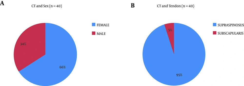
22). CT is more common in females (
2,
8,
18,
20,
21), similar to the current study. The female/male ratio was 2:1. However, Jacobelli argued that CT was equally prevalent in both genders (
8), while Hernandez-Díaz (
15) and Hsu et al. (
23) described a higher prevalence in males.
Chronic rotator cuff disease and CT are associated with work in awkward postures, lifting weights and performing repetitive movements. Recently, Chico-Alvarez (
1) reported that 34.7% of her patients were manual workers and 65.3% administrative. This is in line with the current study, where 40.9% were manual workers and 59.1% administrative.
CT affects the right shoulder (
1,
3,
8,
21,
24), although up to 25% to 30% can be bilateral (
9,
18,
24). In the current study there was greater involvement of the left shoulder (52.3%; n=23), rather than the right one (43.2%, n = 19). Bilateral involvement in 4.5% (n = 2), although most patients (93.18%, n = 41) were right handed. CT most often affects the supraspinatus tendon, then the infraspinatus and subscapularis (
8,
20,
24,
25). In the current series, CT affected mainly supraspinatus tendon (95%, n = 38), and to a lesser extent the subscapularis (5%, n = 2). Similar findings were published by Fernández-Cuadros et al. (
8) and Adamietz (
25).
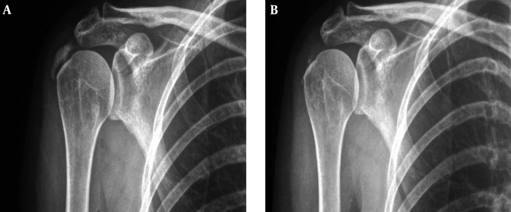
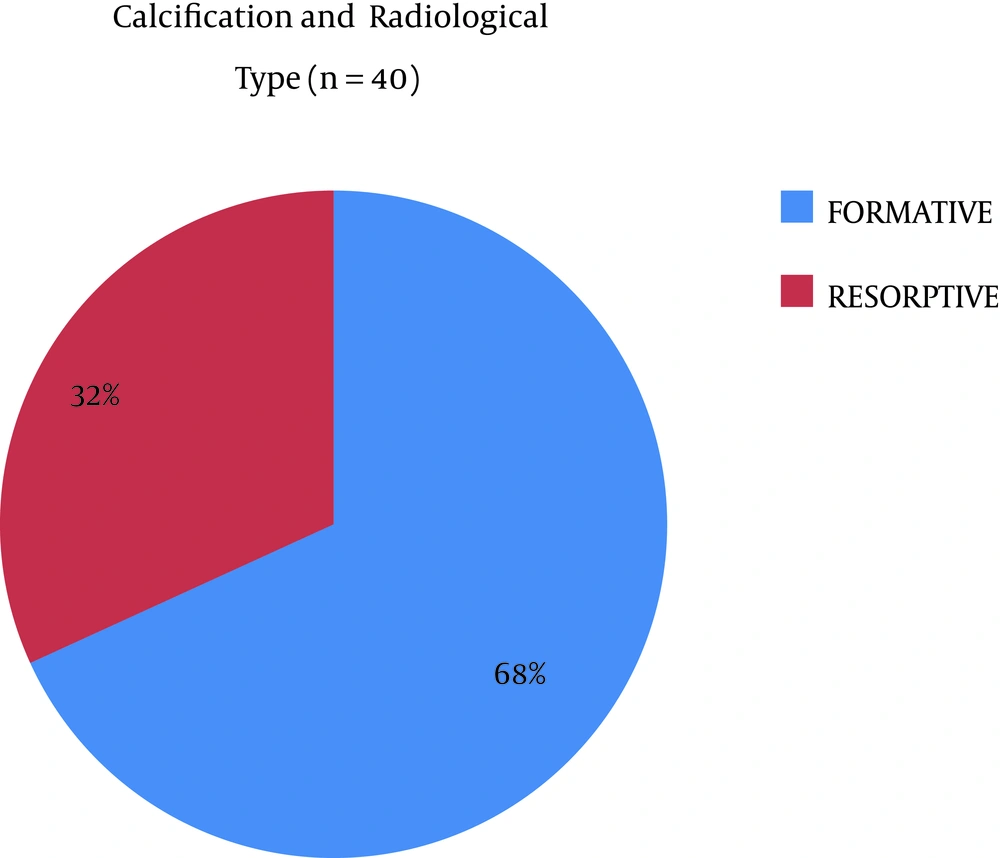
The radiological diagnosis was made by radiography or ultrasound. MRI was only performed for cases suspected with muscle/tendon pathology. The various radiological classifications agreed that type I corresponded to located deposits, well-defined contours, dense texture and homogeneous consistency; whereas type II corresponded to scattered deposits, not-well-defined contours, cloudy consistency and heterogeneous texture1. This radiological characterization was important because type II (resorptive) had a better prognosis than type I (formative) (
8).
CT treatment was controversial. A recent article demonstrated the effectiveness of different rehabilitation techniques to reduce pain but not the calcification (
8). Until a few decades ago, the only option used to treat calcification was acetic acid iontophoresis. However, there are only three classic papers that assess their effectiveness; one favorably (Rioja-Toro et al.) (
3), and two unfavorably (Perron and Leduc) (
13,
14). A recent doctoral study favorably supported the use of iontophoresis on CT (
1).
Iontophoresis is a noninvasive technique that increases the penetration of transdermal substances (drugs) through the skin layers (epidermis, dermis and hypodermis) in a controlled manner and by the application of electric current, based on physical-chemical principles of attraction and repulsion of charges (
26). Its most common applications are: 1) Calcifying tendinitis and myositis ossificans (acetic acid) (
27); 2) Controlling muscle spasms (calcium chloride and magnesium sulfate); 3) Inflammation (dexamethasone); 4) Soft tissue swelling (lidocaine); 5) Acute joint pain in rheumatoid arthritis (zinc oxide) (
26). This technique is cheap based on the economic use of topical medications and since the electrotherapy equipment decreased in size and are more accessible due to low-production-costs (
28).
Applying ultrasound to the skin increases its permeability and facilitates diffusion of different substances into the skin. This transport is called sonoferesis (
29). Low frequency sonophoresis works synergistically with iontophoresis; Le et al. (
30) used ultrasound 10 minutes prior to the application of iontophoresis, with good results.
In 1955 Psaki and Carrol introduced acetic acid iontophoresis as an effective treatment in shoulder CT (
13,
14,
31). Kahn (
31) considered that insoluble calcium carbonate precipitates could become soluble salts of calcium acetate, which could favor the reabsorption of the calcification: CaCO
3 + 2H (CaH
30
2)
2 = Ca (C
2H
3O
2)
2 + H
20 + CO
2. Furthermore, the use of ultrasound for their mechanical and thermal effects could help disintegrate and reabsorb calcification due to increase in local vascularization.
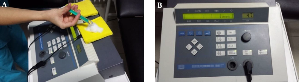
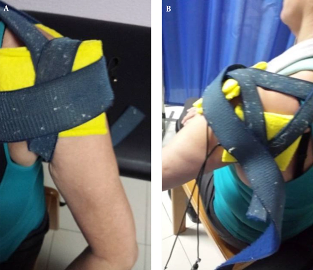
The current study protocol consisted in applying iontophoresis with 2 mL of 5% acetic acid, 4.7 mA × 10 minutes, followed by continuous ultrasound 1 W/cm
2/1MHz x 5 minutes. This protocol differed from those of Rioja-Toro et al. (
3) (3 mL of 5% acetic acid iontophoresis for 20 minutes at 4.7 mA plus ultrasound 1.5 W/cm
2), Perron (5% acetic acid iontophoresis (not specified amount) at 5 mA × 20 minutes followed by ultrasound 0.8 W/cm
2 × 5 minutes) (
13), Leduc (5% acetic acid iontophoresis, 20 mL at 5 mA × 15 - 20 minutes without ultrasound) (
14) and Chico-Alvarez (5% acetic acid iontophoresis (not specified amount) at 4.7 mA for 20 minutes without ultrasounds) (
1).
The average number of sessions in the current series was 19 (from 10 to a maximum of 40 sessions). The criterion to continue or discontinue treatment was the radiological evolution. Perron treated his patients for nine sessions (three sessions/week for three weeks) (
13). Leduc treated them for ten sessions (three sessions/week the first two weeks, and then weekly for four weeks) (
14). Rioja-Toro treated his patients for 40 sessions (five sessions per week) and evaluated them on 20th and 40th sessions (
3). Chico-Alvarez treated his patients in 15 - 30 sessions depending on the radiological evolution (five sessions per week) (
1).
The current study was quasi-experimental pretest-posttestd intervention. Rioja-Toro et al. (
3) conducte a similar experimental pretest-posttest intervention. Perrón (
13) conducted a randomized-experimental-study, similar to those of the Leduc et al. (
14) and Chico-Álvarez (
1).
To the best of authors’ knowledge, the present study supported the highest number of patients; the importance lies in the difficulty of prospectively collect such a number of patients. Perrón (
13) invested four years to find 21 patients; Leduc (
14), three years for 36 patients, but only 27 were used at the end. Rioja-Toro et al. (
3) recruited 34 patients in two years; and Chico-Álvarez (
1) spent a year to recruit 25 patients for each control group. The current study recruited 44 patients in a two-year prospective study.
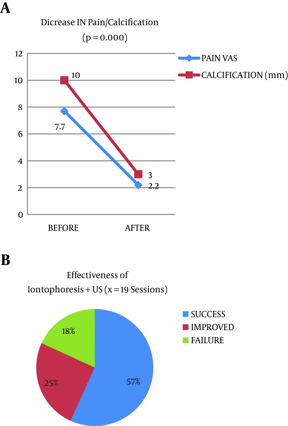
The study achieved a significant decrease in pain from 7.7 to 2.2 points on the VAS scale. This finding was similar to that of Chico-Álvarez (
1), who reduced pain from 5.7 to 2.7. Perrón and Malouin (
13) and Leduc et al. (
14) achieved improvements on pain, but not significantly. Rioja-Toro et al. (
3) reported 54% disappearance of pain and 25.6% improvement in his series. All studies (including the current study) showed that iontophoresis with acetic acid reduced pain in CT of the shoulder.
The current study treatment protocol significantly decreased the size of calcification from 10 mm (beginning) to 3 mm (the end). There was a 56.8% (n = 25) success rate (disappearance calcification 75% - 100%), a 25% (n = 11) improvement rate (decrease in calcification between 25% - 75%), and an 18.2% (n = 8) failure rate (less than 25% decrease). The obtained results were consistent with those of Rioja-Toro et al. (
3), who reported a 13.2% disappearance of calcification and a 46.4% decrease; that was a 59.6% modification of calcium deposits. Hernandez-Diaz reported a success rate of 43.5%, an improvement rate of 52.2% and a failure rate of 4.3% (
15). Chico-Alvarez observed a radiological decrease in calcification from 9.1 mm (start) to 5.7 mm (at the end of treatment), and 3.4 mm in six months follow-up. In the current series, the rapid decline in calcification was probably because ultrasound enhances, the absorption of acetic acid and the resorption thereof. Leduc and Perron obtained radiological improvement of calcium deposits after iontophoresis, although no difference was observed between the groups. These differences were probably due to the lower number of sessions (nine and ten respectively).
In a previous study with a similar sample size (n = 40) authors found that the different rehabilitation techniques could reduce pain but not the calcification (EVA 5.47 to 2.28, improving 3 points) (
8); therefore, authors wondered if pain was associated with calcification, for which a logistic regression study was conducted to see if calcification disappeared and pain diminished in the subjects. The findings of the current study confirmed the hypothesis. This explains that in a similar sample (n = 40) from a similar population (same geographical area), in which iontophoresis disappeared calcification, pain decreased from 7.7 to 2.2; improving 5.5 points on the VAS scale (more pain reduction than the cited article (
8)).
5.1. Study Limitations
An important limitation of the study was the lack of a control group. It was mainly due to the limited number of cases. A 2-year-follow-up was needed to collect such a sample size. As the effectiveness of acetic acid Iontophoresis on CT was accepted for decades and all patients accepted the treatment, it was not ethical to deny the intervention. A pretest-posttest intervention study was applied in this specific situation, to solve the lack of control group, and to give clinical based evidence.
As a contribution to the study, it should be stated that it was difficult to accomplish prospective studies capable of collecting a sample size similar to that of the current experimental study. It was even more difficult to combine clinical-care with teacher-researcher practice. In such studies, the final goal of doctors is the benefit of science, students and ultimately patients. Researchers are urged to conduct similar studies to reproduce the current study and increase consistency and demonstrable clinical-based-evidence, given the encouraging results observed in the study.
5.2. Conclusions
CT is more common in middle-aged working females. It affects the supraspinatus tendon. It is associated with smoking and occupational risks (awkward positions and lifting weights).
Iontophoresis with 5% acetic acid and ultrasound is a safe, simple and inexpensive technique capable to reduce pain and calcification on the shoulder.
The current study showed a level of evidence 2B and grade of recommendation B that allows authors to postulate acetic acid iontophoresis with ultrasound as an effective technique, with a recommended degree of scientific evidence.