


1. Introduction
The fracture of the scaphoid is the most frequent fracture of carpal bones. It is related to a fall over and extended and dorsiflexed hand. Seventy percent of fractures are located on its medial third (waist of the scaphoid). Transverse fractures are more frequent and stable than vertical ones. However, if fracture separation is bigger than 1 mm, fracture becomes unstable. Twenty percent of fractures are proximal and 10% are distal. As vascularization of scaphoid comes from distal side secondary to branches of a radial artery, that would explain why the more common complications of the fracture of the scaphoid are delayed nonunion of fracture and avascular necrosis (1-3).
Common signs of scaphoid fractures are a pain on snuffbox and loss of function on wrist movement. Treatment is conservative (immobilization) and surgical (fixation with a titanium screw). Unstable fractures lead to a 50% pseudoarthrosis. Further complications include a flexed scaphoid, carpal instability, arthrosis, carpal tunnel syndrome, and reflex sympathetic dystrophy (1-3). Because of complications such as the delayed union of scaphoid fracture and pseudoarthrosis, treatment still remains a challenge.
Pulsed electromagnetic fields (PEMF) have been used for decades as an alternative option in delayed bone healing fractures with good results. However, to the best of our knowledge, there are scarce reports, if any, on scaphoid fractures (4, 5). The objective of this case report is to show delayed union fracture as a common complication of scaphoid fracture and to postulate PEMF as an effective treatment option for such a condition, reflected by our clinical and radiological evaluations and supported by a thorough review of the literature.
2. Case Presentation
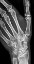
We present the case of a 30-year-old right-handed nurse who fell while skateboarding over her extended and dorsiflexed left hand. After the fall, she felt pain on her hand, but she made rest and took NSAID (non-steroidal anti-inflammatory drug). She did not go to an emergency room for further evaluation. She continued with her normal life. Three months after sports accident and because of persistent pain, she went to an orthopedic surgeon for further evaluation. On examination, the pain was elicited when palpating on snuffbox. An X-ray examination was ordered and a fracture of the middle third of the scaphoid with a line of fracture of 2 mm separation was observed (Figure 1). Because there was already 3 months from the accident, a delayed nonunion fracture of the scaphoid was diagnosed, surgical treatment was postponed, and referral to the rehabilitation department was ordered.

Radiography of the Left Hand Showing a Fracture of a Scaphoid Bone in the Middle Third, with a Line of Separation of 2 mm, Three Months After Sports Fall
In the Rehabilitation department, on examination, the patient remained with a complete range of movement on wrist and fingers, but the pain was elicited on palpation. Orthosis was prescribed in order to immobilize the articulation and PEMF was proposed as a conservative treatment. The patient received 20 sessions of PEMF, using the QS Magneto therapy device®, with a dose of 20Hz-50 Gauss-20 minutes, 5 times a week for 4 weeks. The left hand was placed inside the small applicator, and the solenoid encircled the affected limb. There was no need to take off the orthosis during treatment delivery (Figure 2).
 using the QS Magneto therapy device<sup>®</sup> (C), with a dose of 20Hz-50 Gauss-20 minutes (D).The left hand is placed inside the circled applicator. There was no need to take off the orthosis during PEMF application.")
Patient receiving PEMF (A and B) using the QS Magneto therapy device® (C), with a dose of 20Hz-50 Gauss-20 minutes (D).The left hand is placed inside the circled applicator. There was no need to take off the orthosis during PEMF application.
After 20 sessions, a new X-ray examination was ordered, complete union of fracture was observed, and orthosis was removed (Figure 3). The patient started to recover range of movement and on examination, no pain was elicited after palpation on snuffbox. The patient started using her left hand on activities of daily life; and at one-month follow-up, she remained pain-free.
 and splinting.")
Radiography of the left hand showing consolidation/union of the fracture of a scaphoid bone in the middle third, one month after PEMF (20 sessions, 20Hz-50 Gauss-20minutes, 5 times a week for 4 weeks) and splinting.
3. Discussion
Fractures of the scaphoid are the most common fractures of the carpal bones. Because of the distal perfusion of the bone, there is a great chance to present common complications such as avascular necrosis (up to 41%), delayed union of the fracture (up to 21%), and subsequently early osteoarthritis (up to 32%) (6). For those reasons, treatment of scaphoid fractures remains a challenge. To the best of our knowledge, there are scarce studies on the effectiveness of PEFM in scaphoid fractures and the results are very limited and even controversial (7).
The objective of this case report was to add evidence to the cases presented in the literature and to make a review of such an interesting and challenging topic, suggesting that although used for decades (since 1977), PEFM is still a valid option, inexpensive, and without side effects (8).
Delayed union is the absence of healing by the third month and nonunion is defined as the absence of healing at four to six months after injury (6). This might be due to delay in treatment, inadequate immobilization, localization of the fracture, instability due to the displacement of fragments or additional injury on carpal ligaments (6). In our case report at 3 months follow-up, no healing of fracture was observed; then, delayed union of scaphoid fracture was diagnosed. Once the diagnosis of nonunion was confirmed clinically and radiologically (Figure 1), immobilization with an orthosis and PEFM were prescribed (Figure 2).
Initially, PEFM was used in the treatment of tibial pseudoarthrosis and nonunion with a success rate of 87% (6, 9). Afterwards, Frykman started to use PEMF plus casts in non-united scaphoid fractures with a success rate of 80% (10).
Physical forces have been used in the healing of fractures since the 1970s. These physical forces may be displayed by direct current, PEMF, and ultrasound (6). Inductive-coupled electromagnetic fields have been used in Medicine since 1974 (11). The fundamentals are that physical forces (and PEMF) stimulate osteogenesis and formed callus around the cathode (12, 13).
Although reports on the use of PEMF comes from studies in the 1970s, its use is relatively a newly born option for the treatment of pathological conditions, such as musculoskeletal system disorders (14, 15). In bone healing, PEMF has demonstrated high effectiveness. PEMF enhances endogenous bone and osteochondral repair, incrementing bone mineral density, accelerating osteogenic differentiation in nonunion cells, and controlling the degenerative disease-related pain and cartilage damage (16).
PEFM can penetrate through highly resistant structures such as bone, fat, skin, or even plaster cast, like in our case report. Then, PEFM constitutes a practical exogenous method for cell and tissue modification (17). Electromagnetic fields cause biological changes to the cell environment and restore cell integrity and function. Moreover, PEMF increases the membrane potentials of erythrocytes, increases oxygen delivery, vasodilates blood vessels, and relieves pain without increasing local temperature. PEMF reduces pain and edema after soft tissue injury (15, 18).
PEMF has shown to reduce inflammatory processes having a marked anti-inflammatory effect in several cell lines and in vitro models (16). This would explain the reduction in pain in our case report. The analgesic effect of PEMF therapy might be due to presynaptic inhibition or decreased excitability of pain fibers (19). PEMF is capable of modulating pain, since PEMF may lower the membrane to a hyperpolarization level of about -90mV, then it might block transmission of pain signals (15).
On the other hand, PEMF might modulate the actions of hormones, antibodies, and neurotransmitters surface receptors sites of different cell types, including osteocytes (20). PEMF has anabolic properties. PEMF influences ATP production, increases oxygen and nutrient supply, and improves the removal of waste metabolites via the lymphatic system, and it is responsible for a rebalancing of ions across the cell membrane. All these properties might reduce pain and muscle spasm (21).
All previous anabolic and modulation properties of PEMF have made this modality as a rapid and emerging alternative in the management of bone healing without side effects, unlike drugs (15, 22). In fact, in our case report, the 2 mm nonunion of fracture disappeared and bone healing occurred after 20 sessions exposure to PEMF (5 times/week). The healing of the fracture is in accordance with the studies of Frykman (10) who reported an 80% rate of success, and it also comes in line with the publication of Bora, who claimed a 71% reduction in nonunion (23).
Therefore, PEMF might be considered a viable therapeutic and mini-invasive approach to stimulate bone formation and healing, to control pain, and to counteract the inflammatory pathways triggered by osteolysis (16), as it was observed in our delayed union of the scaphoid fracture.
3.1. Conclusion
PEMF is a safe technique that has shown a promising therapeutic effectiveness in the healing of delayed union of scaphoid fracture, a common complication that becomes a challenge for its difficult management; since untreated, it might lead to nonunion and late osteoarthritis. PEMF could recover delayed nonunion of scaphoid fracture and decreased pain as evaluated clinically and radiologically in our case report.
