


1. Introduction
Lipoma represents the most common soft tissue neoplasm, which is 0.1% - 0.5 % of all benign tumors of the oral cavity (1, 2). The pathogenesis of lipoma is indistinctive and not associated with fat metabolism in the body (1). The buccal mucosa and buccal vestibule are the most common sites for oral lipomas and the tongue, the floor of the mouth and lips are the less common areas, respectively (1). This tumor occurs mostly in the 40-year-old or older patients and has no sex distribution (1). There are many microscopic variants of lipoma such as fibrolipoma, angiolipoma, spindle cell lipoma, pleomorphic lipoma, intramuscular lipoma, and sialolipoma; however, the most common type is fibrolipoma (1).
2. Case Presentation
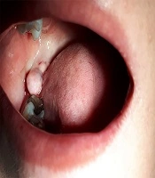
In this study, two cases of fibrolipoma lesions in two women who were admitted at the Oral Medicine Department of Semnan Dental School are reported. The first patient was a 36-year-old woman who had noticed a swelling in her mouth since the year before. Her weight and systematic review were normal. The extraoral examination was not specific, but intraoral examination showed an exophytic mass with a soft consistency, pale pink and smooth, intact surface, approximately 1 × 2 cm in dimensions. It was located in the retromolar pad area on the right side of the mandible (Figure 1). Due to the patient being asymptomatic during this period, she had not received treatment. After obtaining consent, the lesion was removed by electrocautery to prevent bleeding and fortunately, significant bleeding did not occur. The sterile compression pack was sufficient and there was no need for suturing.

The first case of a 36-year-old woman with fibrolipoma preoperative
The second case was a 71-year-old woman who had already noticed the lesion for several years. Type 2 diabetes, hypertension, aspirin consumption, and overweightness were mentioned in history. The extraoral examination was normal, but a 3.5 cm × 1.5 cm exophytic mass was seen intraorally, with a pink, smooth, intact surface located in the pterigomandibular raphe, which had a very soft consistency. Informed consent was obtained and the lesion removed by electrocautery, too. There was no significant bleeding, despite aspirin use, but the suture was closed to prevent probable bleeding.
In both cases after surgery, the specimen was sent to the Oral and Maxillofacial Pathology Department for histopathologic examination, and the result was fibrolipoma (Figure 2). Both patients were asked to refer to the clinic if the lesion was not properly removed or was recurrent, but they have not come back after 6 months.

Buccal fibrolipoma 100×, H & E staining
3. Discussion
The lipoma is a benign soft tissue tumor, composed of mature lobulated adipocytes which are limited by a fibrous band (3). Clinically, alipoma presents as a solitary painless mass which is slow-growing and has a characteristic yellowish color with soft, doughy impression (4). The lesion is typically smooth and non-ulcerated at the surface unless it has been traumatized (4). This tumor is rare in the oral cavity and most seen in the buccal mucosa, lips, and tongue (1-5). Here, we reported two cases, one of them in the retromolar pad area and the other one located in pterygomandibular raphe. Lipomas often present in adults with a mean age of 60 years and are uncommon in children. Lipomas are frequently associated with chronic trauma (3, 4) family history, and diabetes (4). In this report, one of the patients was diabetic. Although numerous histologic variants are defined for lipoma, the most common microscopic variant of oral lipomas is the fibrolipoma (4). Fibrolipoma is characterized by a remarkable fibrous element intermixed with lobules of adipose tissue (5). There are several microscopic variants of lipoma including spindle cell lipoma, intramuscular or infiltrating lipoma, salivary gland lipoma, and myxoid lipoma (3, 6). The consistency of such tumors depends on the depth of the lesion and the quantity of fibrous tissue and it differs from soft to firm (5).
There are several lesions including lymphangioma, angioma, schwannoma, pyogenic granuloma, minor salivary gland neoplasms and liposarcoma, which are considered as the differential diagnosis of fibrolipoma (7). As a consequence of having a greater proliferative rate in fibrolipoma than other types of the lipomas, the accurate diagnosis of this variant is necessary in order to differentiate the lesion from atypical lipomatous tumor/well-differentiated liposarcoma (ALT/WDLS) (7, 8). Moreover, oral liposarcoma and its benign counterpart cannot be distinguished from each other at the clinical examination; hence, precise microscopic examination is obligatory. The differential diagnosis is most importantly based on the presence of lipoblasts in a variable amount in liposarcoma and also a lack of lobular architecture and areas of prominent fibrosis (8).
The treatment of fibrolipoma is entirely surgical (9, 10) but the use of diode laser surgery for oral fibrolipoma has been reported recently, which has several advantages in comparison with conventional blade surgery including the lack of bleeding, no requirement for suture and faster scar healing (7, 9, 10). In these two cases, we used an electro cutter in order to remove the lesions. Although recurrence is rare, shcedulingfollow-up sessions is mandatory (3). Additionally, regressive tissue changes because of the thermal effect of the diode laser are frequently insignificant; therefore, allowing sufficient microscopic examination and accurate diagnosis (9).
3.1. Conclusions
We reported two cases of fibrolipoma that have occurred in infrequent areas such as the retromolar pad area and pterygomandibular raphe. The lesions were removed by an electro cutter with no recurrence at the follow-up sessions. Although fibrolipoma is pretty rare in the oral cavity, there are several lesions that can also have similar clinical features. Therefore, it is recommended that a clinician perform the histopathological examination for the final diagnosis.
