

1. Introduction
Blunt or sharp trauma may cause damages to or may penetrate different parts of the heart (1). Right ventricle trauma is the most common damage, which may be associated with other cardiac cavities (2). Many cases of penetrating trauma have been recorded, such as glass, sharp metals, bullet, and needle, while mainly led to significant damages to the anterior place of the right ventricle (3). Although penetrating trauma may be asymptomatic, but its most common symptoms include dyspnea and chest pain. Since needles are sharp, if left untreated, they can easily migrate to adjacent tissues, leading to hemothorax, cardiac tamponade, and pneumothorax. Even in the absence of migration, it may cause the formation of mural thrombi, which leads to repeated embolization. Rarely, valvular regurgitation may be observed, especially when the needle is embedded in the ventricular septum. Besides, in longstanding cases, it may cause complications such as infective endocarditis. Therefore, removing foreign bodies is essential. However, in most cases, open-heart surgery with CPB is required for removing foreign bodies (4). From 1967 to 2013, there were only 40 reported cases of sewing needle penetration in the heart (4). In the present study, the target case is a rare abnormal needle movement from the right thorax to the right ventricular apex, who referred with pulmonary manifestations of the lung. Following necessary diagnostic tests, a surgery was performed to remove the needle from the patient’s chest, without open-heart surgery and CPB.
2. Case Presentation
The patient is a 3 years old girl who was complaining of right chest pain and mild shortness of breath, while fell on the house carpet two days ago. The pain was in the front of the right thorax, which spread to the right shoulder of the patient, and the breath shortness of the patient became more severe as acting. The patient had no history of heart surgery or any specific disease. In clinical examinations, all vital symptoms were normal, and there was no sign of cardiac tamponade, except reduced sound in the right lung base. Besides, the external object was retained in the midclavicular line between the third rib, which was significant. In the Simple posterior-anterior and lateral graphics of chest radio opacity linear image on the lower part of the heart, the shadow was also significant. Echocardiography was performed to confirm the location of the foreign body and an ocular linear object resembling a large needle was observed in the right ventricular cavity (Figure 1).

Existence of a foreign object in a spiral CT scan
The CT scan spiral lung without contrast revealed a metal linear density image in the lower anterior part of the chest. The CT also showed that the needle pathway is in developing from the anterior part toward the posterior and tangent on the diaphragm. Therefore, first, the surgery was performed on the anterior thoracotomy fourth intercostal of the right side. The external object was not found after removing about 150 CC of blood and clots and lung exploration. Hence, the pericardium on the right longitudinally was opened, and the mediation and heart were explored. After observing the effects of the needle near the right ventricular apex and cutting on the site, the needle came out about 3.5 cm in size and after repairing the ventricle and closing the pericardium partially and placement of the chest tube in the right thorax, thoracotomy was closed. Finally, 3 days after the operation, the patient was discharged from the cardiac surgery department in good general condition (Figure 2).

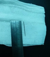
Sewing needle removed from right ventricle after surgery
3. Discussion
Foreign bodies damage the heart and can migrate to adjacent tissues through the venous system or direct penetrating trauma, which mainly occurs in the right ventricle (5, 6). Signs and symptoms of foreign body penetration in the heart depend on the shape, size, and entering the location of the foreign object. However, it may be asymptomatic or include false aneurysm, cardiac tamponade, embolism, conduction disorders, etc. (7). Probable risks of the foreign body determine the necessity of using surgery, such as Pericardial effusion, bleeding, valve damage, embolism, etc. (8). In the current study, the patient was emergently operated due to the abnormal and rapid movement of the needle and the possibility of damaging to valves and heart cavities. Early diagnosis and intervention are always life-saving. These objects should be removed immediately by surgical procedures. Determining the exact location of the foreign body in the heart is important to ensure its complete removal. Moreover, it helps to avoid open-heart surgery and using CPB.
