1. Background
Methylphenidate (MPH), which is also known as Ritalin, is a Central Nervous System (CNS) stimulant. This agent is a common effective drug for Attention Deficit Hyperactivity Disorder (ADHD). This disorder is a highly prevalent psychiatric disease among children (1, 2) with a worldwide prevalence of 5.29% (3). Based on the statistics, the prevalence of this condition is 2-18% in the United States (2). While the short-term use of this mediation is safe in children suffering from ADHD, its long-term impacts on different systems, including the reproductive system, are a matter of concern. Male factors play important roles in the infertility of many populations, imposing a notable financial burden on patients and governments (4).
Reproductive impairments are reported as the adverse effects of several medications.(5) The adverse effects of MPH on the male reproductive system have been reported in several studies. Some of these adverse effects include decreased testes weight, hormone levels, spermatogenesis rate, quality of sperms, and germinal cell function (6-11). The growing prevalence of ADHD and the non-medical use of MPH among the youth (12) have lead male infertility to be a major health problem (4).
2. Objectives
With this background in mind, the current study was performed to investigate the impacts of MPH administration on important parameters of productivity in male rats. These parameters included total body weight, testis weight, spermatogenesis, sperm motility, histopathologic changes, and sex hormone serum levels.
3. Methods
3.1. Study Population
This study was conducted on 54 Wistar male rats from November 2017 to March 2018 at the Animal Laboratory of the Medical Faculty of Mashhad University of Medical Sciences (MUMS), Mashhad, Iran. The rats were randomly divided into three groups of 18. They aged eight weeks with an average weight of 250 g. The rats were obtained from the Animal Laboratory of the Medical Faculty of MUMS. Before and after the study, they were examined by a veterinarian, and none of them had any health issues. In all steps of this study, the animals were treated as per the protocols of animal studies. They were kept in a standard temperature and humidity condition. Hydration and nutrition were adequate, and they were all normal rats.
3.2. Procedures
For 60 days, the control group (group C) received 1 ml of normal saline solution by the gavage method at 6 p.m. The experimental groups, namely low-dose (LD) and high-dose (HD), were gavaged with 2 and 10 mg/kg of MPH, respectively, at 7 p.m. for the same period. Gavaging was implemented using particular syringes in all groups (13).
3.3. Research Evaluations
On the 61st day, rats were taken out of their cages under required conditions, and then weighted and completely anesthetized in an ether container. After obtaining blood samples from the carotid artery in dry tubes, they were instantly sent to a hormone laboratory. In the next stage, the samples were centrifuged, sera were removed, and hormonal levels were measured by special kits. The levels of testosterone, Luteinizing Hormone (LH), and Follicle-Stimulating Hormone (FSH) were measured, as well.
Midline abdominal incision was performed, and the testes and epididymis were excised completely. Subsequently, the two ends of the epididymis were closed, kept in 2 mL of normal saline solution at 37ºC, and rapidly transferred to the laboratory for spermogram assessments, including sperm count and motility. Other reproductive organs, including prostate and seminal vesicles, were also excised. The samples were sent to a pathologist in separate particular containers of formalin 10% solution. The recorded data included the testes weight and histopathologic properties, including edema, congestion, necrosis, maturation arrest, Leydig cell hyperplasia, and decreased spermatogenesis. The study protocol was approved by the Medical Ethics Committee of MUMS. Besides, MPH caused no toxicity to rats.
3.4. Statistical Analysis
The biochemical and histopathologic data were analyzed by SPSS software. Group features were also analyzed using descriptive statistical methods, including central indicators of dispersion and frequency distribution. The normality of data was tested using the Shapiro-Wilk statistical test. Then, appropriate tests were used according to the normal distribution of data. The ANOVA and chi-square tests were used for the ordinal qualitative and morphologic data, respectively. A P value of less than 0.05 was considered statistically significant.
4. Results
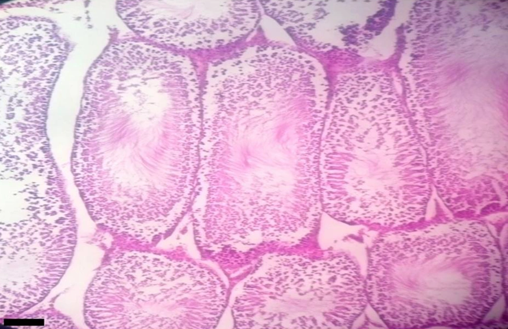
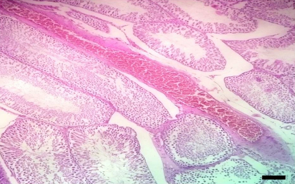
Three groups of 18 rats with an average age of 60 days and a weight of 250 g were used in this study. The results indicated a significant difference between the control and experimental groups in LH and testosterone levels (P = 0.007 and P < 0.001, respectively), but not in FSH. Table 1 presents the differences between the groups. Concerning spermographic variables, the sperm count and motility were significantly higher in both of the experimental groups than in the control group (P < 0.001 for both). Nevertheless, there were insignificant differences between the two experimental groups in this regard. Table 1 tabulates the mean values of the variables. The body weight (P = 0.022) and testis weight (P = 0.021) were lower in the experimental groups than in the control group. The LD group (2 mL/kg) had no significant difference with the control group in terms of the mentioned variables. However, the HD group (10 mL/kg) was significantly different from the control group in this respect (Table 1). In addition, there was no significant difference between the LD and HD groups regarding the body weight and testis weight. Histopathologic studies revealed significant changes in Leydig cell hyperplasia (P = 0.001). These variations included a decrease in spermatogenesis, testis congestion, and necrosis in both of the experimental groups and the control group (P = 0.013, P < 0.001, and P < 0.001, respectively). Nonetheless, no significant changes were observed in maturation arrest and edema (Figures 1 and 2). There were four pathologic stages (stage 0 - 3) for each variable. Table 2 presents the distribution of variables in stages.
| Variable | Groups | Mean ± SD | P Valuea | ||
|---|---|---|---|---|---|
| Between Each Case Group and Control Group | Between LD and HD | Overall | |||
| LH | LD | 0.1951 ± 0.048 | 0.105 | 0.549 | 0.007 |
| HD | 0.2081 ± 0.037 | 0.008 | |||
| C | 0.1689 ± 0.014 | - | - | ||
| FSH | LD | 0.1322 ± 0.065 | - | - | 0.08 |
| HD | 0.1356 ± 0.067 | ||||
| C | 0.08586 ± 0.084 | ||||
| Testosterone | LD | 139.16 ± 58 | 0.04 | 0.2 | < 0.001 |
| HD | 107.83 ± 58 | < 0.001 | |||
| C | 183.66 ± 54 | - | - | ||
| Sperm count, million/mL | LD | 15.77 ± 1 | 0.002 | 0.566 | < 0.001 |
| HD | 11.54 ± 0.7 | < 0.001 | |||
| C | 30.55 ± 1.6 | - | - | ||
| Sperm motility. /mL | LD | 1.44 ± 2.06 | < 0.001 | 0.23 | < 0.001 |
| HD | 0.11 ± 0.32 | < 0.001 | |||
| C | 12.27 ± 3.4 | - | - | ||
| Rat weight, g | LD | 315 ± 35 | 0.717 | 0.15 | 0.022 |
| HD | 299 ± 29 | 0.027 | |||
| C | 328 ± 27 | - | - | ||
| Testis weight, g | LD | 1.42 ± 0.35 | 0.756 | 0.123 | 0.021 |
| HD | 1.33 ± 0.29 | 0.027 | |||
| C | 1.45 ± 0.27 | - | - | ||
aAll P values in this table resulted from the ANOVA test.
| Variable | Groups | Stages | P Valuea | |||
|---|---|---|---|---|---|---|
| 0 | 1 | 2 | 3 | |||
| Leydig cell hyperplasia | LD | 18 | 0 | 0 | 0 | 0.001 |
| HD | 12 | 6 | 0 | 0 | ||
| C | 18 | 0 | 0 | 0 | ||
| Decreased spermatogenesis | LD | 4 | 8 | 6 | 0 | 0.013 |
| HD | 6 | 6 | 4 | 2 | ||
| C | 13 | 5 | 0 | 0 | ||
| Maturation arrest | LD | 18 | 0 | 0 | 0 | 0.191 |
| HD | 16 | 0 | 0 | 2 | ||
| C | 17 | 1 | 0 | 0 | ||
| Edema | LD | 4 | 12 | 2 | 0 | 0.136 |
| HD | 4 | 12 | 2 | 0 | ||
| C | 10 | 8 | 0 | 0 | ||
| Testis congestion | LD | 0 | 18 | 0 | 0 | < 0.001 |
| HD | 0 | 12 | 6 | 0 | ||
| C | 7 | 11 | 0 | 0 | ||
| Necrosis | LD | 16 | 2 | 0 | 0 | < 0.001 |
| HD | 7 | 11 | 0 | 0 | ||
| C | 18 | 0 | 0 | 0 | ||
aAll P values in this table resulted from the chi-square test
5. Discussion
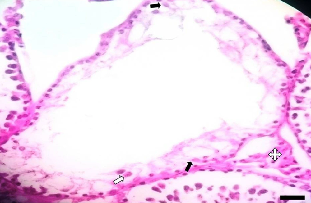
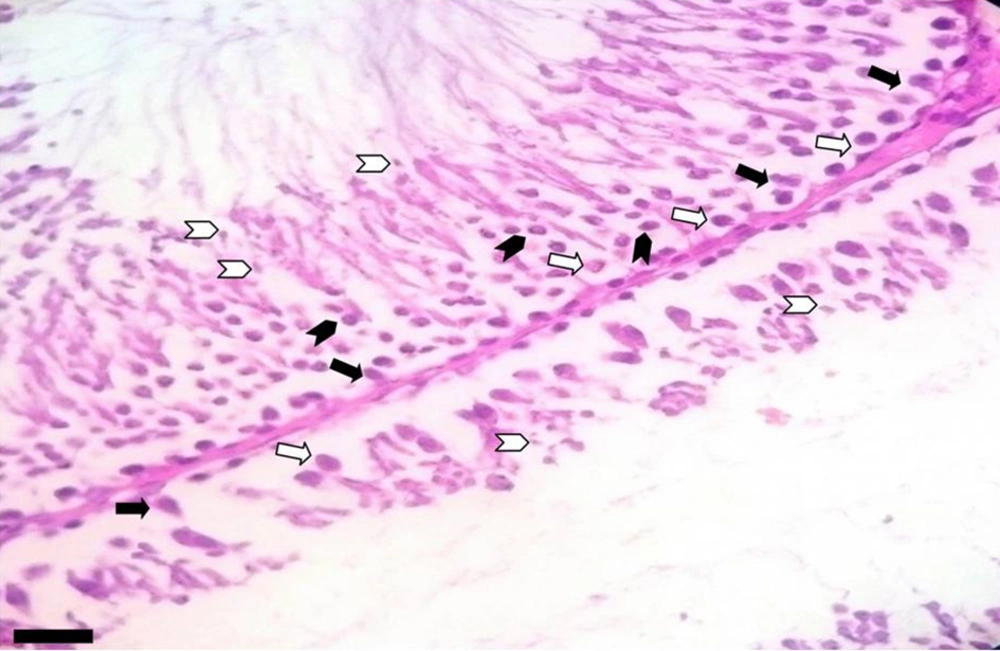
The results of the present study indicated significant differences between the MPH and experimental groups in terms of hormonal, spermographic, and histopathologic characteristics, as well as weight. In this regard, no significant difference was observed between experimental groups regarding LH and testosterone levels, sperm count and motility, Leydig cell hyperplasia, spermatogenesis, congestion and necrosis levels, total body weight, and testis weight. However, there was no significant difference between the control and the experimental groups considering FSH, maturation arrest, and edema levels. The HD and control groups were significantly different in terms of LH levels, body weight, and testis weight (Figures 3 and 4). However, no such difference was observed between the LD and control groups. In addition, there was no significant difference between the two experimental groups in terms of any of the evaluated variables.
The current study revealed a lower body weight in the high-dose MPH group, which is in line with the findings of several studies (7, 14). In this regard, Carias et al. reported a decrease in food intake and body weight in rats treated with high-dose MPH. They also observed a decrease in water intake in both high- and low-dose groups. In the mentioned study, 8 and 30 ng/ml levels were considered as the peak serum concentrations in the low- and high-dose groups, respectively (15). Furthermore, Robison et al. observed that high-dose MPH decreased total weight in male and female rats while this treatment increased food intake only in females. A possible explanation for this result is that the compensation of energy loss resulted from MPH-induced hyperactivity (16). Other trials that treated rats with low doses of MPH reported insignificant weight alterations (9, 17). Similarly, our results indicated a significant difference only in the high-dose group. Based on our findings and those of the previous studies, it seems that there are some dose-dependent relationships between treatment and weight loss (17-19). Similar results have been reported in human studies. Weight reduction is the most common adverse effect of MPH in adults (20, 21). These findings may explain the relationship between obesity and ADHD (22), the role of MPH in the treatment of ADHD-related obesity (23), and the possible mechanism of decreased appetite due to the drug (24). Several pieces of evidence have shown a transient decrease following MPH administration in both humans (19, 25-30) and animals (17, 18, 31), which makes a rebound after treatment cessation (11). In line with the present results, previous studies have demonstrated a decrease in the testis weight, in addition to the prostate and seminal vesicles weight following MPH treatment in rats (11, 32). Teo et al. reported a rebound weight gain in the prostate 30 days after the last MPH administration (18). Montagnini et al. observed no significant alteration 40 days after the experiment (9). According to the blocking effect of MPH on Noradrenaline (NA) and Dopamine (DA) transporters, the presence of DA and NA receptors in testicular tissue may directly affect MPH on the testis (33, 34). Furthermore, germ cell depletion has been mentioned as the possible role player in the literature (35). The results obtained by Fazelipour et al., investigating hormonal changes, namely elevated LH, decreased testosterone, and insignificant changes of FSH, are relatively in line with our findings. They believe that despite the role of reduced testosterone secretion by Leydig cells and the subsequent rise in LH in these changes, the increased liver metabolism of the testosterone-producing enzyme could be the point and the insignificant elevation in FSH reinforces this hypothesis (6, 7, 36). The literature is indicative of the effect of MPH on pulsatile gonadotropin-releasing hormone (GnRH) release (36) and direct impairment of Leydig cells (6). However, there are also reports on the transient negative effect of MPH on testosterone, as well as body and testis weight (6, 37). There are multiple studies indicating spermatogenesis and spermiogenesis impairments, including decreased sperm count and altered morphology (7, 9, 11, 38). In an investigation carried out by Cansu et al. (11), more negative effects were observed in the high-dose group, which is in line with the dose-dependency of some MPH adverse effects. They ascribed this alteration to the increased p53 immunoreactive cell number in the high-dose group, higher testis apoptotic cell count, and lower Transforming Growth Factor (TGF)-β1 activity in high- and low-dose groups. TGF-β1 has both stimulatory and inhibitory effects on cell proliferation and spermatogenesis in rats (39-45). Nonetheless, it is not present in human seminiferous ducts (46). MPH affects dopamine transport, D2 receptors, serotonin, and noradrenaline (31, 35, 47), and D2 and α- and β-adrenergic receptors are expressed in the testis and spermatozoa of rat (33, 34). The toxicity caused during spermatogenesis could lead to apoptosis (48, 49). The increased apoptosis and p53 expression are discussed as the causes of sperm count decrease in some other studies (7, 11, 38). Due to the effect of the GnRH level on FSH (50) and the function of FSH in the primary stages of spermatogenesis (51-53), FSH changes could disrupt the spermatogenesis process. However, no significant difference was observed in the FSH levels in the current study and the one performed by Fazelipour et al. (7). In the present study, Leydig cell hyperplasia was observed in the HD group, which is contrary to the previous findings indicating the decreased Leydig cell count (8) or the absence of significant changes (9). Accordingly, further studies are needed to explain the exact effect of MPH on Leydig cells. Necrosis was another significantly increased parameter in the MPH groups. Necrosis has been rarely evaluated and discussed in the literature. Intraperitoneal injection of 30 mg/kg of hydrochloride cocaine is reported to cause necrosis and decrease the number of testis interstitial cells (54). In a study carried out by Cansu et al., necrosis was not detected (11). The responsible mechanism may be similar to the one explained for apoptosis. This finding also highlights the importance of investigating the cytotoxic effects of MPH. Congestion is also a matter of controversy that was considered in the present study. While congestion was higher in the MPH group, some studies did not detect significant congestion (11) or any associated degrees (7). To indicate limitations and strengths, we investigated a wide range of variables, including hormonal, spermatographic, and histopathologic characteristics, as well as weight. Furthermore, the two LD and HD groups were examined to illustrate dose-dependent changes. Chronic exposure could be another positive point in this regard. Some delayed or rebound effects were not presented at the termination point of medication in the study. Therefore, it is necessary to evaluate rats after medication termination. Additionally, given the probable role of weight loss in sexual dysfunction, this variable should be further investigated. In this respect, changes in weight, not the final weight, may be more accurate to assess the consequences of MPH therapy.
5.1. Conclusions
Sexual hormones, especially, methylphenidate have the potential to cause infertility. These changes may result from the effect of drugs on the central nervous system, including hypophysis or hypothalamus, the direct effect of drugs on the testis, or both of them. These findings suggest that methylphenidate must be used just in indicated patients, and the overuse of this drug can increase the infertility rate in society. Awareness about its adverse effects may decrease the infertility rates.


