1. Background
Following the increased cases of pneumonia of unknown cause in Wuhan, China, in December 31, 2019, a novel coronavirus (severe acute respiratory syndrome coronavirus 2 (SARS-CoV-2)) was identified as the cause of the disease, which has spread rapidly throughout the world (1). SARS-CoV-2 infection was responsible for the disease that was known and named by World Health Organization (WHO) as COVID-19 (2). Until march 31, 2021, a number of 128,104,216 confirmed COVID-19 cases were reported worldwide (3). The number of confirmed patients and deaths in Iran were 1875234 and 62569, respectively (4). The severity of the disease ranges from mild to moderate in approximately 80% of infected patients, and severe and critical conditions are seen in around 20% of the remaining cases. Patients’ clinical manifestations might vary from asymptomatic to patients with symptoms and signs of fever, cough, fatigue, sore throat, shortness of breath, and even respiratory failure and acute respiratory distress syndrome (ARDS) in severe forms of the disease have been reported (5, 6). Severe cases were mostly patients with shortness of breath, respiratory rate (RR) over 30/min, resting blood oxygen saturation less than 93%, and PaO2/FiO2 less than 300 mmHg. When radiographic findings of pneumonia show progression in more than 50% of chest CT scans during 24 - 28 h, it is also considered a severe form of the disease. Critical cases had dyspnea with the necessity for mechanical ventilation, shock, multiple organ failure, and they always required ICU care. The most common chest CT scan findings in COVID-19 include patchy ground-glass opacities (GGOs), consolidations, interlobular septal thickening, crazy paving appearance, fibrotic streaks, and strip-like opacities (7). Comorbidities, like chronic kidney disease (CKD), diabetes mellitus, and hypertension are associated with the severe form of the disease, and CKD increases mortality in COVID-19 patients (8). The immune deficiency associated with CKD makes these patients prone to this highly contagious disease (2, 6, 9). Therefore, they are at higher risk of severe disease and higher mortality (9, 10).
In a study conducted in Spain, mortality was higher than 30% of the general population, and the patients had less fever, weakness, lethargy, and cough (10). Imaging and symptoms of these patients might be different from the general population. Due to the fact that hemodialysis patients are older and they almost always have an underlying disease, they are more likely to be infected, and subsequently, they could be a source for spreading the virus in the community (5).
2. Objectives
This study aimed to determine the clinical and para-clinical findings in hemodialysis patients and compare these characteristics to patients without a history of kidney disease.
3. Methods
3.1. Study Population and Design
This was a case-control, internal multicenter study conducted on 72 COVID-19 patients confirmed by oropharyngeal and nasopharyngeal polymerase chain reaction testing. We collected data from three hospitals affiliated with the Zahedan University of Medical Sciences (ZUMS), south-eastern Iran (Boo Ali Hospital, Ali Ibne Abitaleb Hospital, and Khatam Hospital) from February 2020 to December 2020. The study was approved by the ZUMS ethics committee (IR.ZAUMS.REC.1399.145). During this period, 36 end-stage renal disease (ESRD) patients had the inclusion criteria. During the same period, patients without renal insufficiency who were matched in age and sex and had the underlying disease were included in the study as a control group.
Thirty-six ESRD patients older than 18 years with COVID-19 under hemodialysis (HD) were compared to 36 patients without any history of renal diseases. Patients in the case group were on HD for at least three months prior to admission. Patients in the control group had no history of kidney disease with a glomerular filtration rate of more than 90 percent. The cases and controls were matched according to age, sex, and other underlying diseases. To confirm COVID-19, nasopharyngeal/oropharyngeal swab testing was performed for all patients using SARS-COV-2 real-time reverse transcriptase PCR. Data, including demographic findings, history of comorbidity diseases, vital signs, and oxygen saturation, as well as laboratory tests, such as hemoglobin and white blood cell, lymphocyte, and platelet counts, were collected from the patients’ medical files.
All the clinical, laboratory and low-dose high-resolution computed tomography (HRCT) scan findings were registered in the information forms.
All the cases and controls were evaluated at the time of discharge and two weeks later, and the data from their clinical findings were collected as well.
3.2. Statistical Analysis
Descriptive statistics were used to summarize the data, and results were reported as means and standard deviations. The categorical variables were summarized as numbers and percentages. We conducted comparisons of baseline characteristics, symptoms, laboratory findings, and CT findings between the two groups using a t-test for continuous variables and a Chi-square test or Fisher’s exact test for binary and categorical variables. Statistical analyses were conducted using SPSS v.16 software, and P-values smaller than 0.05 were considered statistically significant.
4. Results
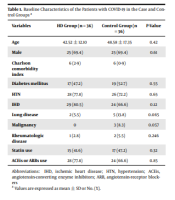
The results of the study showed that the mean age of HD patients and the control group was 42.52 ± 12.10 and 48.58 ± 17.35 years, respectively. In both the case and control groups, 25 (69.4%) patients were male, and the remaining were female. The number and percentage of underlying diseases, including diabetes mellitus, hypertension, ischemic heart disease, pulmonary and rheumatologic diseases, as well as the number of patients using statins, angiotensin-converting enzyme inhibitors, and angiotensin receptor blockers in both the case and control groups are demonstrated in Table 1. Charlson’s comorbidity index in HD patients and controls was 6. As shown in Table 1, there was not a significant difference between the control and HD groups regarding the mentioned factors (Table 1).
| Variables | HD Group (n = 36) | Control Group (n = 36) | P-Value |
|---|---|---|---|
| Age | 42.52 ± 12.10 | 48.58 ± 17.35 | > 0.05 |
| Male | 25 (69.4) | 25 (69.4) | > 0.05 |
| Charlson comorbidity index | 6 (2 - 9) | 6 (0 - 9) | |
| Diabetes mellitus | 17 (47.2) | 19 (52.7) | > 0.05 |
| HTN | 28 (77.8) | 26 (72.2) | > 0.05 |
| IHD | 29 (80.5) | 24 (66.6) | > 0.05 |
| Lung disease | 2 (5.5) | 5 (13.8) | > 0.05 |
| Malignancy | 0 | 3 (8.3) | > 0.05 |
| Rheumatologic disease | 1 (2.8) | 2 (5.5) | > 0.05 |
| Statin use | 15 (41.6) | 17 (47.2) | > 0.05 |
| ACEis or ARBs use | 28 (77.8) | 24 (66.6) | > 0.05 |
Baseline Characteristics of the Patients with COVID-19 in the Case and Control Groups a
Fever was detected in 32 patients in the control group and only 19 cases on HD. Cough and dyspnea were observed in 23 and 24 HD patients and in 36 and 34 patients in the control group, respectively (P < 0.05) (Table 2). There was no statistical difference for other clinical findings, like sore throat, myalgia, anosmia, and vomiting.
| Clinical Presentation | HD Group (n = 36) | Control Group (n = 36) | P-Value |
|---|---|---|---|
| Cough | 23 (63.8) | 36 (100) | < 0.05 |
| Fever | 19 (52.7) | 32 (88.8) | < 0.05 |
| Dyspnea | 24 (66.6) | 34 (94.4) | < 0.05 |
| Sore throat | 20 (55.5) | 23 (63.8) | > 0.05 |
| Vomiting | 5 (13.8) | 6 (16.6) | > 0.05 |
| Myalgia | 15 (41.6) | 17 (47.2) | > 0.05 |
| Anosmia | 4 (11.1) | 5 (13.8) | > 0.05 |
| Respiratory rate (in min) | 18.9 ± 6.1 | 19.1 ± 6.2 | > 0.05 |
| Oxygen saturation (%) | 85 ± 9.7 | 87 ± 9.1 | > 0.05 |
| Hemoglobin (g/L) | 9.1 ± 2.3 | 12.4 ± 2.5 | < 0.05 |
| WBC count | 6.1 ± 4.2 | 7.5 ± 4.8 | > 0.05 |
| Lymphocyte | 2.1 ± 1.8 | 3.4 ± 2.3 | < 0.05 |
| CRP | 48 ± 15.8 | 31 ± 14.6 | < 0.05 |
| Platelets count | 98 ± 25.2 | 130 ± 28.1 | < 0.05 |
Baseline Clinical Presentation and Laboratory Data of Patients with COVID-19 in the Case and Control Groups
Respiratory rate and oxygen saturation were approximately the same in both groups at the time of admission. Hemoglobin, lymphocyte count, platelets count were significantly lower in HD patients, and CRP was significantly higher in this group compared to controls (P < 0.05) (Table 2).
In lung HRCT scan, there were significant differences in dilated vessel and emphysema between the groups (Table 3).
| Radiologic Finding | HD Group | Control Group | P-Value |
|---|---|---|---|
| Ground glass opacification | 21 (58.3) | 14 (38.8) | > 0.05 |
| Consolidation | 9 (25) | 7 (19.4) | > 0.05 |
| Reticular pattern | 6 (16.6) | 6 (16.6) | > 0.05 |
| Mixed pattern | 14 (38.8) | 18 (50) | > 0.05 |
| Anterior | 2 (5.5) | 2 (5.5) | > 0.05 |
| Posterior | 5 (13.8) | 9 (25) | > 0.05 |
| Diffuse | 29 (80) | 25 (69.4) | > 0.05 |
| Bilateral | 32 (88.8) | 34 (94.4) | > 0.05 |
| Unilateral | 4 (11.1) | 2 (5.5) | > 0.05 |
| Central | 7 (19.4) | 12 (33.3) | > 0.05 |
| Peripheral | 29 (80) | 24 (66.6) | > 0.05 |
| Pleural effusion | 16 (44.4) | 9 (25) | > 0.05 |
| Pericardial effusion | 13 (36.1) | 6 (16.6) | > 0.05 |
| Emphysema | 0 | 9 (25) | < 0.05 |
| Bronchiectasis | 1 (2.7) | 0 | > 0.05 |
| Broncowall thickening | 1 (2.7) | 1 (2.7) | > 0.05 |
| Crazy paving pattern | 9 (25) | 10 (27.7) | > 0.05 |
| Reversed halo sign | 4 (11.1) | 1 (2.7) | > 0.05 |
| Dilated vessel | 28 (77.7) | 18 (50) | < 0.05 |
| Airway dilation | 11 (30.5) | 7 (19.4) | > 0.05 |
| Air bronchogram | 1 (2.7) | 0 | > 0.05 |
| Cavitation | 2 (13.8) | 0 | > 0.05 |
| Interseptal thickening | 23 (63.8) | 23 (63.8) | > 0.05 |
| Cyst | 1 (2.7) | 0 | > 0.05 |
| Lymphadenopathy | 3 (8.3) | 0 | > 0.05 |
| Cardiomegaly | 29 (80.5) | 28 (77.7) | > 0.05 |
| Nodule | 17 (47.2) | 11 (30.5) | > 0.05 |
| Subpelural band | 24 (66.6) | 25 (69.4) | > 0.05 |
| Upper zone score | 2.0 ± 1.5 | 2.9 ± 1.1 | > 0.05 |
| Middle zone score | 7.2 ± 3.4 | 6.1 ± 3.6 | > 0.05 |
| Lower zone score | 12.4 ± 5.1 | 9.4 ± 4.1 | > 0.05 |
| Total score | 15.58 ± 11.1 | 13.46 ± 10.8 | > 0.05 |
Radiologic Findings of the Patients
During the course of the disease, out of 36 HD patients, four patients were intubated, and these four patients died (11.1 %). Out of 36 patients in the control group, three required intubation, and two died (5.5%).
5. Discussion
Since the beginning of the COVID-19 outbreak, several studies have been conducted on the prevalence and severity of the disease and the causes that exacerbate the disease. Comorbidities, such as diabetes mellitus, hypertension, and chronic kidney disease, lead to more severe disease (11). Due to the immune system dysfunction in patients with ESRD and using the same place to perform HD, these patients are at higher risk of the severe form of the disease (12). In our study, which was done in Zahedan, Sistan, and Baluchestan province, located in south-eastern Iran, both the control and hemodialysis groups were matched based on their age, sex, and comorbidity (Charlson comorbidity index). Both groups were similar in terms of diabetes mellitus, ischemic heart disease (IHD), lung disease, malignancy, rheumatologic diseases, hypertension, and statin use. Similar to other studies, the common presenting symptoms in our study were cough, fever, and dyspnea in all the patients (9, 13). However, fever, cough, and dyspnea were more prominent in patients without a history of renal impairment than HD patients, which is similar to Collado et al.’s findings (14). This can be due to the immunodeficiency of these patients (15). Compared to the general population, the number of T cells was lower in HD patients. Likewise, serum levels of inflammatory cytokines in these patients with COVID-19 infection were relatively low compared to non-HD patients (12). This causes fewer symptoms, such as fever and cytokine storm in these patients (12, 15). In our study, HD patients had lower hemoglobin, platelets, and lymphocytes and higher CRP than the control group. This can be due to underlying anemia and low platelets in these patients. Also, an inflammatory condition due to dialysis technique causes a high level of baseline CRP in HD patients (16).
In our study, the common finding in HRCT was intercepted thickening, GGO, and mixed pattern with diffuse involvement, and the other findings were cardiomegaly and dilated vessel, in line with other studies (9, 11). GGO was the most common finding in both groups and was more common in HD groups, but this difference was not statically significant. Dilation of vessels was more common in HD patients. Pleural effusion was significantly (P < 0.05) higher in the HD group. In addition to the dilated vessel, it may be caused by cardiac involvement and congestive heart disease in HD patients with COVID-19 (17, 18).
The causes of death in HD patients were cardiac arrest, septic shock, and hyperkalemia, and in the control group, it was cardiac arrest and respiratory failure. Mortality in the HD group (11.1%) was higher than in the control group (5.5%); however, they had fewer initial symptoms. HD mortality in this study was lower than that reported by Kazmi et al. in Pakistan (25.6%) (19) and also Turgutalp et al. in Turkey (16.3%) (18). This could result in the faster diagnosis of patients as well as faster treatment; however, fewer samples can be effective.
Other studies have reported similar results (7). We selected two similar groups for other comorbidities to better determine the effect of dialysis and renal disease and showed that despite the presence of other underlying diseases, except kidney disease in patients, mortality is still higher in HD patients. In another study, Li et al. reported that higher creatinine was associated with higher mortality (20).
In line with another study (18), higher pulmonary and diffuse, and bilateral involvements were associated with higher mortality.
5.1. Conclusions
The results of this study showed that ESRD is an independent risk factor for severe COVID-19. Mortality due to COVID-19 is higher in HD patients.


