A Case of Malignant Peripheral Nerve Sheath Tumour of the Kidney

authors:

avatar Ishwar Ram Dhayal ORCID 1 , * , avatar Shivani Shah 1

Department of Urology and Renal Transplant, Dr. Ram Manohar Lohia Institute of Medical Sciences, Lucknow, UP, India

how to cite: Ram Dhayal I, Shah S. A Case of Malignant Peripheral Nerve Sheath Tumour of the Kidney. Nephro-Urol Mon. 2023;15(2):e133605. https://doi.org/10.5812/numonthly-133605.

Abstract

Introduction:

Malignant peripheral nerve sheath tumor (MPNST) of the kidney is an uncommon neoplasm. We present a rare case of MPNST successfully managed by surgical intervention.

Case Presentation:

A 23-year-old male patient presented with dull aching pain in the right flank associated with hematuria. On examination, an abdominal mass with firm consistency occupied the right lumbar quadrant with the fullness of the right renal angle. Contrast-enhanced computed tomography (CECT) showed heterogeneously hypoenhancing lobulated mass (9.2 × 6.4 × 7.6 cm) arising exophytically from the anterior cortex of the middle and lower poles of the right kidney with necrosis. It was compressing the pelvicalyceal system causing mild hydronephrosis. Kidney function tests revealed normal results. Urine cytology for malignant cells was negative. The patient underwent an open radical nephrectomy. Renal mass was adhered to ascending colon and duodenum and was abutting the liver. The histopathological report suggested malignant peripheral nerve sheath tumors with margins microscopically clear of tumor cells. S-100 and Vimentin were stained positively. The patient has been under regular follow-up (every three months) for the last year.

Conclusions:

If feasible, wide local excision is considered the preferred approach, as prognosis mainly depends on the completeness of surgical resection.

1. Introduction

Rare soft-tissue sarcomas in the extremities, known as malignant peripheral nerve sheath tumors (MPNSTs), are frequently linked to neurofibromatosis (1). Up to 50% of MPNSTs develop in individuals with neurofibromatosis type 1 (NF-1), 10% of MPNSTs are radiation-induced, and 40% are sporadic. The MPNSTs make up 5% - 10% of all soft-tissue sarcomas (2). Adults aged 20 - 50 frequently have MPNSTs, typically associated with the main nerve trunks (3). Most are aggressive, frequently recur, and frequently develop distant metastases (4). The most typical location for metastasis is the lungs (5). The MPNST of the kidney is relatively uncommon (6). We report a rare case of an MPNST of the kidney so that it can enrich the existing medical literature.

2. Case Presentation

An intermittent, dull ache in the right flank that had been present for three months in a 23-year-old man without any known medical conditions was described as having an insidious onset and being non-radiating or referred. The patient also had haematuria, which was on and off for two months and had increased in the last 15 days, associated with the passage of thread-like clots. The patient had a history of fever, which was low-grade, undocumented, and rose over the last four days.

On physical examination, a large abdominal mass of 5 × 4 cm with firm consistency was found, which occupied the right lumbar quadrant with the fullness of the right renal angle. Serum LDH was raised (358 units/L). Urine cytology was negative for malignant cells, and routine urine microscopy suggested 50 - 60 RBCs/HPF. Ultrasound of the whole abdomen showed a large, moderately well-defined heterogeneous echotexture lesion (94 × 87 mm) in the upper-mid pole of the right kidney with internal vascularity and mass effect over the ipsilateral renal pelvis causing moderate hydronephrosis.

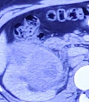
Contrast-enhanced computed tomography (CECT) of the chest and whole abdomen showed the size of the right kidney to be 11.2 × 5.7 cm with a heterogeneously enhancing lobulated mass (9.2 × 6.4 × 7.6 cm) arising exophytically from the anterior cortex of the upper and mid poles of the kidney with internal non-enhancing areas suggestive of necrosis. There was adjacent perinephric fat stranding with no evidence of intralesional fat or increased vascularity. The lesion was hypo-enhancing and was compressing the pelvicalyceal system causing mild hydronephrosis. Superolaterally, it was abutting the inferior surface of the right lobe of the liver with a maintained fat plane. Medially, it was abutting the second part of the duodenum with indistinct fat planes. It was closely abutting the inferior vena cava (IVC) with maintained fat planes (Figure 1). Posteromedially, it was abutting the right psoas muscle with indistinct fat planes. No thoracic metastasis was observed.

Abdomen contrast-enhanced computed tomography (CECT) showing right renal vein dilated with heterogeneous attenuation in the lumen (tumor thrombus) with normal inferior vena cava (IVC)

After preoperative anesthetic clearance, the patient underwent an open radical nephrectomy. Intraoperative findings included a right renal mass of 12 × 8 × 5 cm adhered to the ascending colon and duodenum, abutting liver. Deep vein thrombosis (DVT) prevention was advised post-operatively, along with antibiotics and painkillers. Since then, the patient has received routine follow-ups every three months.

The histological assessment of the biopsy specimen was suggestive of an MPNST. The mass had wavy nuclei in an eosinophilic cytoplasm and spindle-shaped cells organized in bundles and fascicles (Figure 2). The specimen margins were microscopically clear of tumor cells. S-100 and Vimentin were stained positive for MPNST (Figure 3).

Clockwise from top left: Haematoxylin-eosin staining showing the geographic pattern of necrosis, tumor cells arranged in sheets, and ovoid and spindle-shaped cells with hyperchromatic nuclei
Clockwise from top left: Slides showing S-100 stained positive, Vimentin stained positive, and CD56 stained positive

3. Discussion

The MPNSTs are sporadic or radiation-induced aggressive soft-tissue sarcomas that are uncommon and linked to neurofibromas in NF-1 (1). Patients with NF-1 typically appear as a single, rapidly growing, painless mass, several rapidly enlarging masses, or a new onset of pain related to pre-existing plexiform neurofibromas (2). The most frequent parts in which MPNSTs occur include the head and neck area, trunk, and extremities (7). There is rarely kidney involvement. Clinically, it can be challenging to tell various kidney sarcomas apart from the MPNST kidney.

For diagnosis and grading, histopathological and immunohistochemical tests are required (8). The MPNSTs are big (> 5 cm) and tan and grey, with a fusiform appearance. They display the characteristics of spindle cells under a microscope, including a fascicular pattern, varying degrees of mitosis, necrosis, and tumor calcification (9). Benign neurofibroma or schwannanian cells that have not undergone a malignant change could be found. S-100 protein, neuron-specific enolase, and other proteins are detected in MPNSTs (10). In this case of kidney lesion, WT1 staining was performed to rule out Wilms’ tumor (11). Synaptophysin and chromogranin staining was completed to rule out neuroendocrine tumors (12), CK7 and panck for ruling out carcinoma (13), desmin, SMA, and CD34 for ruling out endothelial tumors (14), CD10 for ruling out RCC (15), HMB45 staining for ruling out malignant melanoma (16), and TLAB3 and BcL2 for ruling out synovial tumors (17).

Computerized tomography and magnetic resonance imaging (MRI) are useful imaging methods for determining the size of the tumor and may suggest where the tumor originated (18). Increased uptake on a positron emission tomography scan and the evidence of heterogeneity, necrosis, and bleeding in an MRI could both be used to identify malignant transformation. The three most crucial prognostic markers for MPNSTs are tumor size, local recurrence, and the extent of surgical resection. The only option for treating MPNSTs is a complete surgical resection, which offers the only chance for recovery (19, 20). The MPNSTs are regarded as cancers resistant to radiation and chemotherapy. However, postoperative radiation, advised by an oncology consensus group, plays a part in both disease-free and overall survival (20). The prognosis is still dismal despite multimodal therapy, which includes vigorous surgical resection and adjuvant radiation (19).

3.1. Conclusions

Malignant tumors of the kidney, called MPNSTs, are uncommon. Wide local excision, if possible, has been found to be the best course of action because the prognosis heavily depends on how thoroughly the tumor is surgically removed. It is advised to have postoperative radiation.

References

  • 1.

    Alharbi B. Malignant peripheral nerve sheath tumor of kidney-A case report. Int J Surg Case Rep. 2013;4(10):914-6. [PubMed ID: 23995478]. [PubMed Central ID: PMC3785930]. https://doi.org/10.1016/j.ijscr.2013.07.013.

  • 2.

    Beer TC. Malignant Peripheral Nerve Sheath Tumor (MPNST): An overview with emphasis on pathology, imaging and management strategies. 2012. Available from: https://jdc.jefferson.edu/cgi/viewcontent.cgi?article=1018&context=neurosurgeryfp.

  • 3.

    Ajani MA, Awosusi BL, Nwanji ID. Malignant peripheral nerve sheath tumour in an unusual location of the urinary bladder: case report and review of literature. Afr J Urol. 2021;27(1):104. https://doi.org/10.1186/s12301-021-00204-3.

  • 4.

    Alavi S, Arzanian MT, Nilipour Y. Retroperitoneal malignant peripheral nerve sheath tumor replacing an absent kidney in a child. Case Rep Oncol Med. 2013;2013:627472. [PubMed ID: 24392233]. [PubMed Central ID: PMC3872381]. https://doi.org/10.1155/2013/627472.

  • 5.

    Williams SB, Szlyk GR, Manyak MJ. Malignant peripheral nerve sheath tumor of the kidney. Int J Urol. 2006;13(1):74-5. [PubMed ID: 16448437]. https://doi.org/10.1111/j.1442-2042.2006.01238.x.

  • 6.

    Khandelwal A, Gupta A, Khandelwal K. Malignant peripheral nerve sheath tumor of kidney. Iran J Kidney Dis. 2011;5(6):373. [PubMed ID: 22057067].

  • 7.

    Panigrahi S, Mishra SS, Das S, Dhir MK. Primary malignant peripheral nerve sheath tumor at unusual location. J Neurosci Rural Pract. 2013;4(Suppl 1):S83-6. [PubMed ID: 24174807]. [PubMed Central ID: PMC3808069]. https://doi.org/10.4103/0976-3147.116480.

  • 8.

    Gogate BP, Anand M, Deshmukh SD, Purandare SN. Malignant peripheral nerve sheath tumor of facial nerve: Presenting as parotid mass. J Oral Maxillofac Pathol. 2013;17(1):129-31. [PubMed ID: 23798847]. [PubMed Central ID: PMC3687169]. https://doi.org/10.4103/0973-029X.110708.

  • 9.

    Chitale AR, Dickersin GR. Electron microscopy in the diagnosis of malignant schwannomas. A report of six cases. Cancer. 1983;51(8):1448-61. [PubMed ID: 6402292].

  • 10.

    Smith TA, Machen SK, Fisher C, Goldblum JR. Usefulness of cytokeratin subsets for distinguishing monophasic synovial sarcoma from malignant peripheral nerve sheath tumor. Am J Clin Pathol. 1999;112(5):641-8. [PubMed ID: 10549251]. https://doi.org/10.1093/ajcp/112.5.641.

  • 11.

    Goyal S, Mishra K, Sarkar U, Sharma S, Kumari A. Diagnostic utility of Wilms' tumour-1 protein (WT-1) immunostaining in paediatric renal tumours. Indian J Med Res. 2016;143(Suppl):S59-67. [PubMed ID: 27748279]. [PubMed Central ID: PMC5080930]. https://doi.org/10.4103/0971-5916.191776.

  • 12.

    Kriegsmann K, Zgorzelski C, Muley T, Christopoulos P, Thomas M, Winter H, et al. Role of Synaptophysin, Chromogranin and CD56 in adenocarcinoma and squamous cell carcinoma of the lung lacking morphological features of neuroendocrine differentiation: a retrospective large-scale study on 1170 tissue samples. BMC Cancer. 2021;21(1):486. [PubMed ID: 33933015]. [PubMed Central ID: PMC8088012]. https://doi.org/10.1186/s12885-021-08140-9.

  • 13.

    Selves J, Long-Mira E, Mathieu MC, Rochaix P, Ilie M. Immunohistochemistry for Diagnosis of Metastatic Carcinomas of Unknown Primary Site. Cancers (Basel). 2018;10(4):108. [PubMed ID: 29621151]. [PubMed Central ID: PMC5923363]. https://doi.org/10.3390/cancers10040108.

  • 14.

    Miettinen M. Immunohistochemistry of soft tissue tumours - review with emphasis on 10 markers. Histopathology. 2014;64(1):101-18. [PubMed ID: 24111893]. [PubMed Central ID: PMC7670586]. https://doi.org/10.1111/his.12298.

  • 15.

    Martignoni G, Pea M, Brunelli M, Chilosi M, Zamo A, Bertaso M, et al. CD10 is expressed in a subset of chromophobe renal cell carcinomas. Mod Pathol. 2004;17(12):1455-63. [PubMed ID: 15286660]. https://doi.org/10.1038/modpathol.3800236.

  • 16.

    Gleason BC, Nascimento AF. HMB-45 and Melan-A are useful in the differential diagnosis between granular cell tumor and malignant melanoma. Am J Dermatopathol. 2007;29(1):22-7. [PubMed ID: 17284958]. https://doi.org/10.1097/01.dad.0000249888.41884.6c.

  • 17.

    Hasegawa T, Matsuno Y, Shimoda T, Hirohashi S, Hirose T, Sano T. Frequent expression of bcl-2 protein in solitary fibrous tumors. Jpn J Clin Oncol. 1998;28(2):86-91. [PubMed ID: 9544821]. https://doi.org/10.1093/jjco/28.2.86.

  • 18.

    Sperandio M, Di Poce I, Ricci A, Di Trapano R, Costanzo E, Di Cello P, et al. Malignant Peripheral Nerve Sheath Tumour: CT and MRI Findings. Case Rep Radiol. 2013;2013:517879. [PubMed ID: 23970990]. [PubMed Central ID: PMC3732584]. https://doi.org/10.1155/2013/517879.

  • 19.

    Cai Z, Tang X, Liang H, Yang R, Yan T, Guo W. Prognosis and risk factors for malignant peripheral nerve sheath tumor: a systematic review and meta-analysis. World J Surg Oncol. 2020;18(1):257. [PubMed ID: 32998743]. [PubMed Central ID: PMC7528472]. https://doi.org/10.1186/s12957-020-02036-x.

  • 20.

    Dunn GP, Spiliopoulos K, Plotkin SR, Hornicek FJ, Harmon DC, Delaney TF, et al. Role of resection of malignant peripheral nerve sheath tumors in patients with neurofibromatosis type 1. J Neurosurg. 2013;118(1):142-8. [PubMed ID: 23101443]. https://doi.org/10.3171/2012.9.JNS101610.