1. Introduction
The urachus is a natural embryonic remnant. In adults, it is a fibrous cord potentially located in the midline region between the peritoneum and the transversalis fascia, developing from the bladder dome to the navel (1). In the first trimester of gestation, the urachus is responsible for removing the neonate's nitrogenous waste via the placenta and the umbilical cord (2), but its function in adults is unclear. The urachus opening after birth causes anomalies. Four types of congenital urachal anomalies include patent urachus (~50% of all anomalies), umbilical-urachal sinus (~15%), vesicourachal diverticulum (~3% to 5%), and urachal cyst (~30%), which may be seen in children or adults. These anomalies may mostly close after birth but open again following some pathological conditions (3). Urachal anomalies occur at an incidence of 1 in 5,000 births; of this number, about 2% can be found in adults. It is two-fold more common in males than in females (4). The clinical symptoms are nonspecific and vary between children and adults. The most common symptoms in adults are hematuria, dysuria, and pain. Of course, it is possible to be asymptomatic or associated with dermatitis or abscess of the periumbilical. Most patients with urachal anomalies are asymptomatic unless associated with infection (5). Ultrasonography (US) and a computerized tomography (CT) scan are effective tools for diagnosis. Timely and correct diagnosis of urachal anomalies is vital because they are prone to infection and malignancy (6). The two complications after these anomalies are calculi and cancer (7). According to our best search of the English literature, urachus calculi are a rare anomaly, with only eleven cases reported in adults to date.
2. Case Presentation
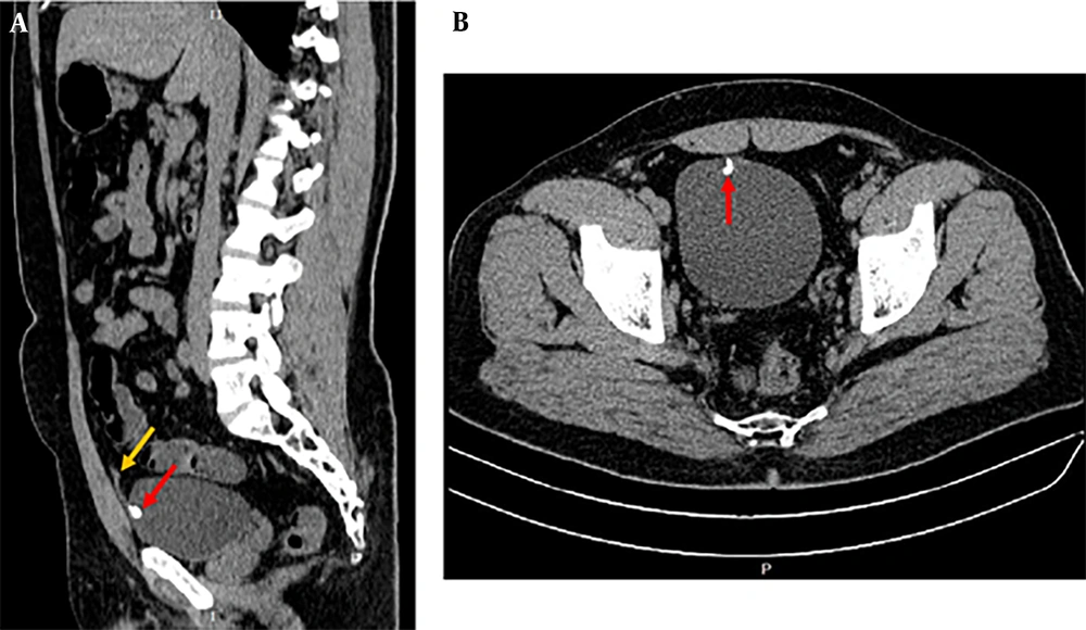
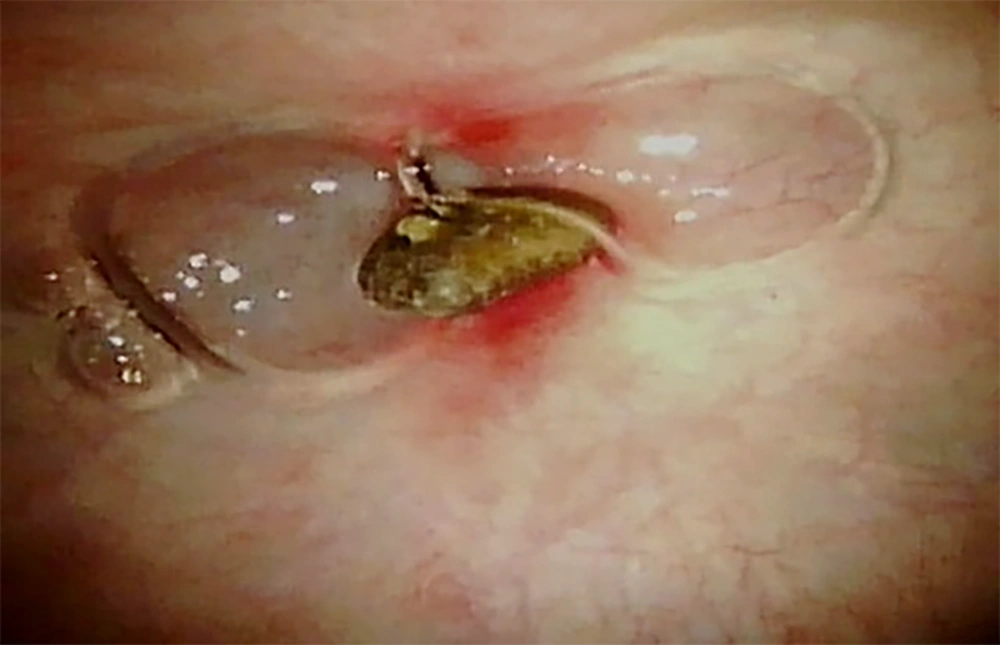
The patient was a 38-year-old man referred to a urologist complaining of abdominal ambiguous pain. He had tenderness in the abdomen wall on a physical exam. Vital signs and laboratory results were normal. He had no weight loss, urinary obstruction, irritation symptoms, hematuria, or medical history. A CT scan of the abdomen and pelvis was requested for the patient. The results of the CT scan in both sagittal and axial axes showed a calculus with a size of 10 mm attached to the anterior and inner wall of the bladder, which was located at the insertion of the bladder to the urachus (Figure 1). Cystoscopy was performed to definitively diagnose and differentiate urachus calculus from bladder calculus. Cystoscopy images (Figure 2) showed that the calculus is connected to the urachus duct, and no trace of tumor or mass was observed in this area. The collection of evidence suggests a calcified urachus. The patient was discharged with only conservative treatment and a mild analgesic.
The images of the computerized tomography (CT) scan in both sagittal (A); and axial (B); axes show a calculus (red arrow) with the size of 10 mm attached to the anterior and inner wall of the bladder, which was exactly located in the insertion of the bladder to the urachus. The yellow arrow shows the portion of the urachus.
3. Discussion
We report an unusual presentation of a symptomatic urachal remnant with calculus just above the bladder in an adult. According to previous studies, we concluded that urachus calculi are a secondary complication of urachus anomalies, including vesicourachal diverticulum, cyst, sinus, and cancer. Infections, bladder outlet obstruction, and urine stasis can also be due to infections. To date, only eleven similar patients have been reported as case reports. The present patient is the 12th case (presented in Table 1).
| Authors | Year | Age | Gender | Symptoms | Diagnostic | Stone Size | Therapy | Test that Leads to Diagnose | Ref. |
|---|---|---|---|---|---|---|---|---|---|
| Rawal and Maldjian | 2022 | 44 | M | Abdominal pain following an accident | Urachal calculus | 7 mm | Conservative treatment | CT | (8) |
| Kato et al. | 2022 | 28 | M | Umbilical drainage | Urachal calculus + inflammation | - | Laparoscopic resection | CT | (9) |
| Seo et al. | 2008 | 58 | M | Abdominal pain + urinary frequency | Urachal cyst + calculi | 1.8 × 1.3 cm + 0.6 × 0.3 cm | Laparoscopic resection | CT + US + cystoscopy | (10) |
| Hladun et al. | 2021 | 73 | F | Hematuria+ mucous discharge in urine | Urachal cyst + calculus + tumour | 4.2 cm | Laparotomy | CT + MRI | (11) |
| Nakano et al. | 2014 | 96 | F | Pyuria+ bacteriuria + fever + loss of appetite | Urachal sinus + calculus | - | Removed through the navel by soaking in sterilized olive oil | CT + physical examination | (12) |
| Rodrigues and Gandhi | 2013 | 45 | M | Abdominal pain | Urachal calculus | - | - | CT | (13) |
| Milotic et al. | 2001 | 73 | F | Nocturia | Urachal cyst + calculi | 5 - 10 mm | Laparotomy | CT + sonography | (14) |
| Diehl | 1991 | 74 | M | Intractable urinary tract infections | Urachal cyst + calculus | - | Surgery | X-ray | (15) |
| Leyson | 1984 | 32 | M | Suprapubic pain after a drinking + nausea | Urachal cyst + calculus | - | Surgery | X-ray | (16) |
| Ozbulbul et al. | 2010 | 60 | M | Asymptomatic | Vesicourachal diverticulum + calculi | - | - | CT + US + MDCT urography | (3) |
| Good et al. | 2022 | 26 | M | Red umbilical nodule + clear malodorous discharge | Urachal sinus + calculus | 1 cm | Surgery | CT + US | (6) |
| Current study | 2023 | 38 | M | Abdominal pain | Urachal calculus | 10 mm | Conservative treatment | CT + US + cystoscopy | - |
Reported Cases of Urachal Remnant Calcification in Adults
In the vesicourachal diverticulum, the urachus communicates only with the bladder dome. This condition results when the vesical end of the urachus fails to close. Vesicourachal diverticulum is asymptomatic in most cases and is usually discovered incidentally on axial CT performed for unrelated reasons, appearing as a midline cystic lesion just above the anterosuperior aspect of the bladder (17). The US readily demonstrates an extraluminally protruding, fluid-filled sac that does not communicate with the umbilicus. This lesion is found in patients with chronic bladder outlet obstruction, and it may be complicated by urinary tract infection, intratracheal calculus formation, and an increased prevalence of carcinoma after puberty (18).
In accordance with our case, Rodrigues and Gandhi indicated a rare calcified urachal remnant similar to a bladder calculus near its insertion into the urinary bladder. They were associated with left-sided renal colic (13). Rawal and Maldjian demonstrated a rare case of urachal remnant calcification with a 7 mm size located at the anterior bladder wall at the embryological urachus insertion on a CT scan. They stated that two important factors that cause urachal calcification are urachal carcinoma and bladder infections, including schistosomiasis and tuberculosis (8).
Sometimes, the presence of urachus cysts leads to calcification of the cyst wall and the formation of calculus (19). For example, Ozbulbul et al. reported a midline cyst exactly above the bladder anterosuperior portion that included two calculi on CT images. They stated that intratracheal calculus formation might be due to chronic bladder outlet obstruction that subsequently leads to complications such as urinary tract infection and the occurrence of adenocarcinoma. The existence of urachal calculus indicates that there was already a connection between the bladder and urachal remnant that permitted urine to flow into the urachal lesion and induced the formation of the calculi (3).
Tanomogi et al. reported a cyst approximately 3 cm by cystoscopy that communicated the umbilicus to the bladder. The patient complained of persistent discharge from the umbilicus for 18 years. Computerized tomography scan and magnetic resonance imaging (MRI) revealed a 5 × 3 cm calculus within the cyst removed by surgery. The authors claimed that > 210 cases of urachal cysts had been presented in Japan, while about 10% were accompanied by calculus (20).
Calculus formation is special for adults, probably due to the long-term stoppage of urine in the urachal remnant, carried out by a crystallization factor including mucus or shed epithelium. Timely diagnosis and treatment of the urachal remnant can prevent complications such as calculus formation or abscess. If this disorder is completely ignored, it may become a silent and chronic source of inflammation, infection, and cancer (7).
Diehl reported a urachal cyst that was communicated with the bladder and contained calculus from the infection's origin. Histological examination showed no evidence of malignancy. The calculus is composed of calcium phosphate (15). Seo et al. showed calcifications within a cyst in front of the bladder in the US. In addition, cystography revealed the existence of calculi of 1.8 × 1.3 cm and 0.6 × 0.3 cm in the pelvic cavity and outer of the bladder. They removed the cyst and its calculi by a laparoscopic incision (10).
It is proven that calcification occurs in 50% - 70% of urachal cancers (21). Urachal carcinomas can produce typical psammomatous calcifications that are detectable on CT. Calcifications in a midline supravesical mass are diagnosed as urachal carcinoma (22, 23).
Hladun et al. reported a rare and aggressive cancer originating from the urachus in a 73-year-old female. The patient was followed up for 12 years, showing the slow growth of cancer without any symptoms, and finally, it became calcified and entered the bladder lumen. After the patient developed haematuria and mucous discharge in urine, an urgent CT scan and MRI showed a 4.2 cm calcified cystic lesion connected to the lumen of the bladder. An open diagnostic laparotomy discovered a calcified tumor originating from the urachal remnant. The tumor, which was 5 × 4 cm, infiltrated the bladder dome (11).
In line with our study, many surgeons believe in a conservative approach, such as antibiotic and analgesia treatment and patient follow-up. They suggest that conservative treatment will spontaneously obviate many lesions and may be especially effective in incidental detection of the urachal remnant (5, 24). On the other hand, some surgeons stated that surgery should be done at diagnosis time since these abnormalities are unlikely to be repaired spontaneously (5, 19).
3.1. Conclusions
Physicians and specialists should notice that the urachus stone is not mistaken for a bladder stone, though rare. Urachus stones require conservative treatments, and the patient must be periodically followed up to ensure symptoms or complications do not occur.


