


1. Background
Urinary stone disease (USD) is a common and clinically important condition (1). A key challenge in the management of USD is the substantial recurrence rate, with approximately half of all patients experiencing a recurrence within five years (2). Effective treatment of large renal calculi should aim for complete stone removal, minimal patient morbidity, maximal preservation of nephron function, and a reduction in the risk of stone recurrence (3). Percutaneous nephrolithotomy (PCNL), using access sheaths up to 30 Fr in diameter, is the established standard of care for managing large kidney stones (4). The two most crucial steps in the PCNL procedure are establishing percutaneous access to the renal collecting system and subsequently dilating the access tract (5). The established best practice, as supported by published research, is to puncture through the posterior renal papilla to minimize the potential for damage to major renal vessels (6).
Historically, accessing the renal collecting system via the fornix callis and renal papilla was considered the sole acceptable approach for PCNL (7). However, contemporary research has demonstrated the feasibility and low complication rates associated with non-fornix access techniques (8). In clinical practice, variations in patient anatomy may preclude ideal papillary access (9). Initial investigations in this patient population suggest that non-papillary access may be associated with shorter procedure times, reduced complexity, and potentially greater ease of execution (10). Contrary to previous assumptions, the risk of hemorrhage in non-papillary access does not appear to be elevated compared to the papillary approach (11).
A study by Sampaio et al. in the early 1992s, supported by subsequent research, indicated that fornix access is associated with a lower incidence of clinically significant vascular injury (12). This is likely due to the proximity of larger segmental arteries to the hilar infundibulum, whereas the smaller arterial branches near the fornix are less prone to substantial bleeding (13). For instance, in one study of 137 patients undergoing non-papillary PCNL, only 9.2% (n = 4) required blood transfusion, and all cases were managed conservatively without serious sequelae (14).
2. Objectives
Given the high volume of patients undergoing kidney stone surgery at our institution, and the absence of comparable research within Iran and specifically at our center, this study was designed to evaluate the impact of access-related complications on patient outcomes. The primary objective was to compare surgical complications between patients undergoing PCNL with favorable papillary access to the calyceal system and those with unfavorable non-papillary access. Specifically, this study investigated the influence of achieving favorable papillary access on surgical outcomes in patients undergoing PCNL.
3. Methods
This retrospective cohort study, approved by the Ethics Committee of Urmia University of Medical Sciences (code: IR.UMSU.HIMAM.REC.1402.112), reviewed the medical records of patients who underwent PCNL between January 2022 and June 2023. A minimum sample size of 103 patients (55 with papillary access and 48 with non-papillary access) was calculated. Data were extracted from the hospital information system for all eligible patients. Collected data included patient demographics [age, sex, weight, Body Mass Index (BMI), comorbidities, and vital signs], pre- and post-operative laboratory values (hemoglobin and creatinine), and documented surgical complications. These complications included intraoperative and postoperative bleeding, pleural injury, colon injury, urinary leakage, and infection. A comparative analysis of these variables was performed between the papillary and non-papillary access groups.
Pleural injury was diagnosed based on fluoroscopic and endoscopic findings, intraoperative observations during upper calyceal access, and postoperative chest X-ray (CXR). Colon injury was identified through intraoperative fluoroscopic imaging and the presence of fecal leakage from the access site. Urinary leakage was diagnosed by persistent urine drainage from the nephrostomy site without diminution in the days following the procedure. Stone-free status was determined via endoscopic visualization [computed tomography scan (CT-scan)] during the procedure and postoperative radiographic imaging. Postoperative infection was defined as the presence of fever and positive cultures, and this information was collected from patient records and laboratory results. Data regarding intraoperative details and postoperative length of stay (typically two days) were also collected.
The history of diabetes and hypertension was ascertained from patients' documented past medical history within their medical file. For patients with a diagnosis of diabetes, all necessary pre-operative interventions were implemented to achieve optimal glycemic control. Similarly, for patients with hypertension or a new diagnosis, appropriate measures were taken to manage blood pressure prior to the procedure. The use of CT, which has become the gold standard for diagnosis and treatment planning, has ushered in the possibility of measuring stone burden in multiple dimensions (15). All procedures were performed by a single surgeon.
Following data collection, patients were categorized into two groups (papillary and non-papillary) based on the access method utilized during the procedure. The papillary access approach was selected because it represents the current standard of care and is considered the safest method, as prior research indicates that fewer vessels in the access path significantly reduce the risk of bleeding (16). From a medical ethical standpoint, it was not permissible to deliberately pursue non-papillary access in a subset of patients. Nevertheless, acknowledging that papillary access is not invariably successful, and despite earnest attempts, some procedures inevitably resulted in non-papillary access, these patients were consequently included in the non-papillary access group. Patient characteristics in each group were then compared.
Inclusion criteria consisted of kidney stones, undergoing PCNL, and complete medical record documentation. Exclusion criteria included incomplete medical records, age outside the 20 - 60-year range, history of malignancy, other renal abnormalities (e.g., polycystic kidney disease, horseshoe kidney), chronic kidney disease, and solitary kidney.
3.1. Sample Sizing Method
The requisite sample size for this investigation was calculated utilizing the following formula, informed by the mean blood loss reported in the study by Hou et al. (17) (1.59 ± 1.01 mL in the papillary group and 4.24 ± 3.79 mL in the control group). Considering a 95% confidence interval
3.2. Statistical Analysis
Statistical analysis was performed using SPSS version 27. Descriptive statistics for quantitative variables are presented as means and standard deviations, while categorical variables are summarized using percentages and frequencies. Data visualization was achieved through the use of tables and figures where appropriate. Univariate comparisons between the two study groups were conducted using appropriate statistical tests. Independent samples t-tests (or the Mann-Whitney U test where appropriate) were used for continuous variables, and chi-square tests (or Fisher's exact test where appropriate) were used for categorical variables. Statistical significance was defined as a P-value of less than 0.05.
4. Results
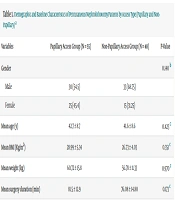
Table 1 shows the demographic and baseline characteristics of the patients undergoing PCNL, stratified by renal access technique: Papillary versus non-papillary. The mean age of the entire patient population was 42.3 ± 8.6 years, with a range spanning from 20 to 60 years. Specifically, the papillary access group exhibited a mean age of 42.7 ± 8.7 years (range: 23 - 60), while the non-papillary access group demonstrated a mean age of 41.6 ± 8.6 years (range: 20 - 60). A two-tailed independent samples t-test revealed no statistically significant disparity in age distribution between the two access groups (P = 0.423).
| Variables | Papillary Access Group (N = 55) | Non-papillary Access Group (N = 48) | P-Value |
|---|---|---|---|
| Gender | 0.140 b | ||
| Male | 30 (54.5) | 33 (68.75) | |
| Female | 25 (45.4) | 15 (31.25) | |
| Mean age (y) | 42.7 ± 8.7 | 41.6 ± 8.6 | 0.423 c |
| Mean BMI (Kg/m2) | 28.99 ± 5.34 | 26.73 ± 4.03 | 0.351 c |
| Mean weight (kg) | 60.72 ± 15.8 | 56.78 ± 11.33 | 0.970 c |
| Mean surgery duration (min) | 81.5 ± 12.9 | 76.08 ± 14.80 | 0.173 c |
| Diabetes | 0.66 b | ||
| Yes | 6 (10.91) | 4 (8.33) | |
| No | 49 (89.09) | 44 (91.67) | |
| Hypertension | 0.62 b | ||
| Yes | 10 (18.18) | 7 (14.58) | |
| No | 45 (81.82) | 41 (85.42) | |
| Stone burden | 0.58 b | ||
| Lower than 2 cm | 9 (16.36) | 10 (20.83) | |
| 2 - 3 cm | 26 (47.27) | 25 (52.08) | |
| Higher than 3 cm | 20 (36.36) | 13 (27.08) |
Abbreviation: BMI, Body Mass Index.
a Values are expressed as mean ± SD or No (%).
b Chi-square test
c Independent sample t-test
The study population comprised 103 individuals, consisting of 63 males and 40 females. Analysis of sex distribution across the access groups indicated no statistically significant divergence (P = 0.140). The BMI was 28.99 ± 5.34 kg/m2 within the papillary access group and 26.73 ± 4.03 kg/m2 within the non-papillary access group. The mean operative duration was 60.72 ± 15.8 minutes for the papillary access group and 56.78 ± 11.33 minutes for the non-papillary access group. The mean patient weight was 81.5 ± 12.9 kg in the papillary group and 76.08 ± 14.80 kg in the non-papillary group. Statistical comparisons demonstrated no significant intergroup differences for BMI, operative time, or patient weight (P > 0.05 for all comparisons).
Table 2 details the postoperative alterations in hemoglobin concentrations within the papillary and non-papillary renal access groups following PCNL. The papillary access group exhibited a mean reduction in hemoglobin of 1.14 ± 1.38 g/dL, while the non-papillary access group demonstrated a mean decrease of 1.10 ± 1.49 g/dL. An independent samples t-test revealed no statistically significant intergroup variation in the magnitude of hemoglobin reduction (P = 0.799).
| Variables | Papillary Access Group (N = 55) | Non-papillary Access Group (N = 48) | P-Value |
|---|---|---|---|
| Mean hemoglobin drop (g/dL) | 1.14 ± 1.38 | 1.10 ± 1.49 | 0.799 |
| Pleural damage (%) | 3 | 2 | 0.640 |
| Colon damage (%) | 0 | 0 | 0 |
| Postoperative infection (%) | 3 | 6 | 0.538 |
| Stone free rate (%) | 89.1 | 89.6 | 0.640 |
| Postoperative urinary leakage (%) | 2 | 3 | 0.923 |
The incidence of pleural injury was 3% in the papillary access group and 2% in the non-papillary access group. Statistical analysis using a chi-square test indicated no significant difference in pleural injury rates between the two groups (P = 0.640). No instances of colon injury were reported in either access group (0% in both papillary and non-papillary). Consequently, due to the absence of variability, comparative statistical analysis was precluded (P > 0.05, demonstrating a lack of observed difference).
Postoperative infection rates were 3% in the papillary access group and 6% in the non-papillary access group. A chi-square test demonstrated no statistically significant difference in infection rates between the two groups (P = 0.538). Stone-free rates were 89.1% in the papillary access group and 89.6% in the non-papillary access group. A chi-square test revealed no significant intergroup difference (P = 0.640).
Postoperative urinary leakage rates were 2% in the papillary access group and 3% in the non-papillary access group. Statistical analysis via chi-square testing showed no significant variation between the groups (P = 0.923), as summarized in Table 2.
5. Discussion
Puncture of the renal collecting system via the fornix of the papilla is generally considered the preferred approach, based on the anatomical distribution of renal vasculature (18, 19). However, the success of papillary puncture is dependent on individual patient anatomy and stone characteristics (16). The traditional view that papillary access is inherently safer than non-papillary access for PCNL is primarily based on cadaveric anatomical studies, lacking robust clinical evidence of increased risk with non-papillary puncture (20). Recent clinical studies (11, 21, 22) are among the few that directly address the safety of non-papillary access. While papillary puncture remains the standard of care within the endourological community, non-papillary puncture is often perceived as high-risk in the absence of more extensive clinical data (23). This study directly compared surgical outcomes in patients undergoing PCNL via papillary and non-papillary access techniques.
Anatomical investigations have demonstrated a correlation between puncture of the upper infundibulum and arterial injury in 67% of cases (24). Arterial lesions were associated with mid- and lower calyceal infundibular access in 23% and 13% of the studied kidneys, respectively (25). The prevailing endourological principle is that minimizing trauma in highly vascularized regions will lead to fewer hemorrhagic complications, thus favoring the papillary approach (26).
This study found no statistically significant differences in BMI, age, or sex between the two groups. Both prospective and retrospective studies have indicated that patient demographics are not significant risk factors for post-PCNL complications (27). Therefore, while age and BMI are generally considered risk factors in surgical procedures, neither this study nor previous research has identified them as statistically significant risk factors for complications following PCNL.
This study's findings regarding complications and blood loss are consistent with existing literature (28), providing clinical evidence supporting the safety of routine non-calyceal punctures. Specifically, this study demonstrated no significant difference in blood loss between papillary and non-papillary approaches. Comparing these results with those of Kyriazis et al. (14), whose study focused on the safety of non-papillary access and reported low transfusion rates (1.5%), the present study's finding of no significant difference in hemoglobin loss further reinforces the feasibility of non-papillary access with acceptable bleeding. Similarly, Kallidonis et al. (10), in a direct comparison of the two methods, also found no significant difference in hemoglobin loss or transfusion requirements, strongly supporting the present study's conclusions and dispelling concerns about increased bleeding with non-papillary access.
Tahra's study, using a robust design and appropriate sample size, found significant differences in hemoglobin drop and transfusion needs between the groups (23). This is consistent with the present study, reinforcing its validity. Notably, despite the increased complexity often associated with these stone types, the mean hemoglobin drop reported in the present study was relatively low, indicating that even in challenging cases, non-papillary access can be performed with controlled bleeding.
Comparing these results with Hou et al. (17), the only study reporting greater bleeding in the non-papillary group, highlights a potential methodological difference. Another study used direct blood measurement (mL/min via the drainage catheter), while the present study used hemoglobin drop. While direct blood measurement may be more precise, it may not perfectly correlate with hemoglobin changes, which is a more clinically relevant metric (29). Variations in surgeon experience and surgical technique may also contribute to the observed differences (30).
The shorter operative time observed in group 2 compared to group 1 (P = 0.027) represents an additional benefit of the non-papillary approach. This reduction in operative time may be attributed to the need for multiple access tracts in some group 1 cases (four cases requiring multiple access) (31). It is also possible that the greater maneuverability of instruments within the infundibular approach facilitates stone removal (32). A study by Wei Gan et al., involving 347 patients treated for staghorn or non-staghorn calculi, reported a mean operative time of 97 minutes and a mean fluoroscopy time of 6.93 minutes (33, 34). Compared to these findings, the present study's non-papillary technique resulted in a substantial decrease in both operative time (56.78 ± 11.33 minutes) and fluoroscopy time (2.67 ± 1.02 minutes). This reduction in operative and fluoroscopy exposure time offers potential advantages for both patients and the surgical team (34).
No statistically significant differences were observed between the two groups in the incidence of complications, including pleural injury, colon injury, postoperative infection, stone-free rate, and urinary leakage. Kyriazis et al. (35) investigated the feasibility and safety of non-calyceal access PCNL in 137 consecutive patients (including 10 with anatomical variations) using fluoroscopic guidance. They reported stone-free rates of 89.2% for single stones, 80.4% for multiple stones, and 66.7% for staghorn stones. The overall complication rate was 10.2%, with a major complication rate of 3.6%. They concluded that non-calyceal access is feasible and safe, achieving high stone-free rates with low complications.
Michel et al. (36), in their review of overall PCNL complications, reported the following prevalence: Urinary leakage (7.2%), fever (21 - 32%), sepsis (up to 4.7%), pleural injury (up to 3.1%), and colon injury (up to 0.8%). The present study's finding of no significant difference in these complications between the two groups suggests that access method alone is not the sole determinant of these complications. Other factors, such as surgeon experience, surgical technique, patient characteristics, and postoperative management, also contribute significantly.
Regarding stone clearance rates, studies such as Kallidonis et al. (10) have demonstrated high success rates with non-papillary access. The present study's finding of no significant difference in stone clearance rates supports this observation, indicating that access method does not influence the ultimate success of stone removal.
Existing literature suggests several potential advantages associated with the non-papillary approach (11, 16). Direct access to the stone location can decrease the need for flexible nephroscopy and shorten operative time (20). Furthermore, the non-papillary approach may reduce the number of access tracts required compared to the papillary approach, as it facilitates greater instrument maneuverability within the collecting system (37). Additionally, establishing papillary access to the renal pelvis can be challenging in the presence of impacted or large posterior calyceal stones (38). In such cases, the non-papillary approach can be particularly advantageous, allowing bypass of the impacted stone and providing access to the pelvis-ureter for stone removal (39). The feasibility and safety of the non-papillary approach have also been explored in the context of mini-PCNL, utilizing an 18 Fr nephroscope with a maximum outer sheath diameter of 22 Fr (40).
5.1. Conclusions
Although no statistically significant differences were reported between papillary and non-papillary access methods for PCNL with respect to hemoglobin loss, postoperative complications, and stone-free rates, these findings suggest that non-papillary access may be a safe and effective alternative to the traditional papillary approach for PCNL, achieving comparable stone-free rates and complication profiles. Further research, including larger-scale, prospective, randomized trials incorporating detailed anatomical and radiological assessments, is warranted to definitively establish the safety and efficacy of non-papillary access for PCNL.
5.2. Limitation of Study
This study has several limitations. The relatively small sample size, cross-sectional analytical design, single-center setting, and omission of potentially influential variables such as surgeon experience and detailed stone characteristics limit the scope of the findings. Future research should address these limitations. Larger sample sizes are recommended to enhance statistical power and improve generalizability. Moreover, no specific similar study was found in the literature review to calculate the sample size, with the only one being the study by Hou et al. (17), comparing actual blood loss (in mL).
Prospective, randomized controlled trials are needed to more definitively establish cause-and-effect relationships. Subsequent studies should incorporate additional variables, including surgeon experience, stone type, size, and precise location, the number of access tracts required, operative time, and surgical costs. Multi-center studies would further enhance the generalizability of the findings. Finally, future research should prioritize investigating the long-term consequences of PCNL with both access methods, including stone recurrence, renal damage, and patient quality of life.
