1. Introduction
Stone formation in the human body is one of the most interesting subjects in medicine. The condition is apparently as old as the human race and has been mentioned in ancient medical treatises of many countries. Among all the locations in the human body, the urinary tract forms the most frequent site for stone formation. Bladder stones, also called bladder calculi, often form when concentrated urine sits in the bladder. As urine stagnates, minerals in the urine form various crystals that might combine to form stones. Bladder stones are very common in people who use an indwelling catheter. The indwelling catheter allows stone crystals to form on the catheter. At routine catheter changes, stone crystals might fall off the catheter and into the bladder. While in the bladder, stones can become progressively larger unless removed. A major problem with bladder stones is that bacteria can grow on them and lead to recurrent bladder infections. Antibiotics can be used to kill the bacteria in the bladder but not in the stones. Another problem with bladder stones is blocking the catheter’s drainage and preventing the bladder from emptying (1).
Urinary bladder calculi vary in size but any calculi of bladder is called giant if it weighs ≥ 100 g. Large urinary bladder calculi are commonly seen in association with recurrent urinary tract infection (UTI), azotemia, and urinary retention. Although the bladder calculi are usually composed of triple phosphate, calcium carbonate, and calcium oxalate, a case report by Becher et al. cited a large bladder calculi composed of uric acid as the major constituent with asymmetrical calcium oxalate and weight of 235 g (2). We report a case of the largest vesical stone burden that was 42 stones weighing 1400 g, developed in a bladder following augmentation cystoplasty.
2. Case Presentation
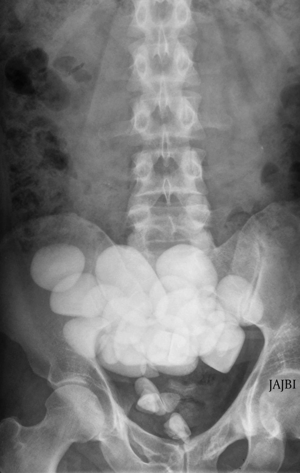
A 25-year-old man presented with a history of recurrent UTI. He had undergoing augmentation ileocystoplasty with cutaneous diversion by Mitrofanoff principle for pelvic fracture distraction urethral defect and rectourethral fistula following road traffic accident ten years ago. His physical examination, including per rectal examination, revealed normal findings. The routine investigations were done and revealed normal hemoglobin level, total leukocyte count of 11 × 109/L, and normal findings on renal function tests (serum creatinine, 97.24 μmol/L; and urea 8.93 mmol/L). Further imaging studies such as ultrasonography, X-ray, and computed tomography of abdomen and pelvic showed the massive stone burden in urinary bladder (Figure 1). The urine macroscopic and microscopic examination showed presence of pus cells with white blood cells. The culture and sensitivity report of urinary sample showed growth of Escherichia coli that was highly sensitive to amikacin, gentamicin, cefoperazone, ceftriaxone, imipenem, and ciprofloxacin.
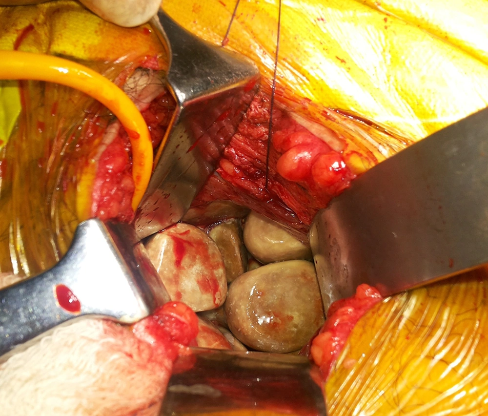
Patient was planned for elective surgery after treating the urinary sepsis. He underwent open cystolithotomy under general anesthesia and a total of 42 stones, weighing 1400 g, were removed. Stone were sent for chemical analysis and they turned out to be of struvite type (Figure 2). Postoperative period was uneventful and he was discharged under satisfactory condition. He was followed up on outpatient department basis and was doing well.
3. Discussion
Bladder stones usually form when substances (such as calcium oxalate) in the urine concentrate and coalesce into hard, solid lumps that lodge in the bladder. Often, several stones form at once. Normally, they are fairly small and are excreted in the urine without complications, but sometimes these stones get trapped in the neck of the bladder. As the time progresses these residues in the urine continue to accumulate and grow into large stone. These stones enlarge sufficient enough to cause various symptoms such as pain, urinary blockage, or infections and thus, requiring surgical intervention. The bladder stones almost exclusively affect the middle-aged and elderly male population. For unknown reasons, these stones are becoming increasingly rare (3).
Bladder stones show great variation in size. Until now, the world’s largest stone was reported by Arthure in 1953 with a weight of 6294 g and was thought to be developed in the bladder diverticulum. Few more cases of large bladder stone were reported in 1921 by Randall, who reported a stone of 1914-g weight. In 1952, Powers and Matflerd reported bladder stones weighing 1410 g. Now a days, due to early presentation and better management, these sizes of bladder stone are seen very rarely.
We reported an interesting case of the largest size of bladder calculi, which developed after augmentation cystoplasty, with a total burden of 42 vesical stones weighing 1400 g (4, 5). Augmented cystoplasty is usually done through the open approach method. This procedure involves the bladder augmentation through the anastomosis of the bowel segment to the native bladder. The most commonly used bowel segment is a detubularized ileum, usually resected from 25 to 40 cm of the ileocecal valve (6).
Stone formation in augmentation cystoplasty or orthotopic neobladder, where bowel is used for the reconstruction, is one of the late but common complications. The formation of bladder stones is one of the most common long-term problems encountered in the postoperative period following the bladder augmentation surgery. Recent literature analysis showed very high (about 40%) incidence of developing bladder stone as a complication to the augmentation cystoplasty. The important associated risk factors are urinary stasis, type of bowel segment used in the reconstruction, and chronic bacteriuria. There is, however, a lower incidence of formation of stones after gastrocystoplasty, which might be due to the lower quantity of mucus production, the lower urinary pH, and the lower incidence of bacteriuria. Although recurrent bacteriuria can be found in more than 75% patients following augmentation cystoplasty, the incidence of symptomatic UTI is low. The bladder calculi are seen approximately five-times more commonly in augmented bladder patients who use intermittent self-catheterization. In patients with Mitrofanoff type channels, these complications are seen almost ten-times the usual (7, 8). Furthermore, in cases where the augmented cystoplasty is combined with ureteric reimplantation, a non-refluxing ureteroneocystostomy should be considered to reduce the risk of upper tract reflux and calculi. The diagnostic methods of bladder calculi diagnosis include plain radiography, ultrasonography, and computed tomography. Satisfactory results can be achieved following surgical intervention such as cystolithotomy or endoscopic cystolithotripsy. Although most of the stones can be removed endoscopically, patients with the large, multiple stones and those with no urethral access require open surgery (9, 10). Similarly, we did open cystolithotomy procedure following which we removed a total of 42 stones weighing 1400 g. These stones were sent to biochemistry laboratory for further chemical analysis in order to know the exact nature of stones, which revealed struvite type stones.
The learning points we gained through this experience are:
• Stone formation is a common complication seen after augmentation cystoplasty.
• They can be easily diagnosed by plain X-ray, ultrasonography, and non-contrast-enhanced computed tomography KUB.
• Surgery is the treatment of choice for removal of these stone.
• Formation of these stones can be prevented by regular bladder wash and good follow-up.

