Logo

Leiomyosarcoma of the Spermatic Cord in a Seventy-Five-Year-Old man

Author(s):
Arash DehghanArash Dehghan1, Ghazal SamiGhazal Sami2,*, Nika EskandariNika Eskandari2
1Department of Pathology, Hamadan University of Medical Sciences, Hamadan, IR Iran
2Student Research Committee, Hamadan University of Medical Sciences, Hamadan, IR Iran


Nephro-Urology Monthly:Vol. 7, issue 1; e24308
Published online:Nov 15, 2014
Article type:Case Report
Received:Oct 02, 2014
Accepted:Oct 28, 2014
How to Cite:Arash DehghanGhazal SamiNika EskandariLeiomyosarcoma of the Spermatic Cord in a Seventy-Five-Year-Old man.Nephro-Urol Mon.2014;7(1):e24308.https://doi.org/10.5812/numonthly.24308.

Abstract

Introduction:

Leiomyosarcoma is a malignant soft tissue tumor that can arise from any tissue containing smooth muscle. Leiomyosarcomas of the spermatic cord are rare tumors of non-testicular origin, which drain into the retroperitoneal lymph nodes and have been reported in less than 150 cases in the literature until now. Radical inguinal orchiectomy and high ligation of the cord is the standard primary surgical procedure in spermatic cord leiomyosarcoma.

Case Presentation:

Here we reported a 75-year-old man who presented with a painless lump in the right hemiscrotum. A right radical orchiectomy was performed. Histopathology confirmed a neoplastic tissue with mesenchymal origin in spermatic cord; further evaluation revealed a leiomyosarcoma of the spermatic cord. The patient was followed up for 1-year and shows no signs of recurrence

Conclusions:

Preoperative diagnosis of spermatic cord leiomyosarcma is difficult and commonly made by histological examination and immunochemical staining.

1. Introduction

Leiomyosarcoma is a malignant soft tissue tumor that can arise from any tissue containing smooth muscle. Leiomyosarcomas arising in paratesticular tissues are extremely rare and approximately 10% of all paratesticular sarcomas are leiomyosarcomas (1). Primary paratesticular tumors constitute 7% to 10% of all intrascrotal tumors (2). Generally these tumors present as asymptomatic, firm, palpable, and slow-growing paratesticular masses (3). These type of lesions are reported in all age groups, but are mostly diagnosed in the sixth and seventh decade of life (4). Radical inguinal orchiectomy and high ligation of the cord is the standard primary surgical procedure (3-6).

2. Case Presentation

A 75-year-old man presented with a history of painless lump in the right hemiscrotum. He had sensation of heaviness in right hemiscrotum. He had no history of trauma, previous hospitalization, specific drug use, or familial disease. Clinical examination findings were normal and no pathologic finding was observed in chest X-ray. Abdominal examination showed no palpable masses or ascites. Local examination of scrotum and inguinal showed a diffused enlargement of right hemiscrotum. The spermatic cord was thick and hard. Tumor markers level such as α-fetoprotein, LDH, and β-human chorionic gonadotropin were within normal limits. The findings of routine blood investigations were normal and there were no specific findings in urinary test. The patient was candidate for right testicular orchiectomy. Upon operating, we discovered a hard tumoral lesion in spermatic cord. The resected specimen of testis, measuring approximately (4 × 3 × 3 cm), had no pathologic changes. A nodular tumor lesion with 9.5-cm length and 4-cm diameter was observed in the spermatic cord. It had monotonous gray-white appearance with no evidence of bleeding or necrosis.

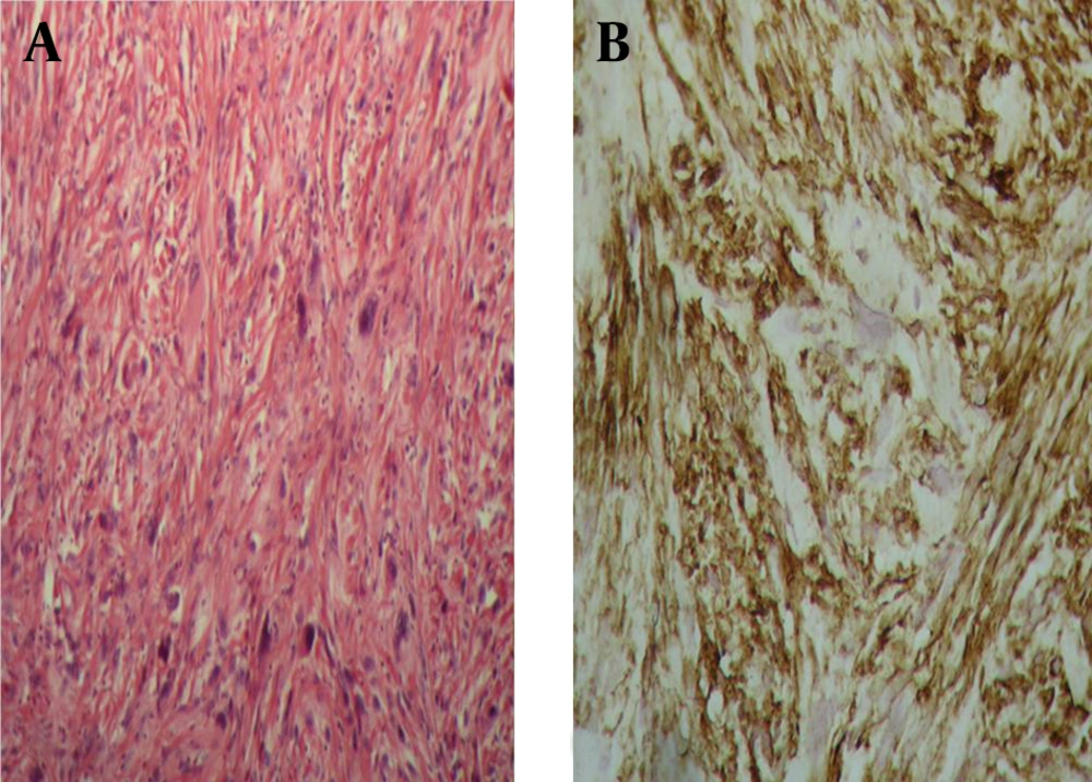
Histopathologic findings showed a neoplastic tissue with mesenchymal origin, which had arisen from smooth muscle cells. Microscopic examination revealed a highly cellular lesion composed of spindle cells with vesicular chromatin. Pleomorphic nuclei were arranged in condensed cellular groups. Neoplastic multinucleated giant cells with bizarre shape were encountered in different areas with two to three mitoses per high power Field (2-3/HPF). Alpha-smooth muscle actin (α-SMA) marker was positive in neoplastic cells. These pathologic features confirmed a leiomyosarcoma of the spermatic cord.

3. Discussion

Leiomyosarcoma constitute 5% to 10% of soft tissue sarcoma. Paratesticular leiomyosarcomas are rare malignant neoplasms originating from scrotum, spermatic cord, or epididymis (7, 8). Leiomyosarcomas of the spermatic cord are uncommon tumors of non-testicular origin, which drain into the retroperitoneal lymph nodes, and have been reported in less than 150 cases in the literature until now (9). This type of leiomyosarcoma arises from undifferentiated mesenchymal cells of the cremasteric muscle and vas deferens. Although this type of lesion is seen in all age groups, peak incidence of spermatic cord leiomyosarcoma is in the sixth and seventh decades of life. Preoperative diagnosis of spermatic cord type is difficult and is commonly made by histopathologic examination and immunochemical staining (7-10). The standard treatment for all types of non-testicular leiomyosarcomas is radical orchiectomy. Our patient had grade 1 spermatic cord leiomyosarcomas. The prognosis after radical orchiectomy is usually good in tumors of grade 1 and 2 (9, 11).

In summary, leiomyosarcoma of the spermatic cord are rare malignant tumors, which are treated with radical orchiectomy, and clinicians should notice them in differential diagnosis of a firm and hard solid mass in the cord. However, a large number of spermatic cord leiomyosarcoma are low-grade hard tumor with good prognosis, long-term follow-up is needed to prevent recurrence and metastasis.
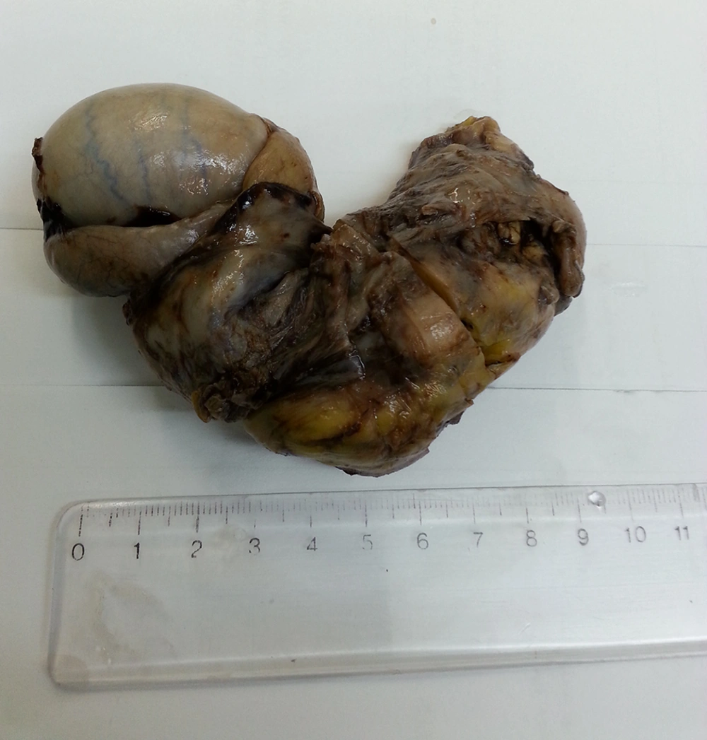
Macroscopic view of the Leiomyosarcoma of the Spermatic Cord
Figure 1.

Macroscopic view of the Leiomyosarcoma of the Spermatic Cord

A) Hematoxylin and Eosin Stain Showing High Mitotic Index. B) Tumor cells showing positive immunohistochemical staining for smooth muscle actin (SMA).
Figure 2.

A) Hematoxylin and Eosin Stain Showing High Mitotic Index. B) Tumor cells showing positive immunohistochemical staining for smooth muscle actin (SMA).

Acknowledgments

Footnotes

References

  • 1.
    Celik O, Unlu G. A rare case: paratesticular leiomyosarcoma. Asian J Androl. 2013;15(6):843-4. [PubMed ID: 23893152]. https://doi.org/10.1038/aja.2013.88.
  • 2.
    Rodriguez D, Barrisford GW, Sanchez A, Preston MA, Kreydin EI, Olumi AF. Primary spermatic cord tumors: disease characteristics, prognostic factors, and treatment outcomes. Urol Oncol. 2014;32(1):52 e19-25. [PubMed ID: 24239475]. https://doi.org/10.1016/j.urolonc.2013.08.009.
  • 3.
    Soipi S, Vucic M, Ulamec M, Tomas D, Kruslin B, Spajic B. Leiomyosarcoma of the spermatic cord with scalp metastasis: case report and literature review. Coll Antropol. 2014;38(2):763-6. [PubMed ID: 25145020].
  • 4.
    Mapulanga V, Mudenda V, Banda L. Case Report on Primary Intratesticular Leiomyosarcoma of the Spermatic Cord. Med J Zambia. 2009;36(4):191-3.
  • 5.
    Enoch S, Wharton SM, Murray DS. Management of leiomyosarcomas of the spermatic cord: the role of reconstructive surgery. World J Surg Oncol. 2005;3(1):23. [PubMed ID: 15860129]. https://doi.org/10.1186/1477-7819-3-23.
  • 6.
    Kyratzi I, Lolis E, Antypa E, Lianou MA, Exarhos D. Imaging features of a huge spermatic cord leiomyosarcoma: Review of the literature. World J Radiol. 2011;3(4):114-9. [PubMed ID: 21532872]. https://doi.org/10.4329/wjr.v3.i4.114.
  • 7.
    Patil V, Verma A, Pattar RS, Nandar S. Paratesticular leiomyosarcoma: a case report. JNMA J Nepal Med Assoc. 2013;52(191):521-3. [PubMed ID: 24907964].
  • 8.
    Galosi AB, Scarpelli M, Mazzucchelli R, Lopez-Beltran A, Giustini L, Cheng L, et al. Adult primary paratesticular mesenchymal tumors with emphasis on a case presentation and discussion of spermatic cord leiomyosarcoma. Diagn Pathol. 2014;9:90. [PubMed ID: 24885500]. https://doi.org/10.1186/1746-1596-9-90.
  • 9.
    Mohammadi Torbati P, Zham H. Epithelioid type of paratesticular leiomyosarcoma: a case report and literature review. Urol J. 2004;1(3):215-7. [PubMed ID: 17914694].
  • 10.
    de Bolla AR, Arkell DG. Leiomyosarcoma of the spermatic cord. Postgrad Med J. 1983;59(693):470-1. [PubMed ID: 6622335].
  • 11.
    Dangle P, Basavaraj DR, Bhattarai S, Paul AB, Biyani CS. Leiomyosarcoma of the spermatic cord: case report and literature review. Can Urol Assoc J. 2007;1(1):55-8. [PubMed ID: 18542764].
comments

Leave a comment here


Crossmark
Crossmark
Checking
Share on
Cited by
Metrics

Purchasing Reprints

  • Copyright Clearance Center (CCC) handles bulk orders for article reprints for Brieflands. To place an order for reprints, please click here (   https://www.copyright.com/landing/reprintsinquiryform/ ). Clicking this link will bring you to a CCC request form where you can provide the details of your order. Once complete, please click the ‘Submit Request’ button and CCC’s Reprints Services team will generate a quote for your review.
Search Relations

Author(s):

Related Articles