1. Introduction
Bladder stones are the most common form of lower urinary tract stone and constitute 5% of all urinary tract stones and 1.5% of all cases admitted to the urology departments (1). In endemic areas, bladder stones develop in children and are mostly found in the setting of poor nutritional and socioeconomic conditions. In contrary, in nonendemic regions, incidence is higher in adults and is usually accompanied with urinary stasis or foreign bodies. The giant bladder stones are very rare (2). Regarding the characteristics of the stone in our case, our report was the first one in Iran and in the world.
2. Case Presentation
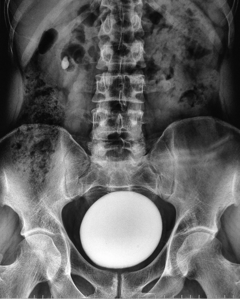
The patient was a 35-year-old man with lower urinary tract symptoms who was referred to the Urology Clinic of Imam Reza Hospital. He did not have gross hematuria or obstructive lower urinary tract symptoms. There was no history of medical and surgical disease and drug history including administration of over-the-counter drugs. Digital rectal examination revealed insignificant finding. In the examination of suprapubic area, a hard mass with no attachment to the abdominal wall or pelvic area was detected. In the primary laboratory tests, results of urine culture were negative and creatinine was 106.08 μmol/L. In urine analysis, 10/hpf of red blood cells, 2-3/hpf of white blood cells, and few oxalate crystals were found. Ultrasonography showed mild bilateral hydroureteronephrosis, and increased bladder wall thickness, and a single large stone with a diameter of 110 mm. In kidneys, ureters, and bladder x-ray (KUB), a round, homogenous, huge density in the pelvic area with smooth edges and another mass with the same density in the right kidney was seen (Figure 1).
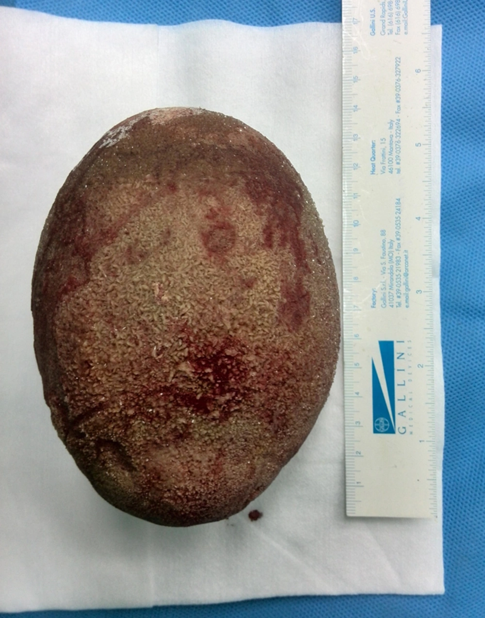
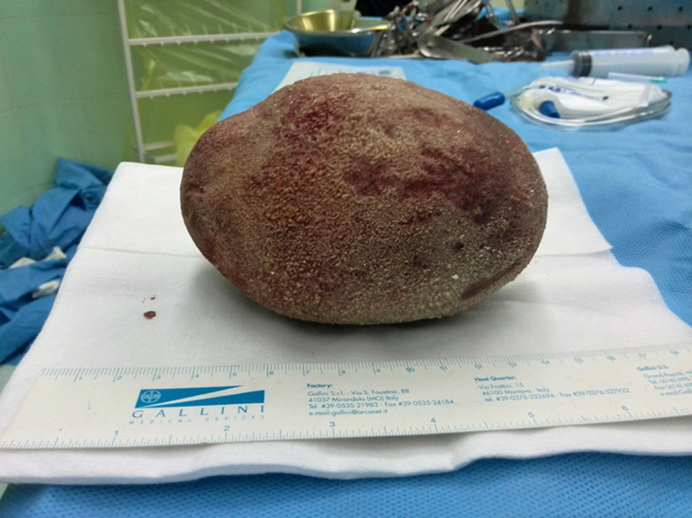
Although the diagnosis of bladder stone was made and we decided to perform open cystolithotomy, cystoscopy was done for rule out of any bladder outlet obstruction (BOO) or urethral strictures. The urethra was normal but introduction of cystoscope into the bladder cavity was impossible due to presence of a huge stone that had occupied all the bladder space. Open cystolithotomy was performed using a midline suprapubic incision and a vertical bladder incision. The size of stone resulted in entrapment and inability to extract the stone by common vesical incisions; therefore, we extended the bladder incision into the posterior bladder wall. The bladder stone weighed 826 g and a dimension of 13 × 10 × 8 cm (Figures 2 and 3). The thick bladder wall was sutured in three layers and a large bore urethral catheter was placed in the urethra. After seven days, the catheter was removed and subsequently, patient’s normal voiding was restored. Stone analysis showed 60% calcium oxalate and 20% calcium phosphate. In laboratory serum tests, PTH value was within normal limits (49.5 ng/L); other results were within normal limits: creatinine 70.72 μmol/L; uric acid 327.17 μmol/L; Na 138 mmol/L; K 4.4 mmol/L; Cl 99.3 mmol/L, Ca2+ 1.2 mmol/L; total Ca 2.40 mmol/L; and phosphorous, 0.87 mmol/L. Magnesium, Ca, oxalate, citrate, and protein in 24-hour urine were within normal limits. Results of venous blood gas test was normal (pH, 7.34; PCO2, 5.9 kPa; and HCO3, 24.5 mmol/L).
3. Conclusions
Bladder stone is mostly single and multiple stones are seen in only in 25% to 30% of cases. These stones are mainly composed of calcium oxalate, calcium phosphate, and ammonium urate. A unique point in our patient was the absence of previous history or current conditions that might predispose him to bladder stone formation such as BOO, urinary tract infection, foreign body, and urologic or pelvic operation. This situation is not compatible with bladder stone epidemiology in Iran as well as industrial countries. Another point was formation of one piece-heavy weight stone that had filled all the bladder space. In majority of reports, the stones were in multiple pieces with lighter weights (3, 4). The bladder stone with the same weight was reported from Nigeria; however, the reported patient had BOO as an underlying etiologic factor (5).
Regarding the composition of the bladder stone and the absence of underlying etiologies such as BOO and UTI, two possible factors might be considered for the development of the stone in this patient. Regarding the patient's socioeconomic status and epidemiology of bladder stone in Iran, nutritional problems seemed unlikely in our patient. Metabolic disorders are not among primary and supplementary laboratory tests. For large-sized bladder stones, all the reports have recommended open cystolithotomy (2). In a few number of reports the first presentation of bladder stones was acute postrenal failure. Nonetheless, in our patient, mild bilateral hydroureteronephrosis was found on ultrasonographic study and serum creatinine was within normal limits (6-8).


