1. Introduction
Gestational choriocarcinoma is most commonly seen in women of reproductive age, generally within the first year after pregnancy. It usually occurs following an intrauterine pregnancy. Although this condition most frequently originates from a molar pregnancy, it may also occur following an abortion or a normal delivery. Patients with intrauterine choriocarcinoma frequently present with abnormal uterine bleeding (1). Less commonly, they may present with amenorrhea and even unusual clinical presentations such as acute abdomen resulting from spontaneous uterine perforation, hematuria, hemothorax, or fetomaternal hemorrhage (1-5). About 30 percent of the patients with choriocarcinoma initially present with metastatic disease and lung is the most common site of metastatic involvement (1, 6). Renal metastasis may result in oliguria, hematuria or massive retroperitoneal hemorrhage (7). Renal metastases are almost invariably preceded by pulmonary metastases. This seems to be the result of dissemination of tumor cells from lung metastasis through the general circulation (7).
Herein, we report a case of choriocarcinoma presenting with prolonged amenorrhea followed by hematuria and left flank pain after a normal vaginal delivery.
2. Case Presentation:
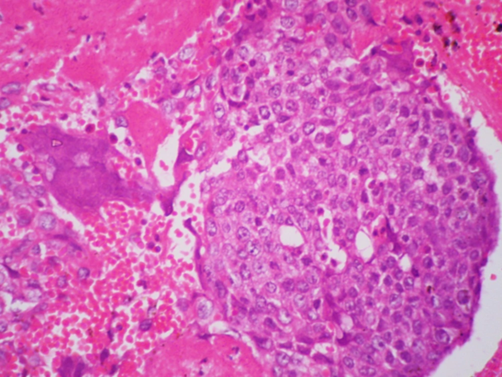
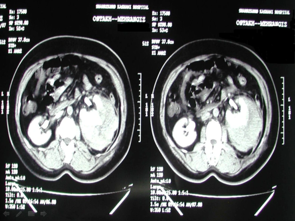
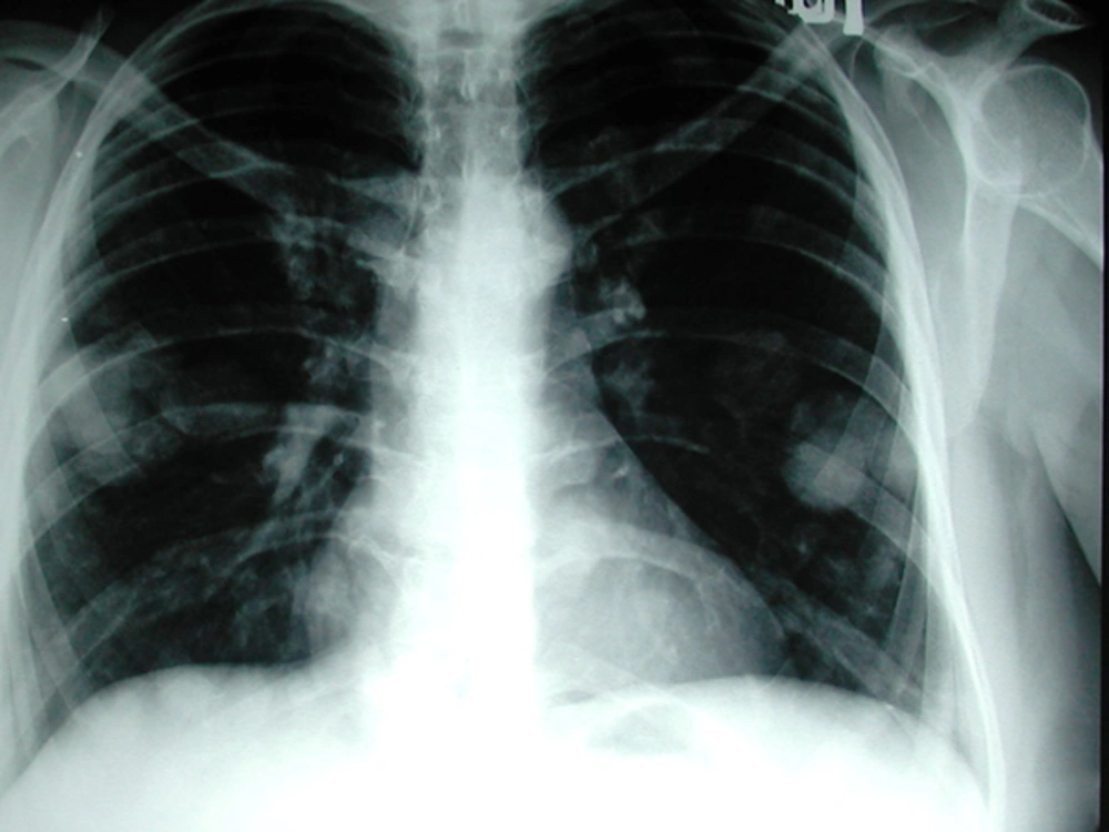
A 43-year-old rural woman (G7L6EP1) presented with intermittent amenorrhea and vaginal bleeding since her last normal vaginal delivery three years ago. Later, the patient complained of a persistent vaginal bleeding for about two months as well as hematuria and left flank pain. The patient underwent endometrial curettage. On microscopic examination of the curettage material, sheets of cytotrophoblasts and syncytiotrophoblasts were seen growing in a plexiform pattern in the background of hemorrhage and necrosis. The trophoblastic cells showed nuclear atypia and mitotic activity (Figure 1). No chorionic villi were evident. The patient was then referred to our center for treatment of choriocarcinoma. At the time of admission in our hospital, the patient had a large uterus and left flank tenderness on physical examination. Serum β-hCG level was 1500 μIU/mL at the time of admission and rised to 10000 μIU/mL before beginning the treatment. Abdominal and pelvic sonography revealed a large uterine mass with myometrial invasion and heterogenous echo pattern measuring 57 × 36 mm in the fundus and body. A 126 × 82 mm mass lesion was also seen in the middle pole of the left kidney. Computerized tomography of the abdominopelvis showed a large hemorrhagic mass in the left kidney confined to the fascia gerota (Figure 2). Two enlarged lymph nodes were also evident in the left renal hilus. Metastases to the liver and spleen were absent. On chest X-ray, multiple nodules were seen in both lungs and in the left retrocardiac space (Figure 3). Upper and lower gastrointestinal endoscopy were normal. Brain imaging revealed no brain metastasis. According to world health organization prognostic index for gestational trophoblastic disease, our patient fitted into the high risk category with a total score of 13 (age score: 1, antecedent pregnancy: 2, interval: 4, β-hCG: 1, largest tumor: 2, site of metastases: 1, number of metastases identified: 2) (8). Chemotherapy with standard EMA-CO regimen (etoposide, methotrexate, actinomycin D, cyclophosphamide, and vincristine/oncovine) was planned for the patient. Her general status was checked weekly by complete blood count, serum BUN and creatinine and liver function tests. Serum β-hCG level was also measured weekly. After receiving five courses of chemotherapy, the patient became febrile and pancytopenic. This condition was successfully managed with G-CSF, leukovorin and antibiotics. After 8 courses of chemotherapy, her serum β-hCG level fell to 3 μIU/mL. She is now in remission and is followed with serial serum β-hCG measurements.
3. Discussion
The case we reported presented with prolonged amenorrhea followed by left-sided flank pain and gross hematuria. Although 50 percent of gestational choriocarcinomas are preceded by a molar pregnancy, our patient lacked such a history and developed choriocarcinoma following a normal vaginal delivery.
Patients with high risk metastatic gestational trophoblastic neoplasia should be first treated by multiagent chemotherapy. Standard EMA/CO regimen is highly effective for treatment of high-risk gestational trophoblastic neoplasia (9). Wang et al. (7) have reviewed 448 cases of choriocarcinoma admitted in their center in a 30 year period. The incidence of renal metastasis was 6.9% in the studied population. These renal metastases were found to be very sensitive to chemotherapy. This may be attributable to the high drug concentration in the renal tissue during their excretion (7).
Adjuvant surgical procedures may be indicated in chemotherapy-resistant high-risk gestational trophoblastic neoplasia. These procedures especially include hysterectomy and pulmonary resection of chemotherapy resistant metastatic foci of choriocarcinoma in the lung. Surgical intervention may also become necessary to control hemorrhage (10). In our case, complete remission of disease was achieved with chemotherapy alone and without need to do any of these surgical procedures.
In conclusion, gestational trophoblastic diseases should be considered in the differential diagnosis of prolonged amenorrhea in patients of reproductive age with a history of prior pregnancy. Moreover, symptoms related to metastatic involvement such as hematuria and flank pain may be among the first clinical manifestations of choriocarcinoma. Metastatic lesions found in any organ including kidneys in parous women of reproductive age should raise the possibility of choriocarcinoma Standard EMA/CO regimen is usually the choice treatment in patients with high-risk gestational trophoblastic neoplasia.


