Multifocal Dysembryoplastic Neuroepithelial Tumor: A Report of a Rare Case

authors:

avatar Farzad Sina 1 , avatar Maryam Poranian 1 , avatar Saeed Razmeh ORCID 2 , * , avatar Jouan Taheri Talesh 1

Iran University of Medical Sciences, Tehran, Iran
Yasuj University of Medical Sciences, Yasuj, Iran

how to cite: Sina F, Poranian M, Razmeh S, Taheri Talesh J. Multifocal Dysembryoplastic Neuroepithelial Tumor: A Report of a Rare Case. Rep Radiother Oncol. 2018;5(1):e74048. https://doi.org/10.5812/rro.74048.

Abstract

Introduction:

Dysembryoplastic neuroepithelial tumor (DNET) is a benign tumor commonly observed in young people and mostly in the temporal lobe. This tumor usually presents with seizure and involves glial and neuronal cells.

Case Presentation:

The current report presents a case of a 25-year-old female, with intractable seizures including complex partials and secondary generalized seizure. The brain magnetic resonance (MR) showed multiple nonspecific hyperintense lesions in the bilateral temporal lobe. The patient underwent surgery, and selective temporal lobectomy was performed.

Conclusions:

Microscopic examination showed multifocal DNET, and the patient was seizure-free after surgery. Although DNET is very rare, the presentation of this case can help with its diagnosis and treatment.

1. Introduction

Dysembryoplastic neuroepithelial tumor (DNET) is a benign tumor commonly observed in the supratentorial cerebral cortex, mostly in the temporal lobe. These tumors involve glial and neuronal cells (1). The epileptic seizure is the common presentation that can cause drug-resistant temporal lobe epilepsy and usually affects young adults (2, 3). Since multifocal DNET occurs rare, it is usually reported as a case study (4). The current report presented a case of DNET and discussed radiological findings and common manifestations that should be considered in patients with intractable epilepsy and temporal lesion.

2. Case Presentation

The patient referred to the hospital with four months history of recurrent seizure that did not respond to any treatment. She also had at least four complex partials or secondarily generalized seizure daily that usually lasted for less than five minutes. The family history of seizure was positive in her son without any lesions in brain imaging. In the physical examination, she was conscious, and the neurological examination of the patient was normal. The brain magnetic resonance (MR) showed multiple nonspecific hyperintense lesions in the bilateral temporal lobe, mostly in the right side, in the white and gray matter in association with mesial temporal sclerosis (Figure 1). Moreover, there was no enhancement after gadolinium. She underwent right frontotemporal craniotomy and selective temporal lobectomy was performed, and the tissue sample was sent for histopathological examination. Microscopic examination showed a nodular aggregate of oligodendroglial-like cells bearing some mucin filled cystic space in between that replaced normal cortical neuron. The tumoral cells had round uniform nuclei, some with distinct nucleoli and perinuclear clear zone, which in some foci surrounded neuronal cells (Figure 2). In the follow-up, the patient was seizure-free after surgery.

The MRI, T2-weighted, axial and coronal showed multiple nonspecific hyperintense lesions in the bilateral temporal lobe in white and gray matter associated with mesial temporal sclerosis (A and B). Axial T1C+ image shows no significant enhancement of the tumor in the temporal lobe (C).
The H&E staining of the left temporal lesion revealed oligodendroglial-like cells, while tumoral cells had round uniform nuclei, which in some foci surrounded neuronal cells (A and B)
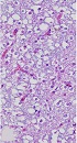
. Additional immunohistochemical findings: synaptophysin immunostain (C), neuronal glial (D), fibrillary acidic protein (GFAP) immunostain (E), and Ki-67 immunostain (F).

3. Discussion

The DNET is a benign, mixed neuronal and glial tumor that usually originates in the supratentorial cortex (5). The prognosis of this tumor is good after surgery and many patients become seizure-free. It often presents with complex partial or secondary generalized epilepsy (6, 7). Sirbu presented a case of DNET associated with sudden unexplained death in epilepsy (8). Most of these tumors are located in the temporal lobe that can be associated with mesial temporal sclerosis. The involvement of the cerebellum, thalamus, hypothalamus, pons, and basal ganglia is also reported in this tumor (9-11). Argyroupoulou et al. reported a case of DNET with cerebellar atrophy and involvement of the thalamus and internal capsule (12). In the brain computed tomography (CT), this tumor is usually hypodense and sometimes is associated with calcification and multicystic appearance. This tumor does not usually have edema and mass effect, but Guduru et al. reported a case of DNET presented with seizure and the tumor was located in the temporooccipital with mass effect and edema (13). In the current case there was an atypical presentation in the brain imaging and a multifocal hyper signal lesion in the temporal lobe in association with cortical dysplasia. Multifocal DNET is an extremely rare presentation of this tumor that can be in association with cortical dysplasia. White et al. reported a case of multifocal DNET presented with temporal lobe epilepsy and concomitant intradural spinal cord lipomas (14). In another report, Andrew et al. presented a case of multifocal DNET with seizure and there was a multiple lesion in supra and infratentorial (15). Multifocal DNET is a rare presentation, and since the tumor is multifocal, the main treatment is the medical therapy (15, 16), however, in the current case, since seizures were resistant to treatment, the patient underwent surgery and the patient was seizure-free.

References

  • 1.

    Davis GA, Kalnins RM, Fabinyi GCA. Dysembryoplastic neuroepithelial tumour and mixed DNET-ganglioglioma: Seizure outcome following surgery. J Clin Neurosci. 1997;4(4):451-6. https://doi.org/10.1016/s0967-5868(97)90034-5.

  • 2.

    Fay-McClymont TB, Hrabok M, Sherman EM, Hader WJ, Connolly MB, Akdag S, et al. Systematic review and case series of neuropsychological functioning after epilepsy surgery in children with dysembryoplastic neuroepithelial tumors (DNET). Epilepsy Behav. 2012;23(4):481-6. [PubMed ID: 22386912]. https://doi.org/10.1016/j.yebeh.2011.12.011.

  • 3.

    Fujimoto K, Ohnishi H, Tsujimoto M, Hoshida T, Nakazato Y. Dysembryoplastic neuroepithelial tumor of the cerebellum and brainstem. Case report. J Neurosurg. 2000;93(3):487-9. [PubMed ID: 10969950]. https://doi.org/10.3171/jns.2000.93.3.0487.

  • 4.

    Labate A, Briellmann RS, Harvey AS, Berkovic SF, Federico P, Kalnins RM, et al. Temporal lobe dysembryoplastic neuroepithelial tumour: Significance of discordant interictal spikes. Epileptic Disord. 2004;6(2):107-14. [PubMed ID: 15246955].

  • 5.

    Burneo JG, Tellez-Zenteno J, Steven DA, Niaz N, Hader W, Pillay N, et al. Adult-onset epilepsy associated with dysembryoplastic neuroepithelial tumors. Seizure. 2008;17(6):498-504. [PubMed ID: 18316209]. https://doi.org/10.1016/j.seizure.2008.01.006.

  • 6.

    Cascino GD. Surgical treatment for epilepsy. Epilepsy Res. 2004;60(2-3):179-86. [PubMed ID: 15380562]. https://doi.org/10.1016/j.eplepsyres.2004.07.003.

  • 7.

    Sandberg DI, Ragheb J, Dunoyer C, Bhatia S, Olavarria G, Morrison G. Surgical outcomes and seizure control rates after resection of dysembryoplastic neuroepithelial tumors. Neurosurg Focus. 2005;18(6A). E5. [PubMed ID: 16048291].

  • 8.

    Sirbu CA. Dysembryoplastic neuroepithelial tumor and probable sudden unexplained death in epilepsy: A case report. J Med Case Rep. 2011;5:441. [PubMed ID: 21899755]. [PubMed Central ID: PMC3183037]. https://doi.org/10.1186/1752-1947-5-441.

  • 9.

    Garrett M, Eschbacher J, Nakaji P. Dysembryoplastic neuroepithelial tumor: A review. Barrow Q. 2008;24:9-13.

  • 10.

    Paudel K, Borofsky S, Jones RV, Levy LM. Dysembryoplastic neuroepithelial tumor with atypical presentation: MRI and diffusion tensor characteristics. J Radiol Case Rep. 2013;7(11):7-14. [PubMed ID: 24421925]. [PubMed Central ID: PMC3888338]. https://doi.org/10.3941/jrcr.v7i11.1559.

  • 11.

    Valarmathi K, Brihadisvarar S, Sathyalakshmi R, Mary LS. Dysembryoplastic neuroepithelial tumour - Report of a rare case. J Evol Med Dent Sci. 2016;5(104):7699-701. https://doi.org/10.14260/jemds/2016/1737.

  • 12.

    Argyropoulou MI, Arvanitis DL, Tzoufi M, Fanis SL, Papadopoulou ZL, Efremidis SC. Dysembryoplastic neuroepithelial tumour and cerebellar atrophy: Case report. Neuroradiology. 2001;43(1):73-5. [PubMed ID: 11214654].

  • 13.

    Guduru H, Shen JK, Lokannavar HS. A rare case of dysembryoplastic neuroepithelial tumor. J Clin Imaging Sci. 2012;2:60. [PubMed ID: 23230542]. [PubMed Central ID: PMC3515966]. https://doi.org/10.4103/2156-7514.102057.

  • 14.

    White RD, Kanodia AK, Sammler EM, Brunton JN, Heath CA. Multifocal dysembryoplastic neuroepithelial tumour with intradural spinal cord lipomas: Report of a case. Case Rep Radiol. 2011;2011:734171. [PubMed ID: 22606558]. [PubMed Central ID: PMC3350284]. https://doi.org/10.1155/2011/734171.

  • 15.

    Yang AI, Khawaja AM, Ballester-Fuentes L, Pack SD, Abdullaev Z, Patronas NJ, et al. Multifocal dysembryoplastic neuroepithelial tumours associated with refractory epilepsy. Epileptic Disord. 2014;16(3):328-32. [PubMed ID: 25204011]. https://doi.org/10.1684/epd.2014.0680.

  • 16.

    Bonney PA, Boettcher LB, Conner AK, Glenn CA, Briggs RG, Santucci JA, et al. Review of seizure outcomes after surgical resection of dysembryoplastic neuroepithelial tumors. J Neurooncol. 2016;126(1):1-10. [PubMed ID: 26514362]. https://doi.org/10.1007/s11060-015-1961-4.