1. Introduction
Nail abnormalities are a rare sign of cutaneous T-cell lymphoma (CTCL), but this presentation is even rarer for B-cell lymphoma, especially diffuse large B-cell lymphoma (DLBCL). Their presentation can be very pleomorphic and misleading. Demonstration of specific infiltration of the nail apparatus by histological examination and B-cell colony has been very rarely reported. Moreover, although systemic therapies of lymphoma are likely to improve specific nail lesions, little is known about the effect of local treatments. Among them radiotherapy is an attractive alternative to use in situations in which the disease has spared lymph nodes or other hematopoietic organs (1). These patients usually present as refractory cases to conventional therapies such as antifungal therapy, local surgery such as nail extraction, etc. However, for definite diagnosis we need to perform a surgical biopsy for histopathological investigation and immunohistochemistry (IHC). The histopathology of the most of reported cases are cutaneous T-cell lymphoma (CTCL) and a few cases with histopathology of primary cutaneous marginal zone B-cell lymphoma (PCMZL), but DLBCL histopathology is very rare. We report the case of a patient with DLBCL histopathology who developed specific nail abnormalities that were confirmed histologically and immunohistochemically, who initially was successfully treated with radiotherapy and then systemic chemotherapy after relapse with extracutaneous nodal involvement.
2. Case Presentation
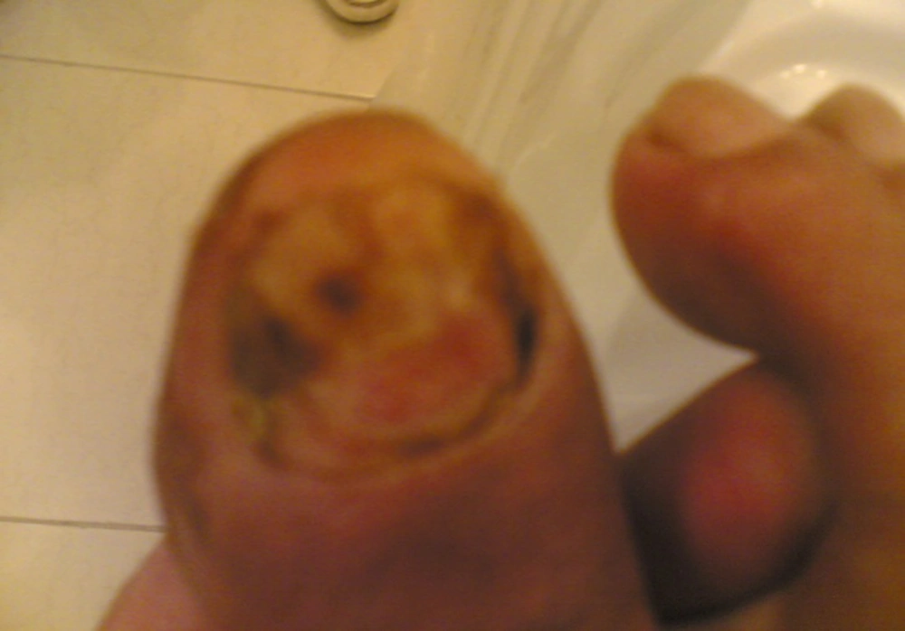
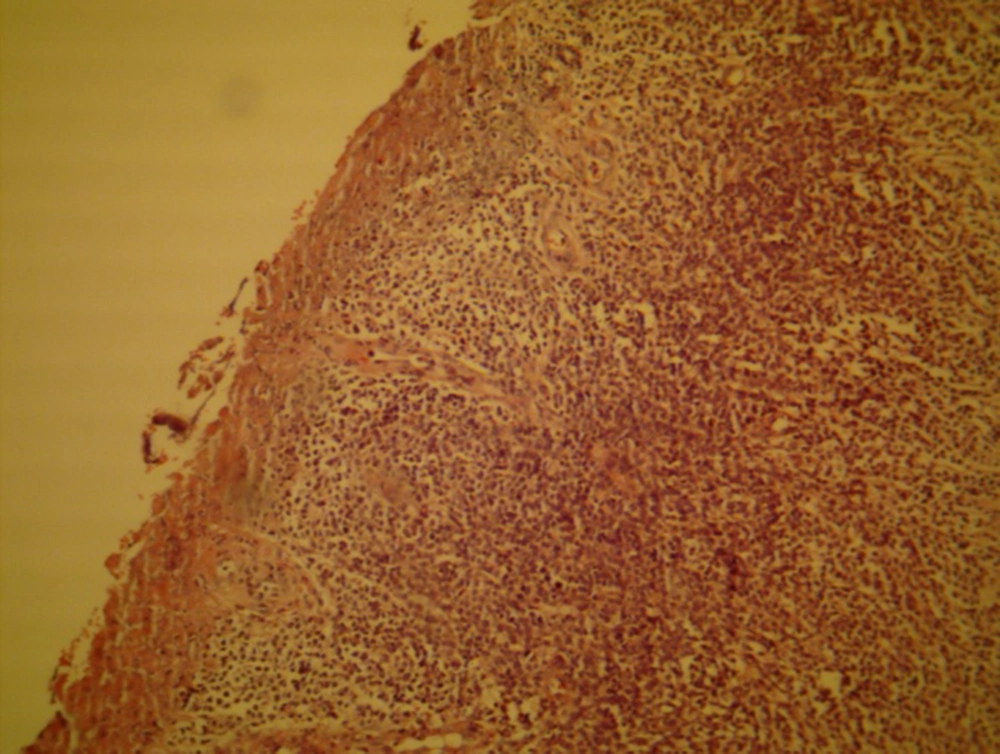
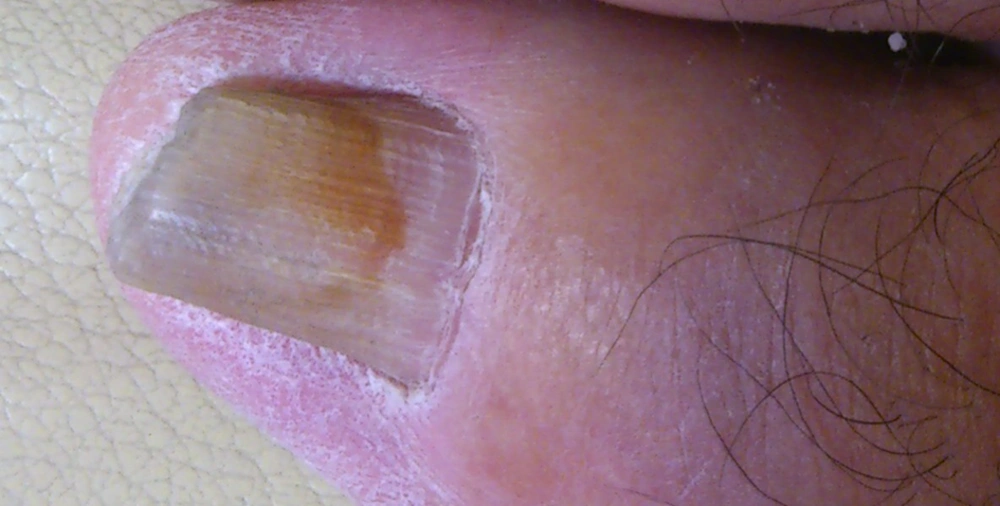
A 74 years old male patient with a history of nail deformity and nail bed ulceration for three years with suspected lymphoma presented at our department (as shown in Figure 1). He had complained of recurrent ulcerative lesions and nail deformity in the big toe for 3 years after minor trauma to the nail, and had been treated with intermittent topical and systemic antifungal medications. The patient had also undergone three nail extractions with local surgery over the last three years, with no improvement. There was no history of weight loss, anorexia, fever or night sweating. A physical examination showed that the nail was aminations were normal. The histopathology report deformed and separated from nail bed with ulcerative lesion on its internal border with mild inflammation (as erythema and tenderness). The rest of physical ex was compatible with DLBCL (Figure 2). According to IHC report: CD45 + /Pancytokeratin-/CD20 + /CD3-/CD10-/BCL-2 +. Laboratory tests such as complete blood count, LDH, and liver and renal function tests were within normal range. ESR at 1 hour was 22 mm, and CRP was 2 +. CT-scan of lungs, abdomen, and pelvis were normal. Bone marrow aspiration and biopsy were also normal. Local radiotherapy was dekivered with 6 MeV Electron and 0.5 cm bolus (3960 cGY/22F). Two months after radiotherapy, an acceptable response was detected. Patient was under routine follow-up every two months. Physical examination showed no abnormal findings up to two years, when a palpable firm semi-fixed non-tender 2 × 2 mass was detected in the proximal anterior aspect of the thigh (on the same side as nail involvement). Physical examination of other organs was normal with normal lab profiles. One month later the patient was visited and at that time an increase in size of the mass to 4 × 3 cm was observed. An incisional biopsy was done and the pathology report and IHC were compatible with DLBCL. CT-scan of lungs, abdomen and pelvis were normal, but in MRI of the thigh a residual enhancing 3 × 3 cm mass was visible. Patient was treated with CHOP regimen (cyclophosphamide 700 mg/m2, doxorubicin 50 mg/m2 vincristine 1.4 mg/m2 and prednisolone 100 mg daily/5 days) for 6 cycles followed by involved field radiotherapy (3600 cGY/20 F). During the last two years he has been under follow-up every two months. In the last visit the nail was normal (as shown in Figure 3) and there was no palpable mass on the thigh. Physical exam of the other organs were also normal with normal lab profile and imaging.
3. Discussion
The term primary cutaneous lymphoma (PCL) is used to define those lymphomas that are present in the skin and are confined to the skin without evidence of extracutaneous disease. PCL is an uncommon entity, with an overall incidence of 1 - 1.5/100,000. About 75% of all PCLs are of T-cell origin, and 25% are of B-cell origin (2). Males had a statistically significant higher IR (incidence rate) of CL than females (14.0 vs 8.2/1 000 000 person-years, respectively; male-female IR ratio [M/F IRR] = 1.72; P < 0.001) (3). Most cutaneous T-cell lymphomas are mycosis fungoides (1). Lymphomatous Unguis involvement with MF was first described as "yellow nail syndrome" of the nails. Yellow discoloration, slowed growth, nail thickening, and increased curvature were also reported in MF, in which tumor infiltration of the nail bed was confirmed histologically and immunohistochemically (4). The three main types of cutaneous B-cell lymphomas in this new classification are primary cutaneous marginal zone B-cell lymphoma, primary cutaneous follicle center lymphoma, and primary cutaneous large B-cell lymphoma. Primary cutaneous marginal zone B-cell and primary cutaneous follicle center lymphoma are indolent types with an excellent prognosis that should be treated primarily with non-aggressive therapies. Primary cutaneous large B-cell lymphoma (PCLBCL) is an aggressive lymphoma (3). PCLBCL usually needs aggressive systemic treatment (1). Primary cutaneous marginal zone B-Cell lymphoma (PCMZL) is a low-grade B-cell lymphoma that originates in the skin (5). Clinically, PCMZL is an indolent disease and has an excellent prognosis. The neoplastic cells express B-cell markers and usually bcl-2 and are negative for CD5, CD10, and bcl-6 (6-8). Most of the patients with MZL present with good performance status and without B-symptoms (9). On the optimal treatment of PCMZL nothing has been published thus far (5). Radiotherapy and surgical excision have been suggested as preferred treatments in patients with solitary or localized skin lesions (5, 10). Radiotherapy doses vary between 2000 and 4000 cGy (20 and 40 Gy) (5). The disease is very responsive to radiotherapy and doses of approximately 30 Gy can result in long-term control in more than 90% of patients (1). Topical mechlorethamine has proved to be an efficient treatment in CTCL (11). An unusual feature of a minority of cases of PCMZL is the association with B. burgdorferi infection, in which case the disease may respond to antibiotics (1). Recent studies show an association between B. burgdorferi infection and a significant minority of PCMZL cases in endemic areas in Europe. In contrast, such an association was not found in American and Asian cases (12). In a study performed by J. Hoefnage on 50 cases of Marginal Zone B-Cell Lymphoma, the majority of the patients (36/50 [72%]) presented with multifocal skin lesions, and 14 patients (28%) presented with solitary or localized lesions. The initial treatment of the patients with solitary lesions consisted of radiotherapy or excision. Cutaneous relapses developed in 19 (48%) of 40 patients who had complete remission and were more common in patients with multifocal disease (5). Development of extracutaneous disease after successful local treatment of PCMZL is rare, and usually presents with nodal involvement. In Hoefnage’s study after a median follow-up period of 36 months, only 2 patients (4%) developed extracutaneous disease with nodal involvement (5). Histological examination at the time of disease progression showed blastic transformation of the tumor cells in the lymph node biopsy specimens. Subsequent courses of chemotherapy including CHOP; dexamethasone, cytarabine, and cisplatin (DHAP); and doxorubicin, cyclophosphamide, vincristine, methotrexate, bleomycin, and prednisone (MACOP-B) ultimately resulted in complete remission of the nodal localizations. According to different studies, lymph node metastases in PCMZL are possible and may require starting a systemic treatment (13). Different studies on PCMZLs emphasized their indolent clinical behavior and excellent prognosis (14). The 5-year disease-specific survival rate exceeds 95%. In contrast to PCMZL, PCLBCL has a much worse prognosis, with a cause-specific survival rate of about 50% at 5 years. Chemotherapy with R-CHOP followed by radiotherapy is usually recommended (1).


