1. Background
The World Health Organization has introduced Leishmaniasis as one of the most important seven tropical diseases all around the world. Leishmaniasis is caused by different species of leishmania. These diseases are transmitted by the bite of infected female sandflies of the genus Phlebotomus in the Old World. There are three basic clinical forms: cutaneous, mucocutaneous, and visceral leishmaniasis (1, 2). It has a wide range of different clinical symptoms on humans and animals. Sandflies have very diverse habitats, but they are found mostly in tropical regions, although in recent years the disease has spread to other areas (3, 4).
About 12 million people from 88 countries suffer from leishmaniasis, of which 16 are among the developed countries of the world. It is estimated that about 350 million people are at risk for various forms of leishmaniasis. The number of new cases is about 2 million per year (5). Iran is located in the endemic areas of leishmaniasis. Two types of cutaneous leishmaniasis including the urban form with dry lesions, human reservoir, and its vector Phlebotomus sergenti is endemic in Tehran, Kerman, Bam, Mashhad, Nishabur and Sabzevar cities. Another form is rural of which the reservoirs are mainly rats and Gerbillinae rodents, main vector is P. papatasi and the cities of Natanz, Isfahan, Shiraz, Sarakhs, Lotf Abad, Kashmar, Baft and Khuzestan province are endemic foci (6, 7). Skin lesions caused by Leishmania may take several years until complete recovery if treatment is not taken. In this case, they can be infected with secondary infections such as yaws and myiasis (1, 8).
Several studies have shown that leishmaniasis in Iran and all over the world is on the rise. There is no certain cure for it and physicians, based on their experiences besides routine drugs, use different treatments and local methods for treatment of their patients. Reports indicate recurrence, poor recovery, or inappropriate treatment of patients (9, 10). Because today Leishmaniasis is a serious health problem in some countries including Iran, there is a need for more fundamental research in order to better understand its epidemiology and disease cycle. Fars province is now a focus of endemic cutaneous leishmaniasis due to the spread of the disease in almost all part of the province.
2. Objectives
This study aimed to investigate some epidemiologic factors of patients with cutaneous leishmaniasis referring to Marvdasht health center, Fars province, Iran. So that they can be used to better understand the main effective factors in this disease and to apply prophylactic approaches in this endemic area.
3. Methods
This descriptive-analytic study was conducted on the epidemiology of cutaneous leishmaniasis from 2010 to 2016. Accordingly, demographic and clinical data of all microscopic approved cases recorded in Marvdasht Health Center, the Infectious Diseases Unit. The routine process of case finding was passive method and in some period of time active case finding was done. Data from people who were not approved microscopically were excluded from the study. Data included demographic information such as age, sex, occupation, place of residence, season, number of lesions, and history of disease.
To analyze the data, descriptive statistics such as mean, standard deviation, percentage estimation and frequency of qualitative data were used. Also, statistical charts such as linear charts were also used. For analysis of variables, independent sample t-test and ANOVA were used and analyzed by SPSS version 16 software. The significance level was 5%.
4. Results
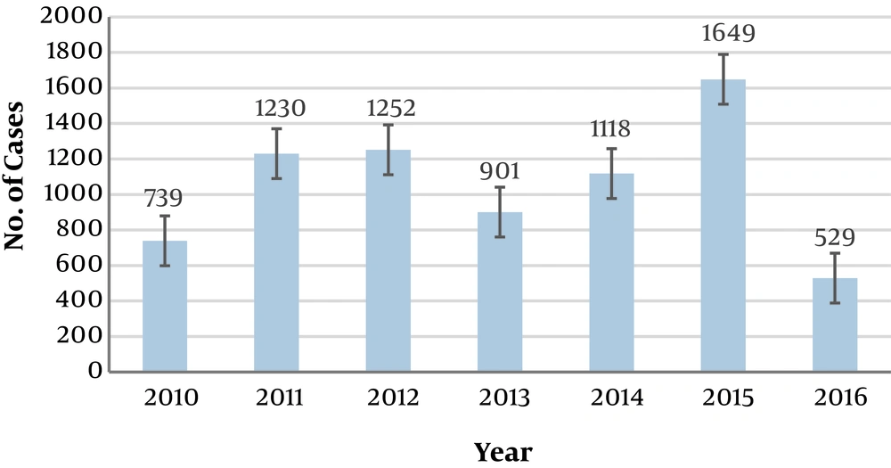
This study was conducted from 2010 to 2016 in Marvdasht, Iran. Accordingly, during the aforementioned years, 7418 confirmed cases of cutaneous leishmaniasis referred to local health centers for receiving treatment (all participants provided written informed consent). During this period of time, most cases of disease were recorded in 2015 (22.2%) and the least cases belonged to 2016 (7.1%) and 2010 (10%) (Figure 1). There were significant differences between year 2016 and the previous years (except 2010) for mean number of cases. And also there were significant differences between mean number of cases of 2015 and other studied years (P value = 0.001). There were no significant differences between number of cases in two sexes (P value = 0.86), 54.4% of the cases were men and 45.6% of cases were women.
In total population, the mean age of patients was 32.9 ± 26.9 years; this was 32.2 ± 28.7 and 33.6 ± 24.7 for women and men respectively. There was no statistically significant difference between the mean age of both sexes (P value = 0.89). The highest number of cases of cutaneous leishmaniasis occurred in the 21 to 30-year-old group, and the least cases were in people over the age of 60 years. 14.6% of cases were in category of less than 10 years old (Table 1). According to the findings, the highest number of cases of disease occurred in autumn and the least occurred in spring (Table 2). Also, the incidence of diseases in rural areas (58.4%, 95% CI: 0.5815, 0.5864) was higher than urban areas (41.6%, 95% CI: 0.4105, 0.4215).
| Age, y | Frequency | % |
|---|---|---|
| < 10 | 1084 | 14.6 |
| 11 - 20 | 954 | 12.9 |
| 21 - 30 | 1695 | 22.9 |
| 31 - 40 | 1346 | 18 |
| 41 - 50 | 1086 | 14.7 |
| 51 - 60 | 713 | 9.6 |
| 60 > | 540 | 7.3 |
| Total | 7418 | 100 |
| Season | Frequency | % |
|---|---|---|
| Spring | 197 | 2.6 |
| Summer | 1013 | 13.7 |
| Fall | 4119 | 55.5 |
| Winter | 2089 | 28.2 |
| Total | 7418 | 100 |
The study of the clinical consequences of lesions in people with cutaneous leishmaniasis showed that, the mean duration of exposure until the onset of clinical signs of the disease was estimated to be 151 ± 55.9 days. Moreover, location of lesions on the face and upper limbs was present 62.8%, on the lower limbs 25.2%, and in 12% of the cases it was observed in both parts. Most lesions were on the hands and the least cases were on the trunk (Table 3). The average number of lesions was 1.588 per person. In 2484 cases only one lesion was recorded, and the least number of lesion was related to the person with 4 and 5 lesions (Table 4).
| Place of Lesion | Frequency | % |
|---|---|---|
| Hand | 2937 | 39.6 |
| Face | 695 | 9.4 |
| Trunk | 166 | 2.2 |
| Foot | 1587 | 21.4 |
| Foot and leg | 284 | 3.8 |
| Trunk and foot | 102 | 1.4 |
| Hand and trunk | 332 | 4.5 |
| Hand and foot | 787 | 10.6 |
| Face and trunk | 528 | 7.1 |
| Total | 7418 | 100 |
| Number of Lesion | Frequency | % |
|---|---|---|
| 1 | 2485 | 33.5 |
| 2 | 1661 | 22.4 |
| 3 | 1112 | 15 |
| 4 | 571 | 7.7 |
| 5 | 573 | 7.7 |
| 6 ≤ | 1016 | 13.7 |
| Total | 7418 | 100 |
In all examined patients, the average size of the lesions was 4.27 cm, with the smallest of 1 cm and the largest of 5 cm wide.
5. Discussion
According to the results of the present study, the number of cases of disease in males was higher than that of females, but there was no significant difference between average age of morbidity in two sexes. In the study of Nilforoushzadeh et al. 2015 in Isfahan, the rate of disease in men (61.8%) was higher than that of women (38.2%) (6). Also, in other studies the percentage of cutaneous leishmaniasis cases in men was reported 64.1% in Ilam (Ilam province), 56% in Andimeshk (Khuzestan province), 61% in Khorasan-Razavi province, 61.8% in Khatam (Yazd province) and 93.8% in Hamedan (Hamedan province). In all mentioned studies, morbidity rate was significantly different between males and females (10-14). But unlike previous studies, in Lamerd city (Fars province), the number of cases in women (51.8%) was higher than men (48.1) (15). The higher morbidity rate of disease in men can be attributed to some reasons such as their higher outdoor activity, less clothing and body coverage (compared to women who wear hijab in Iran), and more contact with vectors.
Our findings showed that the highest number of cases occurred in the age group of 21 - 30 years. In Isfahan, most cases were in the age group of 10 - 30 years and in Andimeshk in the age group of 15 - 24 years (6, 12). Furthermore, the highest number of cases in Khorasan-Razavi province, Khatam (Yazd province), and Hamedan province were in the age group of under 10 and 20 - 30, 10 - 30, 15 - 24 years old respectively (10, 13, 14). Our results are in accordance with results of the above studies and suggest that in both sexes, the incidence of disease in young people is higher than in other age groups.
In the present study, the incidence of disease in rural residents was higher than urban residents. In contrast to our study in Hamadan and Isfahan provinces, the rate of urban cases was more than rural areas (6, 14). The incidence of disease in urban or rural areas, as well as in certain occupations, is due to the difference in the epidemiological cycle of the disease (agents, vectors, reservoirs, and biological differences) in different geographical regions.
In Isfahan, due to the fact that more people live in the cities that are located on or near colonies of gerbil rodents and also phlebotomine sandflies as vectors exist. Therefore, all conditions for the transmission of the disease are available in this endemic urban area. But in our study, due to the fact that the residents of rural areas were more exposed to infected reservoirs and vectors, more cases were reported from these areas.
According to our findings, most lesions were on the hands and the least were on the trunk. And also most of the patients had 1 lesion on their body. In Isfahan, Fars, Khuzestan, and Ilam provinces, their results were completely similar to ours (6, 11, 15-18). In Andimeshk, most lesions were on the hands and feet (12). In Hamedan, most patients had 1 to 2 lesions on their hands and feet (71.6%) (14). Due to the fact that sandflies are not able to bite their host through clothing, lesions are often seen on uncovered areas such as hands, feet and face. Also, the number of lesions on the body indicates multiple exposures to disease vectors and number of bites.
In accordance with other studies, most cases of disease occurred in autumn and the least cases occurred in spring (6, 12, 13, 16, 18). In studied area the activity of sandflies is typically seasonal and there is a main peak of activity in the end of April. Considering that there is usually a 6-month incubation period for cutaneous leishmaniasis, therefore the symptoms of the disease appear in autumn.
The most important limitation of our study was that, some of the leishmaniasis cases did not go to the health centers actively, so in some period of time active case finding was done instead of routine process (passive method) in order to find all cases in these important endemic foci.
5.1. Conclusions
Marvdasht city is an endemic area of cutaneous leishmaniasis in Iran. According to the results, the incidence of disease was higher in men and mostly occurred in rural areas in autumn. As a result, targeted preventive measures (vector control, reservoir control, case finding and treatment, health education, etc.) should be taken by health authorities and decision makers based on the basic information of high risk population and other epidemiological and ecological factors affecting the transmission of disease in this endemic area.
Our results showed that the number of cases reported has fluctuated in recent years. Therefore, it is very important for the health system to implement an accurate and effective continuous monitoring strategy for this important vector-borne disease in this part of the country.
