Correlation Between Breast Tumor Size and Lymph Node Involvement in Patients Referring to the Pathology Department of Rasoul Akram Hospital in Iran

authors:

avatar Maryam Mohammadhosseini 1 , avatar Ahmad Majd ORCID 1 , * , avatar Hamidreza Mirzaei 2 , avatar Mona Farhadi 3 , avatar Nasrin Shayanfar 4

Department of Biology, North Tehran Branch, Islamic Azad University, Tehran, Iran
Cancer Research Center (CRC), Shahid Beheshti University of Medical Sciences, Tehran, Iran
Departments of Microbiology, Karaj Branch, Islamic Azad University, Karaj, Iran
IRAN University of Medical Sciences (IUMS), Tehran, Iran

how to cite: Mohammadhosseini M, Majd A, Mirzaei H, Farhadi M, Shayanfar N. Correlation Between Breast Tumor Size and Lymph Node Involvement in Patients Referring to the Pathology Department of Rasoul Akram Hospital in Iran. Thrita. 2020;9(2):e110659. https://doi.org/10.5812/thrita.110659.

Abstract

Background:

Breast cancer has the highest mortality rate, second to gastric cancer, among Iranian women and is one of the most common cancers in the world. The incidence of breast cancer in women is increasing gradually. Meanwhile, ductal breast carcinoma experiences more increases than other malignancies and is one of the most important health problems.

Objectives:

This study aimed at evaluating women with ductal breast carcinoma because of the significance of pathological factors and their association with breast cancer progression.

Methods:

This retrospective study was conducted using data of ductal breast carcinoma women during the years 2018 and 2019. In this cross-sectional study, demographic data (age, sex, and pathology of breast mass) of 50 patients referring to Rasoul Akram hospital (Tehran, Iran) were gathered. Then, the data were analyzed by SPSS 26 software using the t test and Levene's test. The results were presented using descriptive statistics.

Results:

Fifty patients with ductal carcinoma were assessed based on their pathological information. The examination of factors including tumor size, involvement/non-involvement of lymph nodes, histological grade, and age of patients revealed a significant direct relationship between tumor size and lymph node involvement (P <0.05), while no significant relationship was found with other mentioned factors.

Conclusions:

The prevalence of ductal breast carcinoma in Iranian women is increasing that may lead to death in many patients. Thus, it is necessary to evaluate this disease. In this study, a significant relationship was found in terms of tumor size and lymph node involvement, which can be effective in early diagnosis and prevention of this type of cancer.

1. Background

With the highest mortality rate, breast cancer is one of the most prevalent cancers in the world, so that 502,000 women annually die due to this disease (1, 2). Based on available statistics, breast cancer accounts for approximately 33% of gynecological cancers, with an estimated prevalence of 8 to 10% in different countries. In Iran, accounting for 24.4% of all cancers, breast cancer is the first prevalent cancer in women. Recent studies in Iran have revealed that the rate of breast cancer is 17.81%, which has significantly increased in recent years (1, 3).

Breast cancer is a heterogeneous disease in clinical behavior. Pathological factors like pathology degree, tumor size, lymph node metastasis, histological type, vascular invasion, and cell proliferation rate may be effective in specifying the prognosis and the need for adjuvant therapy (4). Tumor size and lymph node status for most breast cancer patients are bio-indicators of tumor invasion and independent prognostic factors for survival after diagnosis. The tumor size and the number of positive lymph nodes in axillary autopsy are related directly. An association has been observed for decades to date in all major studies performed, with the majority of studies on the BRAC1 mutation (5, 6).

Ductal carcinoma in situ (DCIS/stage 0 breast cancer) mammographically detects about 20% of breast cancers (7). Five percent of cases in women are diagnosed before the age of 40 (8). The factors predicting mortality after diagnosis of DCIS are not known. In particular, preventing invasive recurrences using radiotherapy or extensive breast surgery (mastectomy) has not been suggested to decline the mortality rate of this type of breast cancer. Breast cancer mortality rates are low in women with DCIS, and a large group is required to be studied for a long time to accurately estimate the mortality rate.

2. Objectives

This study aimed at evaluating the association between the pathological specifications of breast cancer patients with ductal carcinoma to reduce the mortality rate in these patients

3. Methods

In this study, 50 patients with breast cancer of ductal carcinoma type were recruited. All patients were equal in terms of gender (female), not receiving chemotherapy and mastectomy. Necessary permission and ethical code were obtained from the Ethics Committee of the Islamic Azad University, North Tehran Branch, to keep the confidentiality of patient data based on the Helsinki Declaration. The patients’ data were regarded as confidential and used only for research purposes.

This was a descriptive-analytical, cross-sectional study. The statistical population included patients with a diagnosis of breast cancer referring to Rasoul Akram Hospital from June 22, 2018, to June 21, 2019. A convenience sampling was used. Data were collected by a field method and completing checklists using patients’ files. The checklist included age, sex, location of the tumor, the extent of tumor invasion to the peripheral organs, stage of involvement of lymph node, besides the metastatic involvement, tumor stage, type of tumor pathology, and tumor size.

3.1. Statistical Analysis

After recording in checklists, raw data were entered into SPSS 26 software and presented by descriptive statistics (mean, standard deviation, number, and percentage). To evaluate the differences between the size of the tumor and involvement/non-involvement of lymph nodes, the independent t test was used. To perform the t test, it was necessary to check the assumption of the homogeneity of variances in the two groups. For this purpose, Levene's test was used.

4. Results

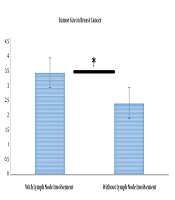
As indicated in Table 1, when lymph node involvement was present, the mean tumor size was 3.44, while it was 2.42 in the absence of involvement. To test this hypothesis, the independent t-test was used, the results of which are presented in Table 2. To assess the equality of variances, the results of Levene’s test were first assessed. As observed in Table 2, Levene’s statistic is not significant for the size variable, suggesting that the condition of the equality of variances was met. Since t = 2.72 and P < 0.05, the difference in tumor size between the two involved and non- involved lymph node groups was significant (Figure 1).

Table 1.

Descriptive Data About Tumor Size in Two Groups With and Without Lymph Node Involvement

Standard Error (SE) of the MeanStandard Deviation (SD)Average Tumor SizenStatus
0.421.923.4421Lymph node involvement
0.211.132.4229Lymph node non-involvement
Table 2.

T-test Results of Two Independent Groups on the Difference in Tumor Size Between the Two Groups With and Without Lymph Node Involvement

VariableConfidence interval of 0.95Standard error of the meanCompare meansSignificance levelDegree of freedom (DF)T valueLevene's test
Upper limitLower limitSignificance levelF value
Tumor size0.141.890.431.010.023482.430.102.72
The difference in tumor size between two groups with and without lymph node involvement

5. Discussion

Breast cancer is the most prevalent cancer among women, with an increasing rate in developing countries. In Iran, breast cancer involves women at least a decade earlier than in developed countries (9). Although specific treatments have been developed, improving the treatment results, approximately one-third of treated patients get involved with the metastatic type (10, 11). Hence, the outcome of initial treatment should be further improved, and more effective treatment strategies should be developed for recurrent and metastatic disease. The association between breast cancer and pathological factors is one of these types of strategies.

Many studies have been carried out in many countries suggesting the significance of tumor size and lymph node involvement in estimating the breast cancer prognosis (12-14). In a study conducted by Fisher et al. (1969) on 2,578 patients with breast cancer, they found an association between the size and lymph node status (5). In 1978, Valagussa et al. studied 716 patients and reported that the survival rate was directly proportional to the size of the initial tumor (15). In the same year, a linear relationship was reported by Smart et al. between the tumor size and lymph node involvement (16). An analysis of data from 24,740 breast cancer cases recorded in the SEER program of the US National Cancer Institute by Carter et al. revealed a linear relationship, as well (17). In 2017, Samavati et al. reported that among malignancies, invasive ductal carcinoma was the most prevalent malignancy in Iran (87.6% of all malignancies) with an increasing rate (18). Thus, given the increased number of patients with invasive ductal carcinoma in our country (Iran), this type of breast cancer was studied, and a direct relationship was obtained between the tumor size and lymph node status, confirming previous studies.

The age under 20 years was not observed in this study, which is in line with other studies (19-21), indicating the importance of breast masses in the ages of 40 to 60 years that are the most malignant masses in this age range (22). The mean age of 50 patients in the present study was consistent with previous studies.

5.1. Conclusion

In this study, we showed a correlation between lymph node status and tumor size in ductal breast carcinoma that may lead to metastasis and recurrence in the disease. The rate of this disease and its mortality is increasing in Iran, so it is suggested that more attention be paid to adopting preventive measures besides early diagnostic methods and assessing more risk factors in this regard.

Acknowledgements

References

  • 1.

    Ranjkesh M, Fathi Azar F, Ghatreh Samani F, Tarzamni MK, Vali Khani E. Evaluation of adjunctive sonography results in screening of women with mammographically dense breasts for early diagnosis of breast cancer. Iranian J Breast Dis. 2017;10(1):7-19.

  • 2.

    Salmaninejad A, Kangari P, Shakoori A. Oxidative stress: development and progression of breast cancer. Tehran Univ Med J. 2017;75(1):1-9.

  • 3.

    Shayan A, Khalili A, Rahnavardi M, Masoumi SZ. The relationship between sexual function and mental health of women with breast cancer. Sci J Hamadan Nurs Midwifery Fac. 2017;24(4):221-8. https://doi.org/10.21859/nmj-24042.

  • 4.

    Allred DC, Harvey JM, Berardo M, Clark GM. Prognostic and predictive factors in breast cancer by immunohistochemical analysis. Mod Pathol. 1998;11(2):155-68. [PubMed ID: 9504686].

  • 5.

    Fisher B, Slack NH, Bross ID. Cancer of the breast: Size of neoplasm and prognosis. Cancer. 1969;24(5):1071-80. https://doi.org/10.1002/1097-0142(196911)24:5<1071::aid-cncr2820240533>3.0.co;2-h.

  • 6.

    Olivotto IA, Jackson JS, Mates D, Andersen S, Davidson W, Bryce CJ, et al. Prediction of axillary lymph node involvement of women with invasive breast carcinoma. Cancer. 1998;83(5):948-55. https://doi.org/10.1002/(sici)1097-0142(19980901)83:5<948::aid-cncr21>3.0.co;2-u.

  • 7.

    Ernster VL, Ballard-Barbash R, Barlow WE, Zheng Y, Weaver DL, Cutter G, et al. Detection of ductal carcinoma in situ in women undergoing screening mammography. J Natl Cancer Inst. 2002;94(20):1546-54. [PubMed ID: 12381707]. https://doi.org/10.1093/jnci/94.20.1546.

  • 8.

    Brinton LA, Sherman ME, Carreon JD, Anderson WF. Recent trends in breast cancer among younger women in the United States. J Natl Cancer Inst. 2008;100(22):1643-8. [PubMed ID: 19001605]. [PubMed Central ID: PMC2720764]. https://doi.org/10.1093/jnci/djn344.

  • 9.

    Sirous M, EBRAHIMI A. The epidemiology of breast masses among women in Esfahan. Iranian J Surg. 2008;16(3).

  • 10.

    Clarke R, Skaar TC, Bouker KB, Davis N, Lee Y, Welch JN, et al. Molecular and pharmacological aspects of antiestrogen resistance. J Steroid Biochem Mol Biol. 2001;76(1-5):71-84. https://doi.org/10.1016/s0960-0760(00)00193-x.

  • 11.

    Libson S, Lippman M. A review of clinical aspects of breast cancer. Int Rev Psychiatry. 2014;26(1):4-15. [PubMed ID: 24716497]. https://doi.org/10.3109/09540261.2013.852971.

  • 12.

    Daly MB, Clark GM, McGuire WL. Breast cancer prognosis in a mixed Caucasian-Hispanic population. J Natl Cancer Inst. 1985;74(4):753-7.

  • 13.

    Paterson AHG, Zuck VP, Szafran O, Lees AW, Hanson J. Influence and significance of certain prognostic factors on survival in breast cancer. Eur J Cancer Clin Oncol. 1982;18(10):937-43. https://doi.org/10.1016/0277-5379(82)90241-3.

  • 14.

    Rosen PP, Saigo PE, Braun DJ, Weathers E, DePalo A. Predictors of recurrence in stage I (T1N0M0) breast carcinoma. Ann Surg. 1981;193(1):15-25. [PubMed ID: 7458446]. [PubMed Central ID: PMC1344996]. https://doi.org/10.1097/00000658-198101000-00003.

  • 15.

    Valagussa P, Bonadonna G, Veronesi U. Patterns of relapse and survival following radical mastectomy.Analysis of 716 consecutive patients. Cancer. 1978;41(3):1170-8. https://doi.org/10.1002/1097-0142(197803)41:3<1170::aid-cncr2820410355>3.0.co;2-i.

  • 16.

    Smart CR, Myers MH, Gloeckler LA. Implications from seer data on breast cancer management. Cancer. 1978;41(3):787-9. https://doi.org/10.1002/1097-0142(197803)41:3<787::aid-cncr2820410301>3.0.co;2-e.

  • 17.

    Carter CL, Allen C, Henson DE. Relation of tumor size, lymph node status, and survival in 24,740 breast cancer cases. Cancer. 1989;63(1):181-7. https://doi.org/10.1002/1097-0142(19890101)63:1<181::aid-cncr2820630129>3.0.co;2-h.

  • 18.

    alizade otaghvar H, samavati S, nafissi N, hosseini M, gholami S. [Evaluation of Breast Masses Prevalence in an Educational Hospital in Iran]. Razi J Med Sci. 2017;24(158):17-23. Persian.

  • 19.

    Afsharfard A, Mozaffar M, Orang E, Tahmasbpour E. Trends in epidemiology, clinical and histopathological characteristics of breast cancer in Iran: results of a 17 year study. Asian Pac J Cancer Prev. 2013;14(11):6905-11. [PubMed ID: 24377624]. https://doi.org/10.7314/apjcp.2013.14.11.6905.

  • 20.

    Ziaei JE, Sanaat Z, Asvadi I, Dastgiri S, Pourzand A, Vaez J. Survival analysis of breast cancer patients in northwest Iran. Asian Pac J Cancer Prev. 2013;14(1):39-42. [PubMed ID: 23534759]. https://doi.org/10.7314/apjcp.2013.14.1.39.

  • 21.

    Soheili F, Alizadeh S, Hassani M, Bastami P. The pathologic assessment of breast masses, before and after Menopause. Iranian J Obstet Gynecol Infertility. 2013;16(68):21-7.

  • 22.

    Nguyen-Pham S, Leung J, McLaughlin D. Disparities in breast cancer stage at diagnosis in urban and rural adult women: a systematic review and meta-analysis. Ann Epidemiol. 2014;24(3):228-35. [PubMed ID: 24462273]. https://doi.org/10.1016/j.annepidem.2013.12.002.