1. Introduction
Septic arthritis is one of the problems of rheumatological and orthopedic emergency medicine (1, 2). The disease is caused by various microorganisms, particularly bacteria, causing destruction of the joint space. Research has shown that certain infections and diseases or the use of some medications can cause the disease to develop (2).
Goldenberg believes that bacteria can enter the joints during different surgeries or via intra-articular corticosteroid injections and thus can cause septic arthritis (3). However, conclusive and specific evidence has not been established for the etiology of this disease (2).
The prevalence of this disease in several countries and in patients of different ages has been investigated. One research article determined that eight out of one hundred patients with more than one of the symptoms, such as fever, pain, and arthritis, who were reported on in 1987, suffered from septic arthritis. Jeng and his colleagues’ study from 1993 to 1995 reported that twenty out of seventy-five cases (27%) who were referred to the emergency department with acute arthritis had been suffering from septic arthritis (4).
In addition, from 2003 to August 2013, twenty-four patients with twenty-five events of septic arthritis were identified at a general hospital in Chile (5).
This disorder causes the most difficulties in the knee and hip joints (3, 6, 7). It causes clinical symptoms, such as pain, swelling, inflammation, stiffness, and a limited range of motion in both active and passive joints (7-9).
Debridement of the necrotic tissue at the site of injury is one way to treat this disorder, but this may be followed by malfunctions of the soft tissue around the joint. Reconstructive surgery is often necessary to cover the damaged areas and reconstruct deep soft tissue in the joints, especially when the knee joint is creating a problem. Various methods of flaps can be used to reconstruct soft tissue defects around the damaged joint. Thus choosing the correct method of reconstruction can lead to the improvement of a patient’s condition and general satisfaction (10, 11).
In this case, a study was done on a patient with septic arthritis in both knees, and the method of soft tissue reconstruction around the knee joint was employed. The soft tissue healing practices were different in each knee, so we are reporting on our case to explain the methods and outcomes of the surgery.
2. Case Presentation
We are reporting on the case of a patient who was treated at Sina teaching and research hospital in 2015 in Tehran, Iran. Three months after surgery, the patient’s range of motion, gait and balance were assessed. In this section, our surgical techniques and reconstruction of the soft tissue of the injured joints will be discussed.
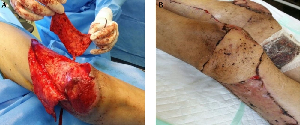
The patient, a 50 year-old man, who had septic arthritis in both knees, was admitted to the orthopedic ward of Sina hospital. He was an addict who had been previously operated on in both knees. After debridement, due to soft tissue defects, a reconstruction consultation and graft operation was performed and this was unsuccessful, so the surgeon decided to repair the soft tissue defects in the left knee with the lateral distal thigh island flap (Figure 1A) and to use the medial head gastrocnemius flap (Figure 1B) method on the right knee to reach a better outcome.
2.1. The Surgical Method for the Lateral Distal Thigh Island Flap
The lateral distal thigh island flap was performed on the patient’s left knee. The flap was taken from a third of distal and lateral thigh. This flap was used to cover the anterior part of the femur and the outside of the knee joint. It measured approximately 18 × 7 cm. The flap was supplied by a large cutaneous branch arising from the first collateral artery of the popliteal artery. The terminal cutaneous branch emerged in the subcutaneous tissue, 10 cm proximal to the knee joint. This branch was the pedicle of the flap.
The flap was outlined on the lateral aspect of the thigh. The emergence of the pedicle was marked 10 cm proximal to the articular space in the depression between the posterior border of the Maissiat’s band and biceps femoris. First, the flap from the anterior, without fascia and areolar tissue, were raised (Figure 2A) and the vastus medialis and fascia lata were retracted. The vascular pedicle arising from the popliteal artery was then exposed and the posterior border of the flap then divided. The flap that had the basis of the pedicle for covering the anterior or lateral knee was then rotated (Figure 2B).
2.2. The Surgical Method for the Medial Head Gastrocnemius Flap
Another method of reconstruction of the damaged soft tissue was used on the right knee the gastrocnemius muscle flap. This method can be recommended as the best way to cover the leg and knee because it is a simple surgical technique and the source of the blood supply to the flap is appropriate. The flap also has more bulk muscle than that of the lateral head.
By performing two separate cuttings and using a separate wide sub-facial technique, this method can be used without serious danger to the skin of patients with two non-arteriosclerotic raised parts of muscles. Attempting to cover one-third of the proximal tibia and the anterior and medial parts of the knee is more common than approaching the lateral part because the muscular body into head of muscle ratio is longer and the arc of rotation of the transfer allows coverage of the proximal third of the tibial, anterior and medial aspects of the knee. The patient is placed in a supine position and their lower limb slightly turned out and placed in a little flexion. An incision is made into the mid-calf, 2 cm behind the posteromedial border of the tibia to begin and the proximal is pulled to the popliteal fossa. Damaging the saphenous vein and nerves should be avoided.
The skin is then cut with deep fascia and a large skin flap is retracted with aponeurosis to the back in order to assess the distance between the two ends of the gastrocnemius. The plane between the soleus and gastrocnemius muscles should be dissected with the fingers. The plantaris tendon is often at this location. The position of the sural nerve in the posterior view gastrocnemius is then determined. It is located between the two heads of the gastrocnemius. The tendon of the medial head is cut down from the distal then and care must be taken about the pedicle. Muscle is dissected from the distal to the proximal and then it can be cut off from its beginning on the femur.
However, the pedicle proximal must not be damaged. This motion will increase the arc of rotation and muscle can be passed under the Gracilis and semi-tendinosus tendons to reach the anterior knee.
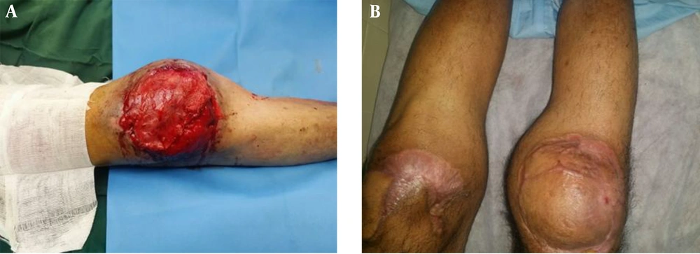
The arc of rotation of the muscle can be increased by releasing the beginning part of the femoral muscle. The cutting the skin can be continued to the lower part of the thigh until the Gracilis, semi-tendinosus, and Sartorius tendons become visible. The medial head of the gastrocnemius separate from the base of this vessel should be passed through the gracilis and semi-tendionosus in order to have the ability to cover the anterior part of the knee (Figure 3A).
2.3. Evaluation and Results
Three months after surgery, both flaps were evaluated and there was not any problem in the color, texture and elasticity of the flaps (Figure 3B). The next step was the evaluation of the active range of motion in the lower extremities and this was performed using a goniometer. The results indicated a full range of dorsiflexion and plantar flexion in both ankles and also full range of motion in the hips. In addition, the movements of flexion, extension, abduction, and adduction were reported in both feet. The range of motion of the knee joints were measured and there were ten degree defects concerning knee extension in both knees.
The assessment of lower extremity muscle strength was also conducted by using manual muscle assessment. The degree of quadriceps muscle strength in both knees was 4+, the hamstring muscles on the right foot was 4+, and 3+ on the left leg. The degree of power in the back of the leg cuff muscles in both legs was 3+. The evaluation of balance was performed via a balance sheet (BBS) and the score obtained by the patient was 26 which represents average balance.
3. Discussion
Muscle flaps are one of the best ways to reconstruct tissue around the joints and bones (12). The use of the flap is valuable because of the possible presence of bone and joint injuries where the blood supply of regional and sub-cutaneous areas is disrupted. Muscle flaps with good internal blood supply resources, configurability and the filling of bony areas represents the best option to reconstruct soft tissue defects resulting from joint and bone injuries.
Smith and colleagues investigated the appropriate surgical techniques to reconstruct soft tissue injury in the lower limb for five years. They reported on sixty patients on which they used three methods of flap, including the gastrocnemius flap method (thirty-five patients), the ipsilateral gastrocnemius transfer method (fourteen patients), and the cross-leg flap (five patients). One of the problems caused by cross-leg flaps was found to be long-term immobilization in the lower limbs and poor body condition. Finally, they concluded that if failure in the transfer ipsilateral gastrocnemius happens, using a free flap is be best way to reconstruct soft tissue (13).
Bashir introduced the gastrocnemius tenocutaneus island flap restoration method and the internal head of the gastrocnemius was used in this, but, in this method, the skin area was placed in the upper end of the lower muscle (12).
Other methods have also been introduced to cover the knees. The posterolateral thigh flap and popliteal posterior-fascial-dermal flap had successful results, but because of the existence of the distal base, they lacked proper nerve sources. Therefore, this would not be a perfect choice to reconstruct soft tissue deficiencies around the knee joint (10).
The saphenous flap is also another way to cover and reconstruct the knee joint and its surrounding tissue, but the complexity of the donor flap creates some problems. In some cases, sensory disorders in the inner part of the leg and the stretching of the scar tissue from the donor part around the joint are difficulties arising from this surgical method (10)
The standard methods for the reconstruction of soft tissue defects in the leg include the gastrocnemius flap for proximal third defects. One of the best reasons to use the gastrocnemius muscle is the unique vascularization of the gastrocnemius muscle (one pedicle to each head), the size of the muscle belly, the fact that it is situated in the dissection field and that its transfer does not affect the function of the spare limb too adversely which makes it particularly suitable for limb-sparing procedures for sarcoma in the region of the knee and popliteal fossa (14, 15).
The reason for using this method in patients was to expose and uncover the bone and joint. Therefore, it is not possible to use skin grafts as this can cause secondary contracture in this area. The use of this flap of muscle tissue provides a suitable platform to create a range of motion in the knee joint.
Concerning the tissue reconstruction of the patient’s left knee, it is important to mention that the joint was reconstructed with skin grafts in the past, but the surgery was not successful. Considering that the patient’s bone was not exposed, a skin flap was used. In this case, there was also the possibility of using a free flap, but it would have required a longer time for surgery and there is always the chance with this method that the flap might have failed and other side effects occurred, so this option should only be considered as the last resort.


