1. Introduction
Wegener’s granulomatosis (WG) is a partially rare and lethal disease, characterized by granulomatous inflammation, and necrotizing vasculitis of small and medium size vessels (1). Its cause is not known. Multiple organs are involved, particularly the upper respiratory tract, the lungs, the kidney and the eyes (2, 3). It affects both males and females. It can begin at any age with a peak during the 5th and 6th decades of life with initial symptoms being non-specific. They mostly include symptoms in the upper respiratory tract, weakness, fatigue, arthralgia and fever without an infectious cause.
Glomerulonephritis (GN) is a common cause of morbidity and mortality. Its expression can be indolent or more frequently, aggressive. Urine analysis has indicated proteinuria and an increased level of urea and serum creatinine. Renal biopsy of patients with Wegener’s Granulomatosis has mostly shown modifications of necrotic, segmentary and focal GN and sometimes in the absence of significant deposits of immunoglobulin and complement and more rarely diffuse necrotic GN with different proliferating modifications.
Skin involvement has been seen in nearly 50% of patients. The initial manifestation of the disease may include deep inflammatory cutaneous plaques, acute or indolent. Other manifestations are palpable purpura, pyoderma-like ulcerations, gingival hyperplasia, inflammatory papule, small ulcers, panniculitis and subcutaneous nodules (4). Generally, the activity of cutaneous affection is related to the systemic disease. Although the histopathology for WG is not pathognomonic for cutaneous involvement.
2. Case Presentation
The case was a 63-year-old female who had presented the first symptoms of Wegener’s disease four years ago: sinusitis and prolonged fever. The otorhinolaryngologic examination rendered evident lesions of the upper respiratory tract. The performance of antineutrophil cytoplasmic antibodies (ANCA) was positive with an initial titre of 1/170 with perinuclear pattern in immunofluorescence. The diagnosis of Wegener’s granulomatosis was established and the patient was treated with prednisolone and cyclophosphamide. The disease improvement was great obtaining a high improvement in biological activity markers (PCANCA decreased to 1/20). Recently, the patient was presented to the emergency department with lower extremities and palpebral edema. She also complained of the appearance of a mild painful ulcer with an erythematous background, located on the middle section of her right arm, which had appeared seven days prior to her referral.
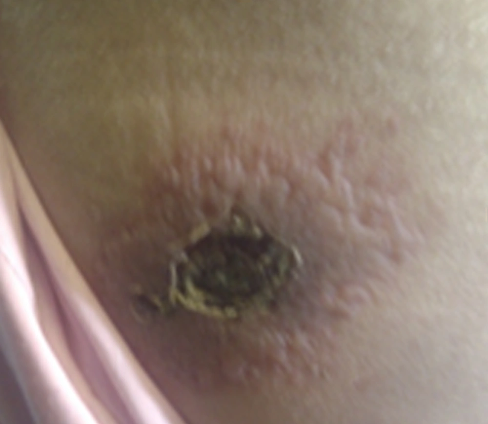
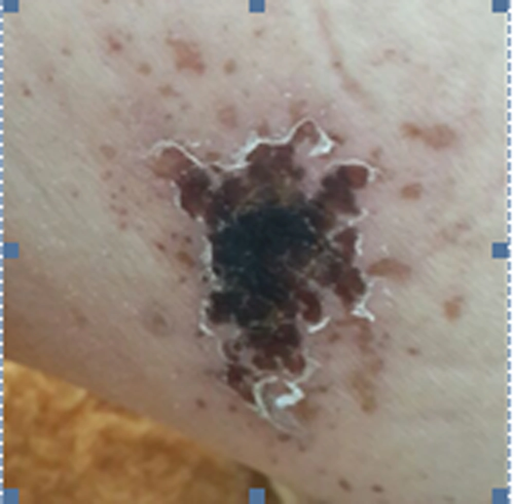
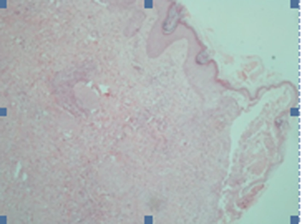
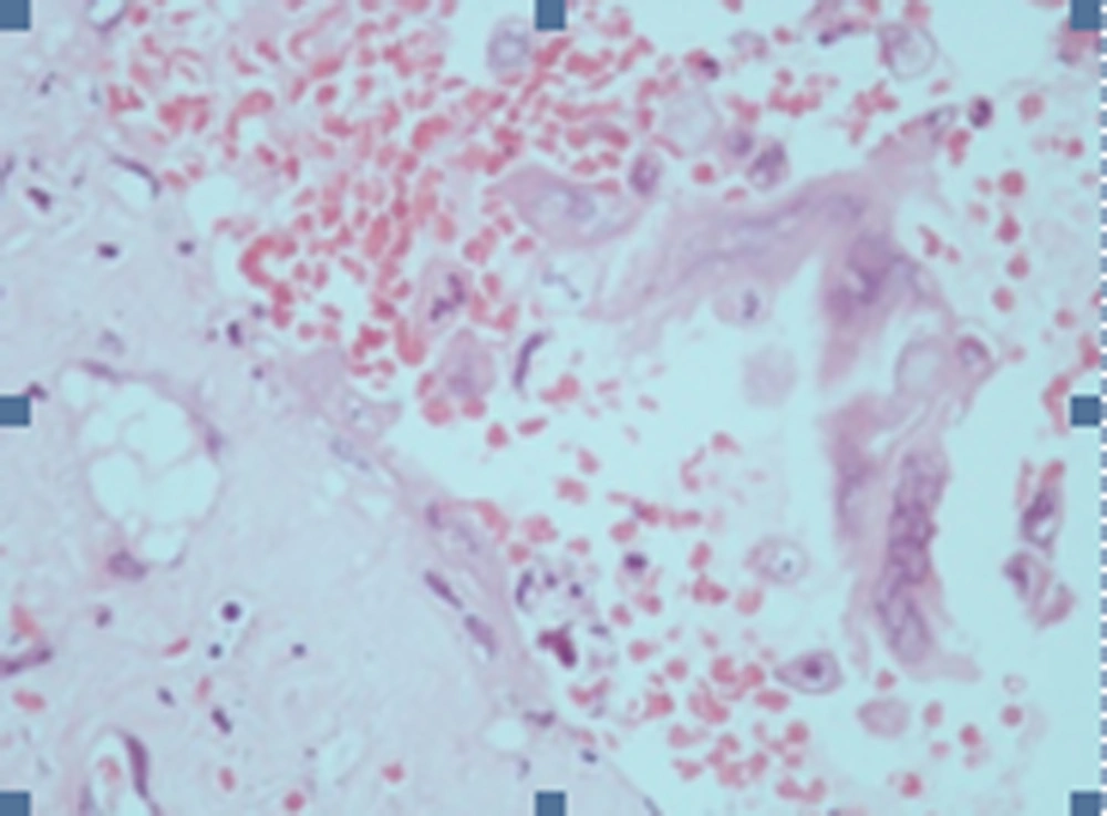
On examination, there was erythema of the arm within which there was a crusted or ulcerated lesion surrounded by small vesicles (Figure 1). Its appearance looked like vasculitis so the patient underwent a punch biopsy. The diagnosis of herpes zoster was confirmed by the biopsy. In the biopsy, ballooning degeneration and herpetic cytopathic changes were evident (Figure 2). Acantholysis or multinucleated keratinocytes formation were obvious (Figure 3). A treatment with acyclovir 10 mg/kg was set for seven days. After two weeks the herpetic lesion evolved greatly (Figure 4). Laboratory tests indicated anemia, increased rate of blood segmentation, proteinuria, low albumin and a high level of serum creatinine. A renal biopsy puncture showed lesions of diffused extra-capillary glomerulonephritis with sclerosis. The patient was followed for about one year. There was no recurrence of herpes zoster, yet two flares of renal disease with rise of creatinine occurred.
3. Discussion
We presented a more unusual case of WG, who had indications of infection of the upper respiratory tract, kidneys and skin yet in a totally uncharacteristic way. Our case was rare due to herpes infection one week prior to disease recurrence and also strange presentation of herpes zoster, which resembled vasculitis. This report could serve as a guide for physicians, informing them that herpes zoster infection could be a prior presentation before WG recurrence and also skin presentation of herpes zoster in Wegener’s granulomatosis could mimic other diseases. The presence of cutaneous presentation of herpes zoster would be explained by the modulation of immunity. Herpes zoster is a painful neurocutaneous disease caused by reactivation of Varicella zoster virus (5). A variety of immunocompromised patients are known to be at increased risk of herpes zoster (6, 7). Wegener’s granulomatosis (WG) is a type of systemic vasculitis characterized by necrotizing granulomatous inflammation (8). Immunosuppressive regimens, especially high doses of glucocorticoids and cyclophosphamides, induce remission in about 80% to 90% of patients (9, 10); unfortunately, disease flares following remission are common. Current standard of care treatments are related to opportunistic infections (9). Cupps et al. reported on the frequent occurrence of herpes zoster among Wegener’s granulomatosis, with the Fauci and Wolff regimen, which includes “initially high doses of glucocorticoids and daily cyclophosphamide”. Recently, shorter courses of cyclophosphamide are employed for remission reduction; however, the risk of occurrence increases. Renal dysfunction and female sex were consistently strong risk factors for herpes zoster in a study by Wung et al. (11). Thus, it is recommended to prevent herpes zoster in patients on treatment for immune-mediated diseases. Another special feature of our case was the discovery of anti-cytoplasmic neutrophilic antibodies namely anti-peroxidase (mPO3) antibodies with perinuclear pattern (P-ANCA) by the immunofluorescence technique. As it has been documented anti-protein 3 antibodies with cytoplasmic antineutrophil cytoplasmic antibod (CANCA) are a characteristic of WG. However, anti-peroxidase antibodies in 5 to 10% of patients with WG are associated with P-ANCA. Another special character of our case was recovery for about four years since the initiation of disease showing good prognosis; however, recurrence with renal involvement indicating a bad prognosis.


