

1. Introduction
The most common congenital heart disease among children is a ventricular septal defect (VSD). The final diagnosis, as well as measuring the size of and spotting VSD using echocardiography during infancy, is of the utmost importance in improving the patient's prognosis. Such patients are at risk of pulmonary hypertension (PH) during infancy (1), which surgery should repair as soon as possible. Some patients suffer from such complications as aortic regurgitation, infectious endocarditis or pulmonary valve stenosis, heart failure, and sudden death (1). The most important factor in the surgical treatment and future health conditions of these patients is how pulmonary artery blood flow is established, and the intracardiac structure of these patients is of secondary importance. Common disorders associated with this disease are patent ductus arteriosus (PDA) and coarctation of the aorta. Thus, in many cases, reducing one of the pulmonary shunts, repair of PDA, and coarctation of the aorta through intervention will lead to preventing pulmonary hypertension and an easier approach to VSD. In these children, PDA can result in heart failure or endocarditis, so it is necessary to close it by surgical and non-surgical procedures such as hemoglobin correction and fluid and sodium restriction, using amplatzer or coil during angiography, indomethacin, nitric oxide synthase inhibitors and ibuprofen synthesis (2-4). In PDAs with a size of less than 3 mm, using coils with a 97% success rate and zero mortality is recommended; in PDAs with a size of more than 3 mm, amplatzer placement with over 98% success rate is recommended; and in PDAs larger than 12 mm, closing the duct with an amplatzer suitable for closing atrial septal defect (ASD) is recommended (5-7). In cases of pulmonary hypertension, the risk of death is higher, with the highest risk of bleeding from the pulmonary artery at the stitching site. Old age, even with no pulmonary hypertension, increases the risk of surgery due to the fragility and calcification of the duct. In the present study, we aim to introduce a case of repairing coarctation of the aorta by stenting, performed in an infant with VSD due to placement of a large amplatzer and its displacement towards the aorta leading to severe coarctation during ductus arteriosus closure. This case report has been approved by the Ethics Committee of Rafsanjan University of Medical Sciences and has the ethics code IR.RUMS.REC.1399.250.
2. Case Presentation
The patient was a one-and-a-half-year-old infant girl, 9 kg, with Down syndrome due to natural childbirth, term weighing 3000 g at birth time, with no family history of heart disease or congenital heart disease, who was brought to the hospital because of congenital heart disease. During her initial examination, a second loud heart sound and a systolic ejection murmur could be heard in the left sternal border. The results of the previous echocardiography indicated the presence of VSD and bidirectional shunt, serious coarctation of the aortic arch with a gradient of 70 mmHg, and pulmonary hypertension observed. The patient had undergone angiography in Shiraz Hospital 5 months ago due to congenital heart disease, and the results indicated VSD, PDA, mild coarctation of the aorta, and pulmonary hypertension. The patient's PDA had been closed during angiography to allow for VSD surgery; however, since the coarctation had not been repaired by balloon aortoplasty before closing PDA and a large amplatzer had been used, the edge of the amplatzer had protruded, leading to more severe stenosis and coarctation of the aorta. Finally, the patient's blood pressure showed no decrease due to the stenosis caused by the amplatzer, so the surgeon could not perform surgery since pulmonary hypertension had not been treated. Hence, the authors of the present article decided to repair the coarctation of the aorta by stenting. The first medications prescribed were captopril, tadalafil, and furosemide (each 1 mg/kg dose), and after two weeks, the patient underwent angiography. Figure 1 shows the steps for placing a stent in the aortic arch. In angiography, a formula stent with a diameter of 10 mm and a length of 30 mm was placed. Using a stent next to the amplatzer and moving the amplatzer towards the pulmonary artery, the coarctation of the patient's aorta was repaired so that no gradient was observed, and pulmonary hypertension fell from 90 to 45 mmHg. One month after the intervention, the infant's general condition, weight, and feeding status were good in the follow-up. In the above patient, whose pulmonary hypertension has decreased after stenting and coarctation repair, in case of proper weight gain, it will be possible to close the VSD with an amplatzer, and there will be no need for open surgery.
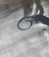
. A, aortogram in lateral view: Stenosis is shown with an arrow; B, aortogram in lateral view: Balloon angioplasty steps and inserting formula stent; C, aortogram in lateral view after stenting: Coarctation repaired.")
Stages of inserting a stent in the aorta by angiography in an infant with ventricular septal defect (VSD). A, aortogram in lateral view: Stenosis is shown with an arrow; B, aortogram in lateral view: Balloon angioplasty steps and inserting formula stent; C, aortogram in lateral view after stenting: Coarctation repaired.
3. Discussion
As the results of the present study showed, failure to repair coarctation with a balloon before closing PDA and using a large amplatzer caused the amplatzer edge to protrude, leading to more severe stenosis and coarctation of the aortic arch in the patient and then preventing pulmonary hypertension from dropping. The surgeon could not perform surgery to close the VSD. Five months after closing the PDA, the patient was brought to the hospital with severe coarctation and pulmonary hypertension. Coarctation was repaired successfully by stenting. The number of cases where surgical procedures are used to close the PDA has gradually diminished because interventional procedures have replaced them and are limited to hard cases and cases where patients have various cardiac disorders requiring corrective surgery, pulmonary hypertension, or cases of premature infants. PDA closing technique using a venous and arterial procedure which can be done using an amplatzer and coil has fewer complications such as bleeding, embolism, dysrhythmia, and length of hospitalization (4). However, complications such as embolism, leakage, and protrusion into the aortic artery have been reported during the placement of the tools (8).
Stenting is a new method used in the treatment of heart diseases, such as coarctation of the aorta, because of the following factors:
More skillful interventionists; various experiences in using different stents; easier access to work tools and procedure facilities; gaining acceptance of non-surgical procedures among parents; preventing multiple surgeries due to disease recurrence after surgery; and daily advances in technologies of designing and manufacturing more suitable stents that can be used at a younger age (9).
3.1. Conclusions
Diagnosis and treatment of VSD during infancy is very important in the patient's health and needs to be repaired by surgery. An effective factor in these patients' treatment and health condition is how to establish pulmonary artery blood flow, which the proper placement of an amplatzer can achieve. The occurrence of coarctation of the aorta following improper amplatzer placement can be repaired by stenting during angiography.
