1. Background
Cavum septum pellucidum is an essential marker for the identification and evaluation of the neural axis of the fetus, and this is a marker between the output of the septum pellucidum as a fluid-filled space. Evaluation of the characteristic of cavum septum pellucidum is recognized as an important component of embryonic ultrasound in the second and third trimesters (1, 2). The septum pellucidum is a thin, black, two-lined line extending from the front of the trunk to Geno and the corpus callosum rostrum to the upper fornix. The septum pellucidum begins to form at 10 - 12 weeks of pregnancy and develops into adulthood at 17 weeks. This is a closed cavity in the brain seen in the midline in the transverse view between the two layers of the septum pellucidum and separates the lateral ventricles. In recent decades, evaluation of the cavum septum pellucidum has been recommended as part of the standard examination required in fetal morphology guidelines. Embryonic development of cavum septum pellucidum is associated with corpus callosum, and the absence of cavum septum pellucidum is particularly associated with a wide range of neuroanatomical malformations such as lethal cases (in some cases of hydranencephaly and alobar holoprosencephaly) and non-lethal cases (schizencephaly, porencephaly, encephalocele, severe hydrocephalus, and syntelencephaly). In fact, evaluation of cavum septum pellucidum and its absence allows the detection of very mild cases and easier diagnosis of septoptic dysplasia (3). The frontal horns are the anterior portions of the lateral ventricles that lie on the sides of the cavum septum pellucidum. According to Practical Parameters published in 1985 by the Ultrasound Institute of Medicine, American College of Obstetrics and Gynecology and Radiology Association, cavum septum pellucidum is examined in fetal screening examinations (US) in biparietal diameter view (4). Frontal horns are typically imaged, and the interpreter must be familiar with their appearance. The studies’ front horns are categorized as triangular or butterfly (5-7). These abnormal morphological features of frontal horns, such as enlargement, square-shaped, spear-shaped, and splaying, may be associated with congenital intraventricular abnormalities such as holoprosencephaly (6) and schizophrenia. In addition, because cavum septum pellucidum and FH are close together, FHs can provide clues to assess cavum malformations. A previous study indicated that CSP should be seen in 100% of cases from 18 to 37 weeks, but this was not observed in other clinical experiences (8, 9). Although previous studies have evaluated FH sizes in neonates (10), few studies have investigated the natural size range of Cavum septum pellucidum in fetuses (11).
2. Objectives
Since the size range of cavum septum pellucidum varies among different ethnic populations and also the necessity of measuring it due to its association with brain anomalies and also because there has been no study in the Iranian population so far, we decided to measure the cavum septum pellucidum in the second and third trimester and determined its spectrum and compared it with the normal and abnormal reference ranges of cavum septum pellucidum in populations of other countries.
3. Methods
The present study was a descriptive-analytical study on all pregnant women in the second and third trimesters of pregnancy referred to the sonography clinic of Ali Ibn-e Abitaleb of Zahedan in 2018. A sampling method was available, and 500 fetuses were evaluated. The researcher performed the ultrasound for pregnant women in the second trimester (18 - 20 weeks) and third (36 - 38 weeks) after explaining the conditions and goals of this study and taking written informed consent at the clinic. Exclusion criteria included intrauterine fetal death (IUFD) oligohydramnios, known fetal abnormalities, multiple pregnancies, and maternal chronic diseases such as diabetes and hypertension. For all pregnancies, gestational age was calculated on the basis of LMP and ultrasound (in the second trimester based on BPD and in the third trimester based on FL). All ultrasounds were performed by an operator with at least two years of experience in pregnancy ultrasound and an ultrasound device (Philips). Ultrasound measurements were performed using a 3 - 5 MHz transducer and trans-abdominal method. Finally, the size of the cavum and BPD, and FL (depending on gestational age) were measured for each pregnancy, and the result was recorded in the information form.
3.1. Ethical Consideration
Written informed consent was obtained from all participants. The current study has been approved by the Ethics Committee of Zahedan University of Medical Sciences (code: IR.ZAUMS.REC.1399.053).
3.2. Statistical Analysis
The data were entered into SPSS software for statistical analysis. SPSS software (version 21) was used for data analysis. Descriptive statistics, central and dispersion indices, charting, tables adjustment, and frequency distribution were determined. An Independent t-test was used to compare quantitative data. The significance level was considered as 0.05.
4. Results
As shown in Table 1, the descriptive findings indicate that the mean age of the mothers under study was 26.36 years old.
| Age (y) | Min | Max | Mean ± SD | Middle |
|---|---|---|---|---|
| Value | 14 | 48 | 6.66 ± 26.36 | 25 |
Table 2 shows the mean cavum septum pellucidum size in the second trimester of pregnancy was 3.71 ± 0.81 mm and 6.11 ± 1.09 mm in the third trimester, and the overall value was 6.11 ± 1.09 mm. It was at the highest value in the third trimester (Table 2).
| Trimester | N | Min Size (mm) | Max Size (mm) | Mean ± SD | Middle |
|---|---|---|---|---|---|
| Second | 249 | 2 | 6 | 3.71 ± 0.81 | 3.8 |
| Third | 251 | 2.59 | 9 | 6.11 ± 1.09 | 6.1 |
| Total | 500 | 2 | 9 | 4.91 ± 1.53 | 4.80 |
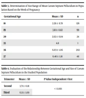
According to the results of Table 3, it was observed that the highest mean of cavum septum pellucidum in the population was at 37 weeks of pregnancy, and the lowest mean of cavum septum pellucidum in the population was at 18 weeks of pregnancy. It can be concluded that as the gestational week increases, the size of the cavum septum pellucidum also increases.
| Gestational Age | Mean ± SD | n |
|---|---|---|
| 18 | 3.59 ± 0.78 | 126 |
| 19 | 3.81 ± 0.82 | 99 |
| 20 | 3.92 ± 0.84 | 24 |
| 35 | 4.8 | 1 |
| 36 | 6.03 ± 1.06 | 202 |
| 37 | 6.46 ± 1.18 | 48 |
Table 4 shows a significant difference between the two groups. The mean cavum septum pellucidum size in the second was significantly less than in the third trimesters of pregnancy (P < 0.001) (Table 4).
| Trimester | Mean ± SD | P Value Independent t-Test |
|---|---|---|
| Second | 3.71 ± 0.81 | < 0.001 |
| Third | 6.11 ± 1.09 |
5. Discussion
In this study, 500 fetuses were evaluated. In the ultrasound examination, the mean cavum septum pellucidum size was 4.91 ± 1.53 mm, and the mean cavum septum pellucidum size in the second and third trimesters was 3.71 ± 0.81 mm and 6.11 ± 1.09 mm, respectively. In a detailed examination in weeks 18, 19, 20, 35, 36, and 37 of pregnancy, the mean cavum septum pellucidum size was 3.59 ± 0.78, 3.81 ± 0.82, 3.92 ± 0.84, 4.8, 6.03 ± 1.06 and 6.46 ± 1.18 mm, respectively. Falco et al. (in Italy) stated that the mean cavum septum pellucidum of the fetus in the second and third trimesters by ultrasound was 5.3 ± 1.7 mm (mean ranging from 2 to 9 mm) (8). Therefore, the cavum septum pellucidum has a mean size of less than 6 mm in the second trimester of pregnancy and is often larger than 6 mm in the third trimester of pregnancy. The present study was compared with the Hu study, which examined fetal cavum septum pellucidum in 2019 (California) (12); these studies show that the size of the cavum septum pellucidum in the second trimester of pregnancy is about 3 mm, while in the third trimester reaches about 6 mm; also this study was compared with the study of Jou et al. on ultrasound measurements of cavum septum pellucidum (13), the mean size of cavum septum pellucidum in the second trimester of pregnancy in both studies was about 3 to 4 mm. In the study of Serhatlioglu et al. in 2003 (Turkey), the mean septum size in the second trimester was 3.1 ± 1.5 mm and 0.5 ± 1.4 mm in the third trimester (14), respectively. In the present study, the mean size of cavum septum pellucidum in the third trimester was 11.6 mm. Consequently, the size of the cavum septum pellucidum in the third trimester is larger than the study by Serhatlioglu et al. in Turkey (14). In the Ruth Grundahl et al.’s study (Frankfurt) in the neurological examination of fetuses of diabetic and normal mothers, the median size of the cavum septum pellucidum was 4.3 (3.3, 3.5) mm; this assessment occurred in the third trimester (41 weeks of pregnancy) (15). But in the present study, the mean size of cavum septum pellucidum in the third trimester was 6.11 mm. This indicates that our study’s mean size of the cavum septum pellucidum is higher than that of Ruth Grundahl et al. study (15). Given that the sizes are always related to the operator and the accuracy of the used devices, it can be argued that this size difference is justified (16, 17), but in most studies that are mentioned, measurements are close to our study.
5.1. Conclusions
In general, the size of the Cavum septum pellucidum in our study was 4.91 ± 1.53 mm. The size of the cavum septum pellucidum is associated with increasing gestational age.
