1. Background
The clinical success of endodontic treatments is associated with a suitable crown restoration. Proper restoration provides esthetic, bears the occlusal load, maintains the remaining tooth structure, and prevents microleakage of oral cavity bacteria into root canals (1, 2). Numerous studies have confirmed the importance of coronal seals for endodontically treated teeth, and there can be no doubt about this (3, 4).
Bonding to normal dentin has always been challenging because of its organic components and dentin tubules containing fluids and a variety of compositions. Endodontic treatment exacerbates this challenge due to changes in dentin's mechanical and physical properties or inhibition of restorative resin polymerization (2).
Irrigants, intracanal dressings, and sealers are an integral part of root canal treatment. Irrigants may lead to dentin changes, including dentin and collagen solubility or dehydration of dentin. Sealers also modify the wettability and reactivity of the dentin surface. Irrigant or sealer residues in dentin or dentin tubules may reduce the bonding and inhibit the polymerization of adhesives (5-9). Some studies have suggested a one-week delay in bonding after endodontic treatment to eliminate the negative effects of irritants. However, this delay time is not always possible in clinical conditions, and sometimes the tooth should be repaired immediately after endodontic treatment (10).
Epoxy resin-based sealers are widely used in endodontic treatments due to their favorable biological and physicochemical properties. The presence of residual materials on the dentin surface significantly reduces the bond strength of adhesive systems to pulp chamber dentin (2, 8, 9). Several materials and strategies have been investigated to remove the sealers residuals from dentin (1, 8). Roberts et al. showed that cleaning the AH Plus contaminated dentin using Endosolv R has no negative effect on the tensile bond strength of the adhesive to dentin and is similar to the uncontaminated control. At the same time, eucalyptol reduced bond strength in the study by Topcuoglu et al. (11, 12). The study by Zang et al. demonstrated that ultrasound could remove root canal sealer residues and improve the microtensile bond strength when combined with acetone (13). Tian et al. showed that the bond strength of a self-etch adhesive decreased significantly for the dry cotton group. Still, ethanol and KC (a surfactant-based cleaner) restored bonding performance after cleaning, with no significant difference from the control (14).
Despite different methods and materials to remove the sealer, their efficiency has yet to be determined. Variations in the results of irrigants, sealers, and dressing effects on the dentin bond strength and the provision of extensive commercial bonding systems have necessitated further study in this area.
2. Objectives
This ex vivo study examined the effects of different sealer removal methods on dentin's Single Bond Plus micro-tensile bond strength exposed to different endodontic irrigants.
3. Methods
3.1. Tooth Selection and Dentin Preparation
After approval by the Ethics Committee (IR.ZAUMS.REC.1397.152), 45 sound-extracted mandibular third molars were collected. The soft tissues were removed with a curette. All specimens were examined under a 10x magnifying glass (Carl Zeiss; Jena, Germany) to exclude those with fracture lines, wear, and decay. The teeth were stored in 0.2 % thymol solution at 4°C for no longer than two months before use.
Teeth were vertically mounted in autopolymerized acrylic resin in cylindrical plastic containers (25 mm diameter and 20 mm high). To expose flat dentin surfaces, the teeth were sectioned 3 mm below the occlusal surface using a slow-speed saw (400 rpm) with a diamond disk under constant cooling. Then, the occlusal surface was flattened by a silicon carbide abrasive paper (Grit-600) until the dentin occlusal surface aligned with the floor. Specimens were checked under an optical microscope with a 20x magnification to ensure uniformity of the dentin surface and the absence of enamel residue.
3.2. Application of Materials and Sealer Removal
The teeth were randomly divided into three groups (n = 15), and dentin surfaces were irrigated as follows:
(G1) control group: Distilled water for 20 minutes.
(G2) sodium hypochlorite (NaOCL) group: 5.25% NaOCL for 20 minutes renewing the solution every 1 minute, followed by distilled water for 1 minute.
(G3) chlorhexidine (CHX) group: Initial irrigation with 2% CHX followed by distilled water for 1 minute.
Prepared AH Plus endodontic sealer per the manufacturer's instructions was applied to all exposed dentin surfaces (except for the uncontaminated control group) with a thickness of 1 mm for 5 minutes. Then, each group was randomly divided into four subgroups (SG) according to the sealer removal methods:
SG1: No root canal sealer (control)
SG2: Moist size 2 cotton pellet
SG3: Ethanol 95% saturated size 2 cotton pellet
SG4: Round diamond bur
Removal of the sealer from the dentin surfaces continued until the surface was visibly clean.
3.3. Adhesive Procedures and Micro-Tensile Test
All dentin surfaces were rinsed with distilled water. A two-step, total-etch adhesive system, single bond plus (3M, ESPE, USA), was applied according to the manufacturer’s instructions.
An automatrix system was placed on the tooth. The composite build-up was produced with the Filtek Z250 (3M ESPE, St. Paul, MN, USA) using an incremental technique to a height of 3 mm, and polymerization was provided by applying holagen light cure (Coltolux 75, Colten Uhaledent, Inc., USA) for 20 seconds.
The specimens were stored in distilled water at 37°C for 24 hours. Then they were sectioned occlusogingivally into serial sticks approximately 1 mm thick using a slow-speed diamond saw (IsoMet; Buehler, Dusseldorf, Germany) under water coolant. A total of 6 - 11 sticks were obtained from each tooth. A digital slide caliper (Tchibo; Hamburg, Germany) was used to check the stick thickness. The specimens were individually fixed to a metallic device with a cyanoacrylate adhesive so that the resin–dentin interface remained without any contact for the micro-tensile test. The metallic device was coupled to a testing machine STM-20 SANTAM (Santam, Iran), and the sticks were subjected to a micro-tensile strength at a crosshead speed of 1 mm/min. The maximum stress at failure was recorded in terms of megapascals (MPa). Failure analysis of specimens in each group was performed with a stereomicroscope (Olympus, Munster, Germany) at 30x magnification. Failure modes were classified as follows: Adhesive failure at the resin-dentin interface; mixed failure; cohesive in resin; and cohesive in dentin.
3.4. Statistical Analysis
Data were analyzed using IBM SPSS for Windows version 22 (IBM Corp, Armonk, NY, USA). Shapiro-Wilk test was used to test the assumption of normal distribution. Comparison between experimental groups was performed using Kruskal-Wallis H and Dunn’s post-hoc tests. The accepted level of significance for all tests was P < 0.05.
4. Results
Table 1 shows the studied groups' mean ± standard deviation (SD) micro-tensile bond strength. Analysis of data showed no significant difference between CHX (14.851 MPa), distilled water (12.619 MPa), and NaOCl (13.051 MPa) when wet cotton was used for cleansing (P = 0.127). However, there was a significant difference between the groups in dentin cleansed with ethanol (P = 0.003). The amount of bond strength was significantly lower in distilled water (6.448 MPa) compared to both CHX (12.083 MPa) and NaOCl (11.743 MPa). In addition, a significant difference existed between the irrigants when dentin was cleared with the round bur (P = 0.004). In this context, the CHX group (20.477 MPa) had a significantly higher amount of bond strength than distilled water (10.647 MPa) and NaOCl (11.175 MPa).
Abbreviations: CHX, chlorhexidine; NaOCL, sodium hypochlorite; SD, standard deviation.
1 In each row, mean values with different capital letters were statistically significant (Dunn's post-hoc test). In each column, mean values with different lowercase letters were statistically significant (Dunn's post-hoc test).
2 The values in the table are mean ± SD bond strength.
3 Kruskal-Wallis H test.
There was no significant difference between the bond strength of different dentin cleansing methods when dentin was treated with NaOCl (P = 0.218). However, the use of round burs for cleansing CHX-treated dentin significantly improved the bond strength compared to the other two methods. Moreover, using ethanol for cleansing normal saline-treated dentin significantly reduced the bond strength compared to the other two methods.
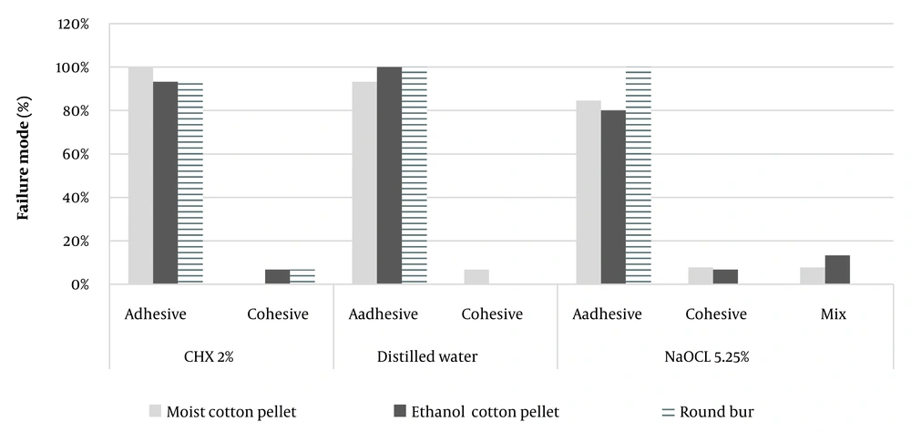
Figure 1 shows the data of the fracture model distribution based on stereomicroscope observations. The common fracture pattern in all samples was adhesive. The number of cohesive and mixed samples was very low.
5. Discussion
The present study tried to investigate the effect of three methods of sealer removal on the dentin bond strength exposed to two common irrigants (i.e., simultaneous irrigants and removal of sealer) by imitating clinical conditions.
AH Plus is an epoxy resin-based sealer used in most endodontic treatments. Lee et al. showed that epoxy resin root canal sealers adhered to dentin and gutta-percha more than other sealers (15). Therefore, this sealer was used in this study and removed from the dentin surface with a moist cotton pellet, ethanol-saturated cotton pellet, and a round bur. Currently, there is no standard sealer removal protocol. Different solvents are used to remove sealers. Solvents such as ethanol, amyl-acetate, acetone, chloroform, halothane, ethanol, xylol, eucalyptol, orange-oil, and Endosolv R have been studied, and none of them was able to completely remove the sealer (1, 8, 11, 12, 16). It seems that the effect of solvents on dentin is related to the presence or absence of a smear layer. In the absence of a smear layer, the solvents increased the mineral content of dentin (calcium and phosphorus levels), affecting the bond strength of the adhesive resins (17).
On the other hand, some solvents solubilize any lipids in dentin or residual odontoblasts. These lipids may deposit on the dentin surface and interfere as a waxy film with the bond (18). Ethanol was the only solvent used in the current study; the smear layer produced from dentin etching was also present on the dentin surface. In this study, removing the sealer from distilled water exposed dentin using ethanol-saturated cotton had the lowest bond strength. Ethanol is routinely recommended as the chemical solution to remove the sealer from the pulp chamber. However, it cannot completely remove sealer residues from the dentin (8). In their assessment with SEM, Kuga et al. (19) showed that 95% ethanol and isopropyl alcohol were ineffective in the cleaning of dentin impregnated with AH Plus sealer. 95% ethanol contains water in its composition, while epoxy resin is hydrophobic. Therefore, the sealer solubility is reduced and cannot completely be removed (20). in the study by Bronzato et al., the use of ethanol to remove sealer did not affect the dentin bond strength. The type of sealer and adhesive system used in their study was different from the present study (1).
This study also showed that using a round diamond bur to clean the sealer from dentin irrigated with chlorhexidine significantly improved the dentin bond strength compared to the control and sodium hypochlorite groups. However, in the control group, ethanol significantly reduced the bond strength, although ethanol did not affect the bond strength of dentin irrigated with NaOCL and CHX.
The negative effect of all endodontic irrigants on the bond strength of adhesive restorations is a rejected hypothesis (10). The results of the CHX effect on the bond strength of adhesive resins to dentin are contradictory (10, 21, 22). Some studies have claimed that CHX has no effect on the interactions of resin systems with dentin due to its non-oxidizing nature (23, 24). Chlorhexidine was used for a short time and at a high concentration (2% versus 0.12%), and self-etch adhesive systems were applied in these studies (22). In another study, CHX significantly reduced the bond strength of total-etch adhesive systems (10). Some studies showed that the bond strength of CHX irrigated dentin with a total-etch system was improved, which was attributed to the inhibition of matrix metalloproteinases -2, -8, and -9, which are collagen-degrading enzymes (21, 25, 26). Kazemi-Yazdi et al. reported that pretreatment with 2% CHX had no negative effect on the micro-tensile bond strength in Clearfill SE Bond. This is a mild self-etch two-step adhesive system with a mild acidic functional monomer, 10-MDP, which agrees with the current study results (27). Also, Fernandes et al. stated that the pre-application of 2% CHX did not reduce the immediate micro-tensile bond strength of a universal adhesive system (single bond universal) (28).
The difference in the bonding materials' monomer content is one reason for the difference in results. The monomer of 2-hydroxyethyl methacrylate (HEMA) present in the single bond has a low molecular weight and an excellent wetting ability, leading to the re-expansion of the shrunk collagen network and an increase in resin infiltration. In contrast, diurethane-dimethacrylate (UDMA) and dipentaerythritol penta acrylate monophosphate (PENTA) monomers in some other bonding systems have a high molecular weight, resulting in a decrease in the diffusion capability of bonding to the demineralized dentin and, as a result, to the reduced adhesive bond strength (2).
Although the results of this study showed no significant effect of NaOCl on the strength of bond materials, other studies have shown that NaOCl has a negative effect on the bond strength due to the oxidation of dentin components and formation of reactive free radicals, which results in the inhibition of adhesive polymerization and decrease in the bond strength (10). In Abo-Hamar's study, NaOCl significantly increased the excite bond strength (7). The difference between these results can be attributed to the application time of NaOCL and the type of bonding system. Prolonged use of NaOCl may increase the bond strength of the solution by increasing the depth of deproteinization and formation of a reverse hybrid layer (7). Dikmen et al. evaluated the effects of different antioxidant treatments (accel, noni-fruit-juice, and proantho-cyanidin) on the micro-tensile bond strength of a self-etching adhesive system (single bond universal adhesive) to NaOCl-treated dentin. They maintained that the micro-tensile bond strength in the NaOCl group was significantly lower than all other groups (29). Our study, it was tried to imitate clinical conditions by applying the irrigant for 30 minutes and the clinical use method (irrigation rather than immersion of dentin); (the common maximum time of the irrigant application during endodontic treatment is 30 minutes (30).
In this study, adhesive failure was the most common type of defect, indicating a poor bond between resin and dentin. The possible explanation for this type of failure is the effect of irrigants and residual sealers on the bond strength of the adhesive to dentin, compared to the adhesive to resin. The results of this study are consistent with Mokhtari et al. (30) and inconsistent with Bronzato et al. (1) (their most frequent failure was the mixed type).
As a limitation of ex vivo studies, the relationship between micro-tensile testing and the clinical condition of adhesives is doubtful; however, micro-tensile tests are ideal for evaluating the effect of laboratory variables. These tests yield better stress dispersion over small surfaces than conventional bond strength tests. Therefore, failures mainly occur in the adhesive. Cohesive failures do not show the real bond strength value, while they reflect the properties of materials. These failures can form due to faults in the sample arrangement along the equipment's long axis (10).
Endodontic treatment and final restoration are not separate entities; instead, they are two constructive elements of a single concept called the endodontic-restoration continuum (31). The long-term clinical success of composite restorations in root canal treatments is influenced by several factors such as the endodontic irrigant and its application time, sealer type, solvent type, sealer removal method, type and composition of adhesive materials and presence or absence of a smear layer and smear plug.
5.1. Conclusions
Given the limitations of this study, it seems that dentin pretreatment with CHX and cleansing of dentin impregnated with AH Plus sealer through round bur, in contrast to dentin pretreatment with normal saline and the removal of sealer with ethanol-saturated cotton, can increase the micro-tensile bond strength of the single bond plus adhesive system to dentin. There was no significant difference between sealer removal methods in dentin pretreatment with sodium hypochlorite. Further studies are needed to investigate the effect of natural and artificial solvents for the removal of various sealers and canal irrigants on bond strength. SEM studies can be carried out on different generations of bonding agents in root canal-treated teeth.

