Introduction
Hydatid disease is a parasitic infection caused by Echinococcus granulosus. In humans hydatid disease is caused by the larvae of E. granulosus [1]. It is seen worldwide and is endemic in some areas, such as the Middle East, Australia, Greece, Turkey, and Iran [1-3]. Dogs and some wild carnivores are definitive hosts. Eggs are passed in the faeces and eaten by the intermediate hosts (sheep, cattle, human), and larvae encyst in the liver [4, 5]. The diagnosis of liver hydatid cyst depends on clinical suspicion, ultrasonography, and computed tomography [5, 6]. Treatment of liver hydatid cyst varies from surgical intervention to percutaneous drainage or medical therapy in patients with liver cysts that are inoperable [6, 7]. Surgery is still the treatment of choice and can be performed by the routine or laparoscopic approach [7]. We report a 33-year-old woman who was diagnosed as a patient with liver carcinoma and multiple huge hydatid cysts.
Case Presentation
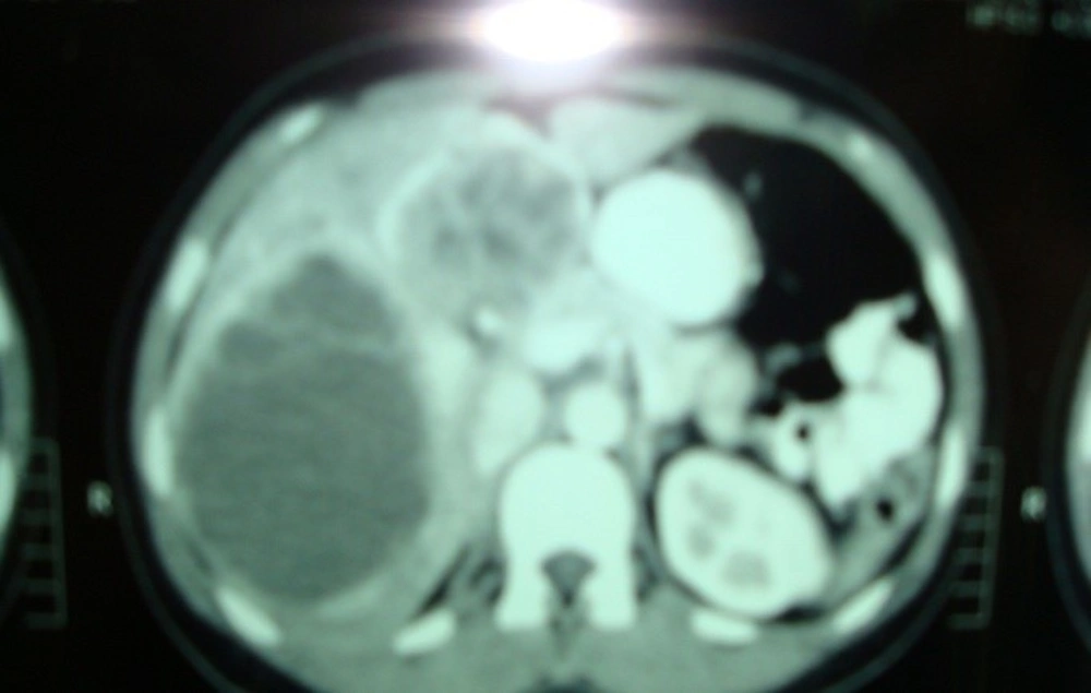
A 33-year-old woman referred to Boo-Ali hospital (Zahedan, Iran) with pain in the RUQ and hepatomegaly. Although, the abdominal pain was the initial problem, on clinical examination, her abdomen was rigid and tense. There was no other complaint and her general health appeared good. She had not any medical history in recent years. Liver ultrasonography revealed 5 cystic masses which 2 were huge (8.5 and 12 cm) with mixed echogenic characters in the liver. Also, a hyperechoic mass found in the liver and she was referred for computed tomography scanning. The liver CT-scan also revealed the cysts which 2 were large cystic masses with septations (Fig. 1).
The normal liver parenchyma was replaced with these cystic septated masses. In many parts of the liver, parenchyma was not visible. Because of the nature of the mass a tentative diagnosis of hydatid cyst was made. Blood sample for serologic test (ELISA), alpha-fetoprotein and viral infection was drawn. Tests for HBV, HCV infections were negative and liver enzyme was more than 2.5 upper limit of normal. We started albendazole 400 mg twice in day and then, she was referred to surgeon. But, the patients asked to visit by a surgeon in other city. Then, she went to another medical center for treatment. Biopsy from hyperechoic mass had been revealed liver malignancy. Later, we missed her to know more about her illness.
Discussion
Hydatid disease is a zoonotic infection caused by the tapeworm of the genus Echinococcus [1, 2]. Morbidity is usually secondary to free rupture of the echinococcal cyst, infection of the cyst, or dysfunction of affected organs. Most symptomatic cysts are larger than 5 cm in diameter [5-7]. Parasite load and the size of the cysts determine the degree of symptoms. A history of living in or travelling to an endemic area can help diagnosis. The liver is the most common organ involved, followed by the lungs [5-7]. Previous studies have reported huge hydatid cyst in the liver but, co-morbidity of hepatcarcinoma and multiple huge hydatid cyst has not reported, to now [2, 5, 6]. The diagnosis of hydatid cyst of the liver depends on clinical suspicion. Ultrasonography and computed tomography scanning, the most important diagnostic tools, are helpful for diagnosis of complications and planning treatment [4, 5, 6]. Although, serology tests are helpful for detection of infection, but the negative test cannot roll out the diagnosis. Since some of the cystic liver lesions have a malignant potential, all patients with liver mass should carefully evaluate by an experienced team to receive an appropriate treatment [4]. Hydatid cyst of the liver must be treated surgically [5, 7]. Albendazole 10 mg/kg/day for 3-6 weeks before surgery should be given to sterilize the cyst and continue for at least 6-8 weeks after surgery to clear up any spilled hydatid fluid containing live scolices. [6, 7]. Chemotherapy is indicated in patients with primary liver or lung cysts that are inoperable [8]. Albendazole is administered in several 1 month oral doses (10-15 mg/kg/day) separated by 14 days intervals [5]. Mebendazole is also administered for 3-6 months orally in dosages of 40-50 mg/kg/day. Then, patients should monitor for adverse effects of agents with a CBC count and liver enzyme test [5-7]. In conclusion, we should mind the hydatid cyst when a patient has a liver mass and has a history of living in or travelling to an endemic area.
