Introduction
The association of papillary muscle rupture with right coronary or left circumflex artery infarction has been clearly demonstrated [1]. Complete transaction of anterior papillary muscle is less common and occurring in 25% of cases as it has dual blood supply, from the first obtuse marginal originating from left circumflex (LCX) and from the first diagonal branch (D1) of left anterior descending coronary artery (LAD) and single blood supply of anterior papillary muscle from single diagonal artery is exceedingly rare phenomenon. Papillary muscle rupture is usually seen in relatively small area infarction, as in our case [2]. In this case anterior papillary muscle rupture caused by single legion of the D1 artery. We assumed that the anterior papillary muscle has been per fused exclusively by D1 artery.
Case reports
A 42-year-old man without history of hypertension or diabetes presented to our hospital with a 2 day history of dyspnea. Physical exam revealed a pulse of 120 beats/min and blood pressure of 60/40 mmHg.
The patient was anuric. On auscultation a 2/6 apical systolic murmur that radiated to axilla was heard. The patients were in cardiogenic shock and had tachypnea and diffuse inspiratory crepitating rales. The ECG revealed sinus rhythm, lateral Q waves and chest X-ray revealed infiltrates consistent with acute pulmonary edema. Results of initial tests of cardiac enzymes included creatine phosphokinase (2133 U/L), and a creatine phosphokinase, myocardial band (MB) level of 165 U/L and a troponin level of 2 mg/mL. Progressive respiratory failure and hypotension developed and the patient required mechanical ventilation. Despite high dose administration of adrenaline and dobutamine, intraaortic balloon pumping was necessary to stabilized hemodynamic state.
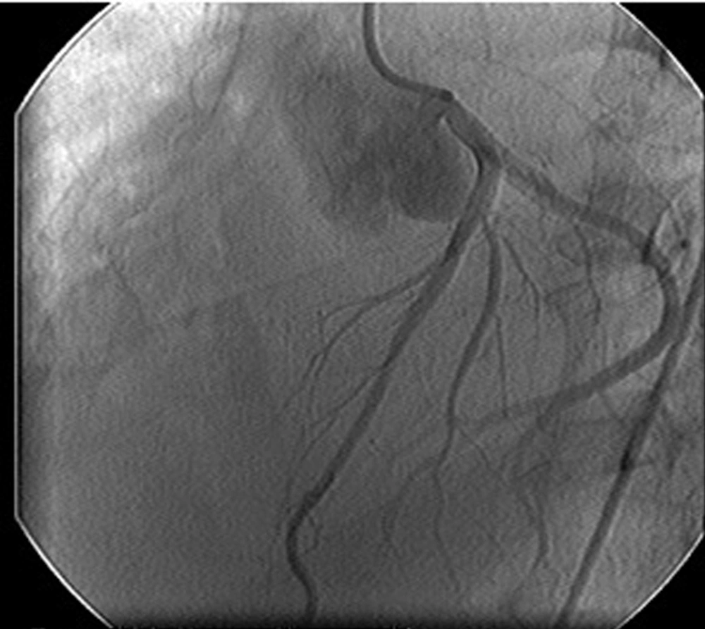
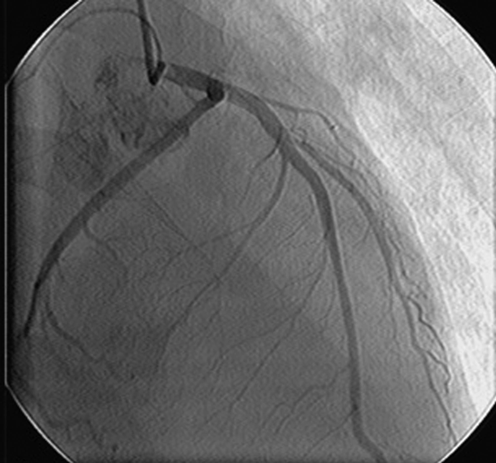
Coronary angiography demonstrated stenosis of D1 artery. The right coronary artery (RCA) and LCX and LAD were normal. The pulmonary artery systolic pressure was 80 mmHg. Transthoracic echocardiography showed anterolateral hypokinesis with an ejection fraction of 30% and sever mitral regurgitation. There was an evidence of mobile masses in left atrium and anterior mitral valve leaflet was prolaptic and flail.
His subsequent course was complicated by acute renal failure. Since most acute cases of muscle rupture (MR) causing an unstable hemodynamic state requiring emergency operation, we decided on immediate mitral valve replacement surgery with coronary artery bypass grafting.
We performed a median sternotomy and cardiopulmonary bypass by arterial cannulation aorta and venous cannulation of both the superior and inferior vena cava. After clamping of the aorta and antegrade and retrograde injection of cardioplegia, left atrium was opened. Intraoperative exam showed complete rupture of anterior papillary muscle. The prolapsed anterior leaflet and necrotic anterior papillary muscle was excised completely but posterior papillary muscle and corda were preserved. After mitral valve replacement (MVR) with carbomedics 29 mechanical (M) prosthetic valve, coronary artery bypass grafting (CABG) to the diagonal branch with saphenous graft was performed.
His pathologic exam demonstrated acute coagulation necrosis of anterior papillary muscle that was per fused by one single artery. Immediately after transfer to ICU, peritoneal dialysis was started. The patient made a slow post operative recovery and was weaned from IABP and inotropic agents during the following 7 days and acute renal failure was recovered after 22 days and the patient was discharged on 30th day after surgery.
Discussion
Severe MR complicating acute MI is an important cause of hemodynamic instability and cardiogenic shock. Nonrandomized series that have reported favorable outcome after early MVR have led to recommendation that early surgery is appropriate in such patients [3]. Arterial supply to papillary muscles consist of small coronary vessels derived from the large epicardial artery because the poster medial papillary muscle has single blood supply from PDA, it is involved in rupture of 6-12 times more frequently than the anterolateral muscle which has a dual blood supply from D1 and LCX [4]. The anomaly of single blood supply to anterolateral papillary muscle from D1 with dominant LCX is exceedingly rare and we have found only one case in literature review that patient had diminutive LCX system. Difference in patterns of vascular supply, collateralization, and connective tissue are possible explanation for different clinical sign and symptoms [5].
We assumed that the anterior papillary muscle was per fused by single D1 artery. The postero medical papillary muscle is supplied solely by RCA and is thus more susceptible to ischemic rupture than the anterolateral muscle which is generally supplied by both diagonal branches and LCX coronary artery. For the same reason postero medical papillary muscle rupture is much more common with inferior myocardial infarction than with anterior infarction [6]. Partial rupture in which only one or more heads of a papillary muscle are separated from the ventricular wall is more common and is likely to be less catastrophic than is complete papillary muscle rupture. In many series, post-MI patients with papillary muscle rupture have been found to be older and less likely to have triple vessel disease, than are those with sever MR unrelated to papillary muscle rupture [7].
To our knowledge, the present is second to document that a single obstruction of the diagonal artery branch led to an antrolateral papillary muscle by an occlusion of only one diagonal artery. In pervious case that was reported by Wada et al. [7], the patients anatomy (hypoplastic and diminutive) LCX system, increased the possibility of papillary muscle rupture but in our case LCX was not hypoplastic and was dominant. Papillary muscle rupture carries a poor outcome that may be improved by surgical correction [8]. This case describes complete transaction of papillary muscle resulting in sever MR and contributing to cardiac shock and renal failure in an young man with a late (24 h) presentation of acute MI. Coronary angiography demonstrated single vessel disease (D1) (Fig. 1). The finding of a new systolic murmur in the post infarct setting should raise the suspicion of mechanical complication including the papillary muscle rupture that must be distinguish from acute VSD (Fig. 2) [7]. Two dimensional and Doppler transesophageal echocardiography (TEE) are useful in the detection of papillary muscle rupture by direct visualization the rupture as a mobile mass appearing in the left atrium during systole or in the left ventricle during diastole [3]. The patient suffered protracted cardiac shock and early TTE with technically limited views demonstrated severe MR and did not display other features suggestive of papillary muscle rupture.
Treatment includes aggressive medical therapy with inotropic drug and intra-aortic balloon pumping (IABP) as a bridge to surgical treatment. The post operative course was eventful by renal failure that was responding to peritoneal dialysis, and patient was discharged on 30th days of surgery. In conclusion papillary muscle rupture following acute MI and D1 artery occlusion is a very rare but serious complication warranting immediate recognition and aggressive medical and urgent surgical intervention.

