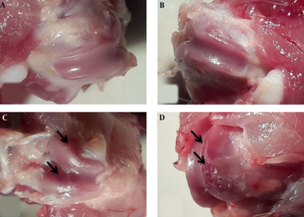
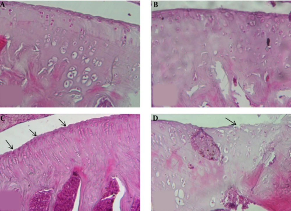
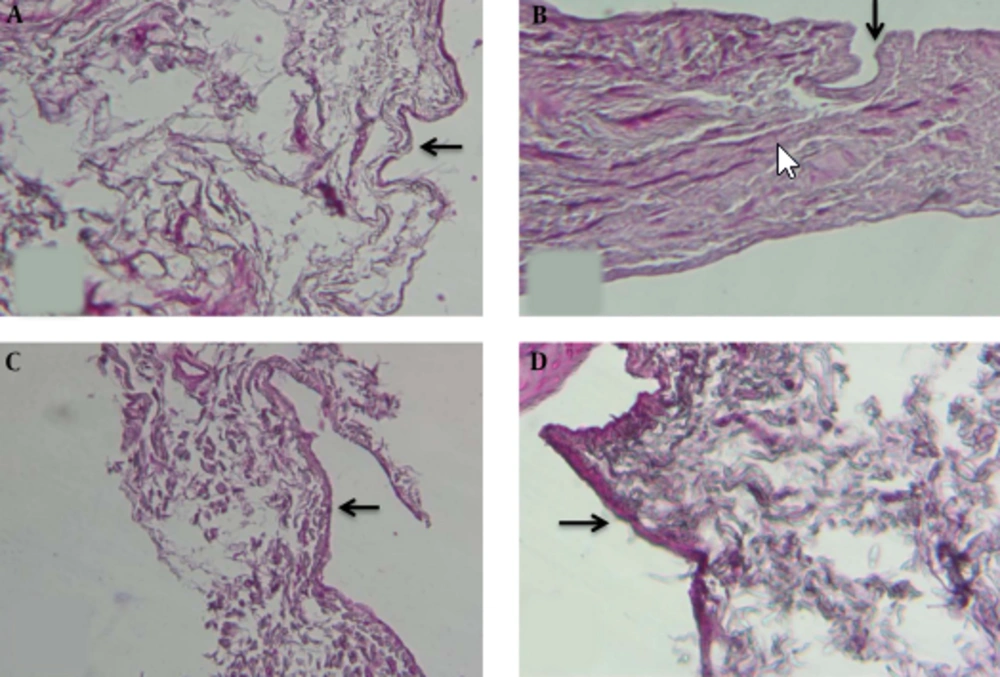
The current study results showed that moderate running exercise could significantly affect the cartilage parameters such as cartilage and synovial layer thickness in female runner rats in comparison with those of the control group and male runner rats. These changes lead to OA occurrence in female runners more than male runners.
Recently, athletes and coaches show great interest in physiological tests to track changes in their training status, predicting their performance, and selecting proper training programs (
20). Generally, physical loading is a double - edged sword. The proper running-induced joint loading remains undetermined (
21). In the current study, it was suggested that moderate - intensity running damaged the articular cartilage, and therefore, it should be considered as a strenuous exercise, particularly for females.
In this regard, Ni et al., revealed the intensity - dependent impact of treadmill running on the lubricin metabolism of articular cartilage in rats (
22). In another study, they showed different cartilage responses by changing the running intensity. Their findings showed that running load might be within and above the upper safe limit during running with low - to - medium and high intensities, respectively (
15). Chondrocyte number and cartilage thickness reduced insignificantly and significantly in male and female runner groups, respectively, versus the controls. This finding was in line with the results of some previous studies. For instance, Ni et al., showed that high - intensity exercise reduced chondrocyte number, cartilage thickness, and glycosaminoglycan (GAG) level (
23). Some studies also reported similar findings on beagle femoral cartilage following a running exercise (20 km/day or 40 km/day) during 15 weeks (
24,
25).
In contrast, mild or moderate running improved cartilage GAG content in dogs (
26) and humans (
27). In other studies, cartilage integrity could be maintained via low - to - medium - intensity running. It was clearly demonstrated that chondrocyte count, cartilage thickness, GAG content, and collagen level significantly increased during running with low - to - medium intensity, suggesting some positive effects on cartilage integrity (
9,
28,
29).
Moderate - intensity running may cause antiapoptotic effects on an intact cartilage and increase the chondrocyte count (
7). However, the results of some previous studies on high - intensity exercise showed a decline in GAG content. In the current study, the loading on cartilage, induced by moderate - intensity running (which caused OA - like changes) exceeded the upper safe limit. The cartilage extracellular matrix may be damaged due to excessive mechanical stress, and the balance in chondrocytes is shifted in favor of catabolic activity over anabolism (
30); these changes degrade both proteoglycans and collagen fibrils (
31).
Gender differences in knee cartilage are examined in few studies. The obtained results revealed that females showed more cartilage changes in a moderate - intensity exercise, compared with males with the same body weight. Some studies indicated that differences in knee cartilage volume were significant in adults aged 26 - 61 years. A previous study, based on radiographic assessments, revealed that males’ cartilage volume were 33% - 42% larger than that of females in normal cases (
7).
Faber et al., showed that gender differences did not significantly affect cartilage volume after body weight and height adjustments (
13). Ding et al., specified that body and bone size had significant effects on this difference, which was in agreement with some speculations, suggesting that cartilage volume had a direct relationship with bone size (
12,
32,
33); consequently, males have a thicker cartilage than females (
3,
34,
35).
However, bone size differences were not adjusted in these studies. In contrast, another study found no gender - related effects after adjustments for the tibial head diameter and body weight (
36). The current study showed some significant changes in rats with the same body weight and age. Therefore, other parameters may describe the gender differences. Nevertheless, these measures are highly reproducible and reflect the joint structure and surface. Therefore, growth factors and sex hormones are the most important factors to explain this difference (
37).
4.1. Conclusion
Clearly, males and females should follow different exercise protocols. The current study results showed that the high exercises were more harmful in females than males. The current study results showed moderate treadmill exercise could lead to mild OA both in male and female rats, while histological changes in female runners were significantly more than male runners.