1. Background
The wound is gap or discontinuity in the epidermis or dermis that occurred following a traumatic or pathological changes in the skin or body [1]. Skin ulcers, caused by different reasons and divided into different types. Wound classification based on depth is divided into two types: The superficial and deep wound. Another classification is based on the length of wound that divided into two, acute and chronic wounds groups [2-5]. Wound healing is the most important biological processes, including repair and production of new tissue [6]. It’s activated a cellular response and contains a complex pathophysiological process including several cellular and biological sub processes, e.g. inflammation, angiogenesis, and collagen confession [7]. Angiogenesis is a landmark of wound healing that provides oxygen, nutrients, and blood-borne cells to the site of tissue injury. In inflammation phase, the vessels constrict and platelets activate the coagulation and fibrin following its creation. Macrophages are important cells in the inflammatory phase which stimulates wound healing. Inflammation phase started at the third day after injury and the maximum of damage observed at the third week. Collagen confession phase begins immediately and lasts for months. In this phase, collagen synthesis begins by fibroblasts and done stimulation of macrophages [8-12]. Inflammation maintenance and scarce vessel formation comprise the most noticeable causes of delayed wound healing [13]. On the other hand, wound fibrosis or anomalous increase of collagen in the wound that lead to an unpleasant scar [14]. Recent research has shown that several combinations are used for wound healing such as Acetic acid, Hydrogen peroxide [15]. Common plant extracts, e.g. Grape seed, Lemon, Rosemary, and Jojoba, have been employed for wound healing and longevity increase. All of these plants have a communal property, e.g. producing combinations with phenolic structure [16]. These phytochemicals ordinarily respond with some combinations such as oxygen free radicals and other macromolecules in order to neutralize free radicals and initiate biological special effects [16].
Medicinal plants have been used traditionally in the treatment of disease. Satureja Khuzestanica is established antioxidants that used in medicinal plants. Satureja khuzistanica belonging to the dark one native plants of lamiaceae and is widely grown in the southern part of Iran [17]. Carvacrol is one of the most important compounds Satureja Khuzestanica. Carvacrol is a wide spectrum of antimicrobial activities against bacteria. When bacteria are exposed to carvacrol, ATP, and intracellular potassium leak from the cell membrane and the bacterial cell dies [18]. Carvacrol is insoluble in water and soluble in alcohol and ether and properties anti-inflammatory, analgesic and anti-microbial [19]. Satureja Khuzistanica is utilizing for its anti-oxidant, anti-lipid, anti-inflammatory, and anti-bacterial effects and might similarly play a role in preventing intra-abdominal adhesion. The major of these properties could be attributed to the plant’s polyphenol, in particular, catechin combinations [20-24]. Anti-oxidant combinations of Satureja Khuzestanica could increase collagen dimensions and hence heal the wounds [25]. These combinations (e.g. epigallocatechin gallate) have also been used as a cause for keratinocytes reproduction and distinction [26]. Also, its anti-fibrogen effects have been established in some animal models [27]. Satureja Rechingeri (Labiatae) is described as a new species from Iran, and its relationship to S. khuzistanica. Satureja rechingeri is belongs to the Lamiaceae family, perennial plant and very aromatic, with wooden base, the branching base, stem is completely covered with long trichomes, basal leaf is circular. The upper leaves are gradually toward the top of the folded side. The chemical composition of the essential oil is carvacrol [28]. Taken to gather, Satureja Khuzistanica and Satureja Rechingeri have anti-oxidant and anti-inflammatory properties and may enhance wound healing process.
2. Objectives
The main goal of the present study was to examine effect of ethanolic extract of Satureja Khuzestanica and Satureja Rechingeri on postsurgical wound healing process.
3. Materials and Methods
All experiments were performed in agreement with principles of laboratory animal care. Male mouse were obtained from Razi herbal medicine research center. Mice were euthanized by excessive doses of ketamine HCl (50 mg/kg) and xylazine (5mg/kg) (Pharmacia and Upiohn, Erlangen, Germany) [29] in accordance with the protocols approved by the Lorestan University Animal Care and use committee. Every attempt was made to minimize the number of animals used and their pain.
3.1. Extract Preparation
Maceration method was employed to make the extract. For this purpose, 700 g Satureja Khuzestanica and 700 g Satureja Rechingeri was transported into an Erlenmeyer, 1 Liter methanol 98% (Nasr Co. Iran) was added and the solution was left at laboratory temperature. Forty eight hours later, the extract was filtrated through a filter paper and the grind was squeezed to discharge. Finally, the extract was concentrated by rotary evaporator [30].
3.2. Animals and Study Design
In this experimental study 80 male NMRI mice weighing 25 - 30 g were used. They were randomly divided into four groups (N = 20 per group). The animals were acclimatized for one week to the condition of our laboratory before the commencement of the experiment. The animals were exposed to 12 hours light and 12 hours dark cycle at a room temperature of 22°C. The animals had free access to standard laboratory chow and water ad libitum. The studies were performed during the same time in all groups. The rats were grouped as follows:
Group 1: control group (without surgery),
Group 2: placebo group,
Group 3: Satureja Khuzestanica-treated group,
Group 4: Satureja Rechingeri-treated group.
The rats were anesthetized once intraperitoneally with ketamine HCl (50 mg/kg) and xylazine (5 mg/kg) [10] in accordance with the protocol approved by the animal care and use committee and prepared for surgery. Circular wounds were made in three layers of skin with 10 mm diameter in all three layers (dermis, epidermis, and hypodermis). Specimens were taken at 3rd day, 7th day, 14st day, and 22th day for microscopic examinations. Sham group received ethanolic extract. Satureja Khuzestanica-treated group and Satureja Rechingeri-treated group received daily topical applications of traditional Satureja Rechingeri (1 g/day) and Satureja Khuzestanica (1 g/day). On the 3th, 7th, 14th, and 22th days of operation, five mice separate from each group and subsequently a segment of treated skin was removed and fixed with 10% formalin solution. Specimens embedded in paraffin after fixation, dehydration, clearing and infiltration. The paraffin tissue blocks were cut with a microtome at 5 μm, stained with hematoxylin and eosin (H&E). Sections studied according Asadi et al. [31], method’s included; 1) epithelial regeneration; 2) granulation tissue thickness, 3) angiogenesis assessment of wound site for the rank of healing. 2.10. Data analysis. Data analysis was performed by SPSS 16, Kruskal-Wallis and Friedman test (for histopathology analysis). Sig < 0.05 was considered as statistically significant.
4. Results
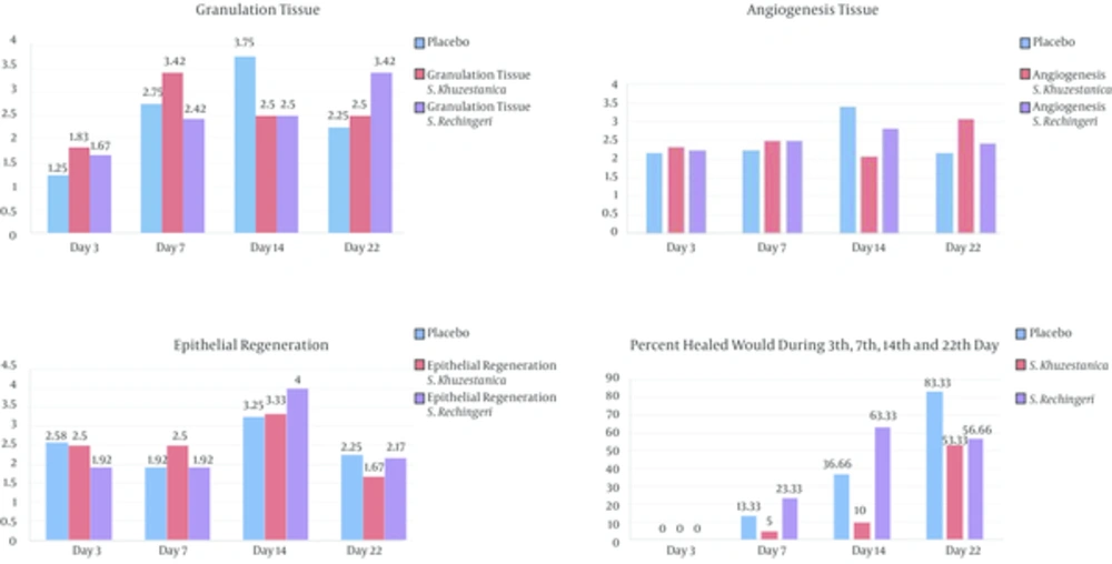
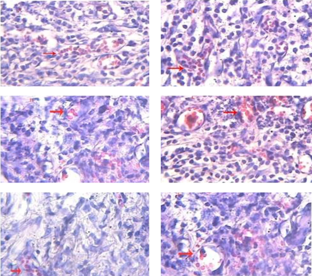
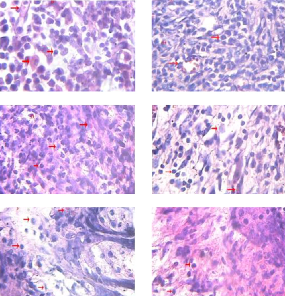
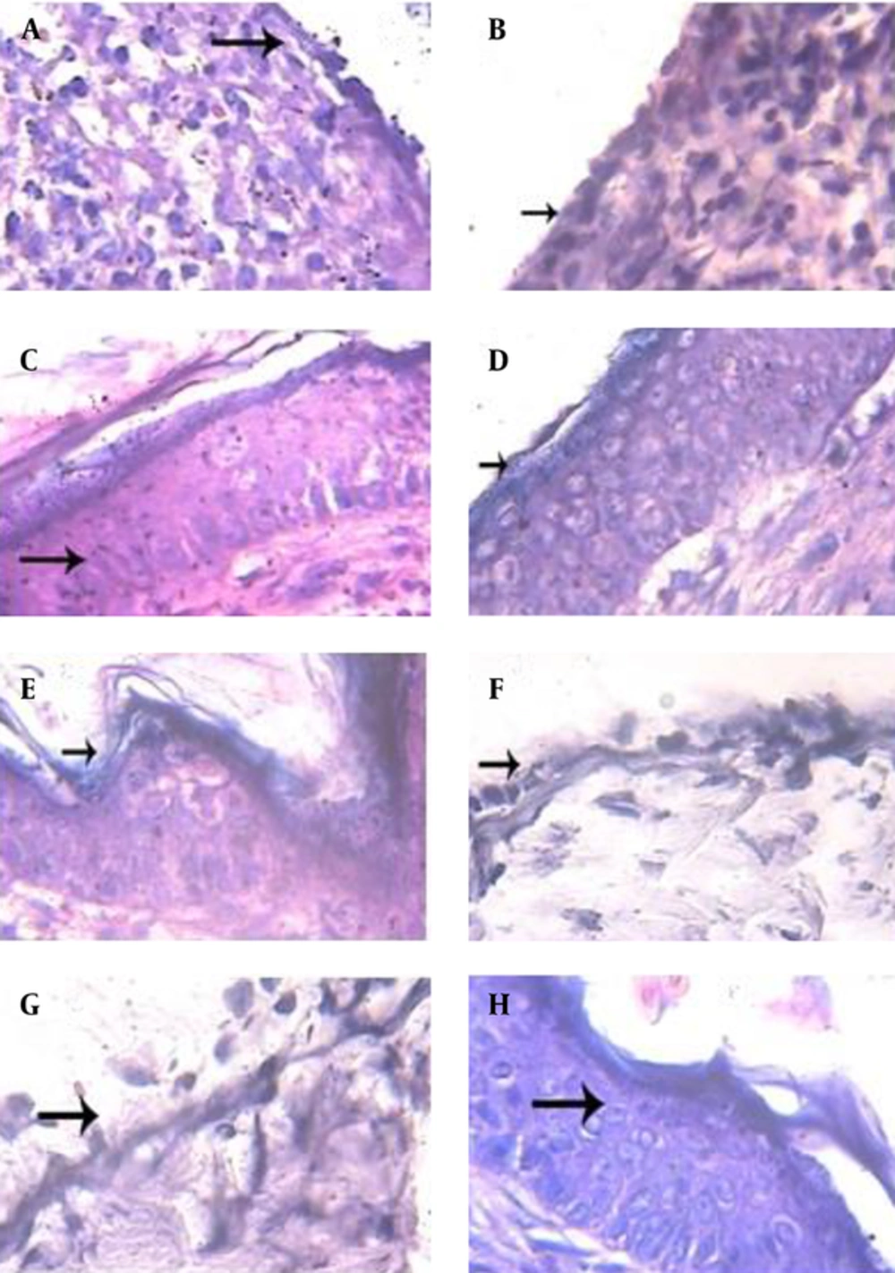
Microscopic features of wounds on day 3: Compare H and E staining sections in the study groups showed that Satureja Rechingeri treated group and Satureja Khuzistanica treated group haven’t effect on the wound healing in the comparison with placebo (Figure 1). Study epithelial regeneration, granulation, and angiogenesis in the sections tissue on day 3 of Satureja Khuzistanica group than sham group and Satureja Rechingeri group (Figure 1).
Microscopic features of wounds on day 7: Compare H and E staining sections in the study groups showed that Satureja Rechingeri treated group has best effect on the wound healing in the comparison with placebo and Satureja Khuzistanica treated group (Figure 1). Satureja Rechingeri ethanolic extract can decrease granulation, increase epithelial regeneration, and angiogenesis. Satureja khuzistanica ethanolic extract can increase granulation, epithelial regeneration, and angiogenesis (Figure 1).
Microscopic features of wounds on day 14: Compare H and E staining sections in the study groups showed that Satureja Rechingeri treated group has best effect on the wound healing in the comparison with placebo and Satureja Khuzistanica treated group (Figure 1).
| Score | Epithelial Regeneration | Granulation Tissue Thickness | Angiogenesis |
|---|---|---|---|
| 1 | Little epithelial organization | Thin granular layer | Altered angiogenesis (1 - 2 vessels per site) |
| 2 | Moderate epithelial organization | Moderate granular layer | Few newly formed capillary vessels (3 - 6 vessels per site) |
| 3 | Complete epithelial organization | thick granular layer | Newly formed capillary vessels (7 - 10 vessels per site) |
| 4 | Complete epithelial organization | Very thick granular layer | Newly formed and well-structured capillary vessels (> 10 vessels per site) |
Satureja Rechingeri ethanolic extract can decrease granulation and angiogenesis and increase epithelial regeneration. Satureja khuzistanica ethanolic extract can decrease granulation and angiogenesis and increase epithelial regeneration (Figures 1 and 3).
Microscopic features of wounds on day 22: Compare H and E staining sections in the study groups showed that Satureja Rechingeri treated group and Satureja Khuzistanica treated group haven’t effect on the wound healing in the comparison with placebo (Figure 1). Satureja Rechingeri ethanolic extract can increase granulation and angiogenesis and decrease epithelial regeneration. Satureja khuzistanica ethanolic extract can increase granulation and angiogenesis and decrease epithelial regeneration (Figure 1).
5. Discussion
Wound healing is a complex process of rehabilitate cellular structures and tissue layers in injured tissue [7]. Wound healing include of different phases such as contraction, granulation, epithelization and collagenation [32, 33]. Research showed that wound healing involved in three phases: inflammation, proliferation, and tissue remodeling with scar formation. The inflammatory phase is described by hemostasis and swelling. Proliferative phase is result from by epithelialization, angiogenesis, and collagen confession. In the maturation phase, the wound undergoes act of contracting resulting in a smaller amount of apparent mark with a scar tissue [34]. Granulation tissue which is creating in the final part of the proliferative phase is primarily composed of fibroblasts, collagen, and recently small blood vessels [35]. Several effects of Satureja Khuzestanica and its compounds have been already examined, indicating that this plant, in traditional medicine, has medical uses including, antibacterial, anti-inflammatory, and antioxidant. In a study that showed vitamin C in addition to Satureja Khuzestanica is effective in preventing abdominal adhesions during the adhesions process [36]. Also, research has shown that Satureja Khuzestanica is effective in preventing atherosclerosis, atherosclerosis is a direct correlation with angiogenesis, that the present study is approving [36, 37]. Therefore probably Satureja Khuzistanica with high antioxidant property cannot be useful in wound healing. The high granulation at the last days indicates that the wound isn’t healing yet and healing process is delayed or wound healing mechanism at the granulation process is stopped. The high angiogenesis at the last days indicates that the wound isn’t healing yet and healing process is delayed, because the epidermis Satureja Rechingeri scores lower than the control group confirmation of the delay caused by this medicine plant [38]. Microscopic observations indicated that the formation of a large scar in Satureja Rechingeri treated group than the control group, causing severe adhesion to the surrounding tissues and create a barrier to the migration of epithelial cells and the resulting delay repair of satureja rechingeri treated group compared to the control group. Satureja Rechingeri may also prevent the wound from being wettish which is suitable for regeneration [39]. Despite the active ingredient antioxidants in plant Satureja Khuzestanica, the results indicate inadequate effect of ethanol extract of this plant in wound healing. Therefore, due to the inefficiency of the ethanol extract of leaves Satureja Khuzestanica healing, its use as a medicinal plant used in traditional medicine and is not recommended. As regards Satureja Rechingeri plant until the fourteenth day will be healing, probably, a short treatment period and should be used only for a specified period and for the better performance is recommended use with humid bandage and this needs to be investigated further.


