Fulltext
The full text of this article is available on the PDF file.
Zahedan Journal of Research in Medical Sciences
The Official Journal of Zahedan University of Medical Sciences
Authors
Background : Hyperglycemia is the principal factor responsible for microvascular complications of diabetes. Diabetic nephropathy is a serious and common complication leading to end stage renal disease. The exact molecular mechanisms of high glucose-induced toxicity on renal cells are still incompletely understood. Therefore in the present study, glucose-induced toxicity was studied in HEK (human embryonic kidney) cells as an in vitro model for diabetic nephropathy.
Materials and Methods : First, the viability of HEK 293 cells exposed to glucose was measured by MTT (Methyl Thiazolyl Tetra-zolium) assay. Caspase-3 activity was determined spectrophotometrically using enzyme specific substrate. Moreover, the alteration in expression of Bax, Bcl2 and caspase-3 were measured by Western blotting.
Results : The results showed that high glucose significantly reduced cell viability after 48 h (p<0.05). In Western blot analysis, the ratio of Bax/Bcl-2 protein expression in cells treated with high glucose was significantly increased compared to controls (p<0.001). The activity of caspase-3 was significantly increased in treated cells compared to control (p<0.01). Moreover, high glucose exposure induced a significant decrease in protein level of procaspase-3 (p<0.01), indicating conversion of pro-form into the mature caspase.
Conclusion : On the current data, it could be concluded that high glucose can cause HEK cell death, in which apoptosis plays an important role possibly by the mitochondrial pathway through higher expression of Bax pro-apoptotic protein and also by activation of caspase-3 related pathways.
© 2013, Author(s). This open-access article is available under the Creative Commons Attribution 4.0 (CC BY 4.0) International License (https://creativecommons.org/licenses/by/4.0/), which allows for unrestricted use, distribution, and reproduction in any medium, provided that the original work is properly cited.
Vakili Zahir N, Abkhezr M, Khaje Piri Z, Ostad SN, Kebriaezade A, et al. The Time Course of JNK and P38 Activation in Cerebellar Granule Neurons Following Glucose Deprivation and BDNF Treatment. Iran J Pharm Res. 2012;11(1):e125795. doi: https://doi.org/10.22037/ijpr.2011.1038
Malekinejad H, Moradi M, Fink-Gremmels J. Cytochrome C and Caspase-3/7 are Involved in Mycophenolic Acid- Induced Apoptosis in Genetically Engineered PC12 Neuronal Cells Expressing the p53 gene. Iran J Pharm Res. 2014;13(1):e125408. doi: https://doi.org/10.22037/ijpr.2014.1382
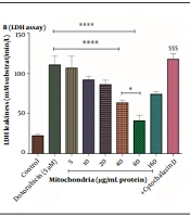
Seydi E, Andalib M, Yaghoubi S, Fakhri A, Yuzugulen J, et al. Mitochondrial Transplantation Alleviates Doxorubicin-Induced Toxicity in Rat Renal Cells. Iran J Pharm Res. 2024;23(1):e146033. doi: https://doi.org/10.5812/ijpr-146033
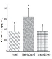
Samiei A, Behpoor N, Tadibi V, Fathi R. The Effect of High Intensity Aerobic Exercise on Levels of Galectin-3 and Protein Kinase C in Diabetic Male Rats. J Clin Res Paramed Sci. 2020;9(1):e80362. doi: https://doi.org/10.5812/jcrps.80362
Sadeghi F, Etebari M, Habibi Roudkenar M, Jahanian-Najafabadi A. Lipocalin2 Protects Human Embryonic Kidney Cells against Cisplatin–Induced Genotoxicity. Iran J Pharm Res. 2018;17(1):e124768. doi: https://doi.org/10.22037/ijpr.2018.2172
Last Update: 1 month ago
Last Update: 1 month ago
Last Update: 1 month ago
Last Update: 7 hours ago