1. Introduction
Dental avulsion is a severe injury in which the tooth is totally displaced out of its alveolar socket. This condition, occurring mostly within the age range of 8 - 11 years, accounts for 0.5 - 3% of all dental injuries (1). Therefore, some factors in this age range (i.e., growth spurt) may increase the development of dental complications (2, 3).
The management of avulsed teeth by parents and dentists affects the healing of the tooth replantation. The status of periodontal ligaments (PDLs) and dental pulps is a crucial determinant for improving the affected teeth (4). The length of extra-oral time and storage medium are salient in determining the status of PDL (5). For instance, during the extra-alveolar time of 5 - 60 min in a physiologic storage medium (e.g., milk), PDL may remain viable but lose the ability to be regenerated. Replacement resorption (ankylosis) is a possible result of an extra-alveolar time of > 60 min because the PDL is necrotic (6, 7).
Numerous studies have reported the delayed replantation of an avulsed permanent tooth. In contrast with several studies on ankylosis (8, 9), there are few investigations on continued root formation and PDL regeneration (10). If root canal infection occurs in this situation, microbial toxins can penetrate into the PDL via dentinal tubules and trigger another type of resorption, known as external inflammatory resorption. However, there is a need for replanting an avulsed tooth for a child even in case of poor prognosis (11). This case report investigated the avulsed maxillary permanent incisors replanted after seven days and reported the clinical and radiographic observations collected over a 3-year follow-up.
2. Case Presentation
The case in this study was a 9-year-old girl referred to the Pediatric Department of Ardabil School of Dentistry, Ardabil, Iran, after an incident, which resulted in dental trauma. The trauma had happened in a car accident one week before referral. Following the injury, the avulsed teeth were kept dry in an open plastic container. The patient’s mother did not report any symptoms of cerebral involvement (e.g., amnesia, headache, unconsciousness, vomiting, dizziness, visual disturbances, and cognitive impairments) after trauma.
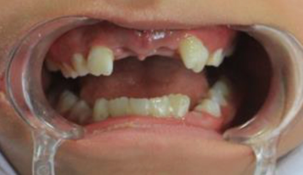
As can be seen in Figure 1, the intraoral examination revealed mixed dentition and avulsion of both maxillary central incisors (i.e., teeth 11 and 21). The case under study was in good general health status, and there was no observation of contraindications for replantation. The avulsed teeth had intact structure with dry periodontal tissue on the root surfaces and closed apical foramina.
To complete the examination, periapical and panoramic radiographic examinations were performed, the findings of which revealed no alveolar bone wall fracture or other hard tissue injuries. After informing parents about the possible risks of delayed replantation, the roots of the avulsed teeth were scraped gently, and the necrotic PDL tissues were removed from the root surfaces.
The access cavity was prepared, and the root canal debridement was carried out for both teeth extra-orally with hand K-files (Dentsply, Maillefer, Ballaigues, Switzerland). In addition, 2.5% sodium hypochlorite and normal saline solution were used as root canal irrigants. The canals were dried with sterile paper points, and the teeth were soaked in 2% sodium fluoride gel for 20 min. Following the application of local anesthetics (i.e., lidocaine 2%, 1.5 mL), the alveolar sockets were carefully debrided with physiologic saline solution to remove any coagulum, granulation, or pathologic tissue.
In the next stage, the teeth were replanted and fixed through the application of a flexible splint (0.7-inch wires) and acid-etch composite resin technique for a period of four weeks. The canals of both teeth were filled using calcium hydroxide. In order to reaffirm the correct repositioning of the teeth, another periapical radiograph was performed.
Systemic antibiotics were prescribed for one week (penicillin V), and oral hygiene instructions were given to the patient. Accordingly, she was recommended to use chlorohexidine (0.1%) mouthwash twice a day for one week and follow a soft food diet for up to two weeks. The splint was removed after four weeks. The intra-canal dressing was renewed by 1-month intervals.
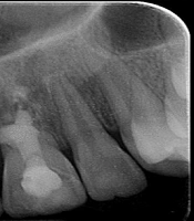
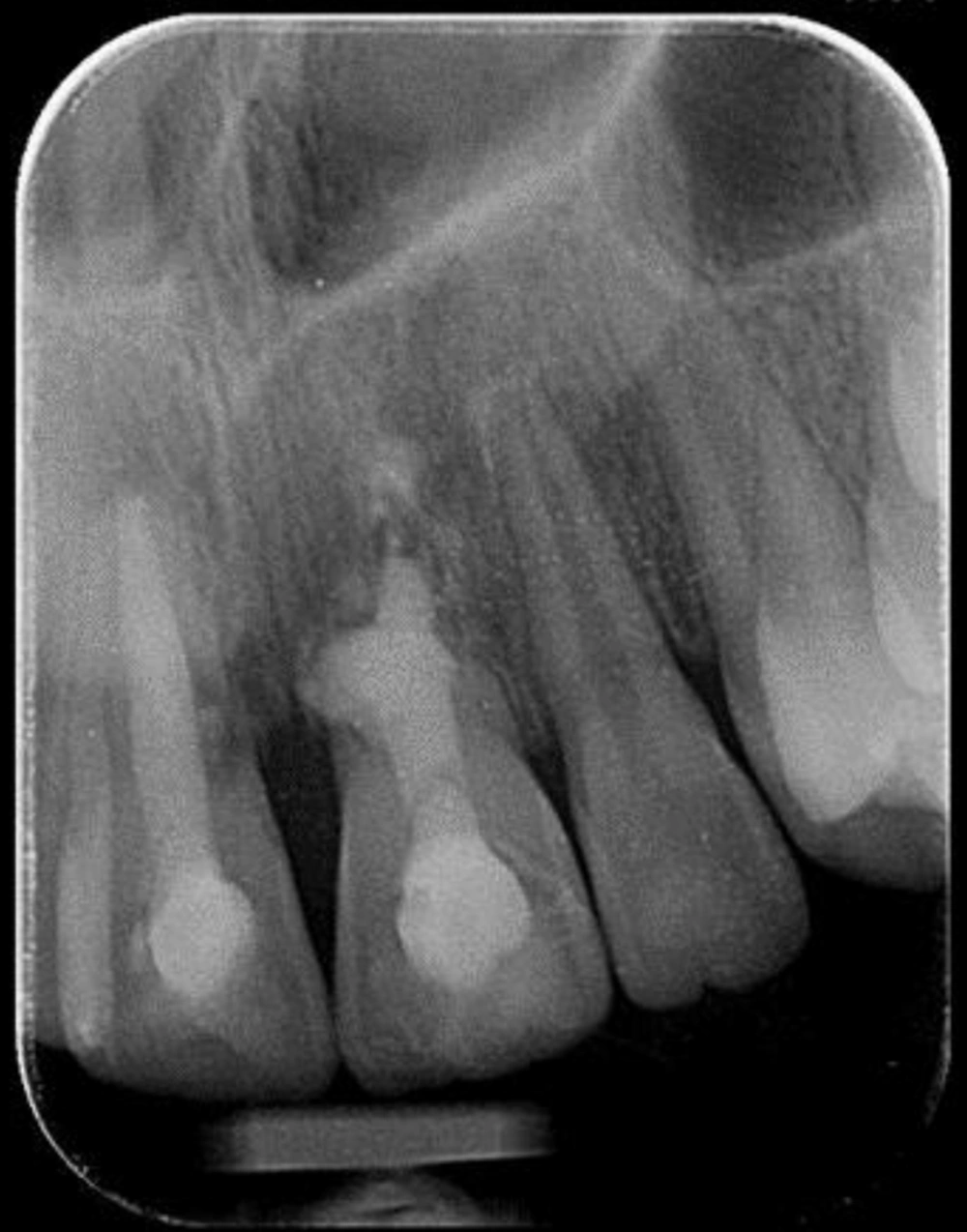
The 2-month follow-up revealed the clinical and radiological signs of external root resorption in tooth 21. In the follow-up, a percussion test of the two avulsed teeth demonstrated a change in the percussion sound due to ankylosis. After the 6-month follow-up, the calcium hydroxide was replaced by mineral trioxide aggregate (MTA) because the tooth was in a stable, functional position. As shown in Figure 2, the intra-oral periapical radiographs of tooth 21 indicated a severe external root resorption replaced by the bone.
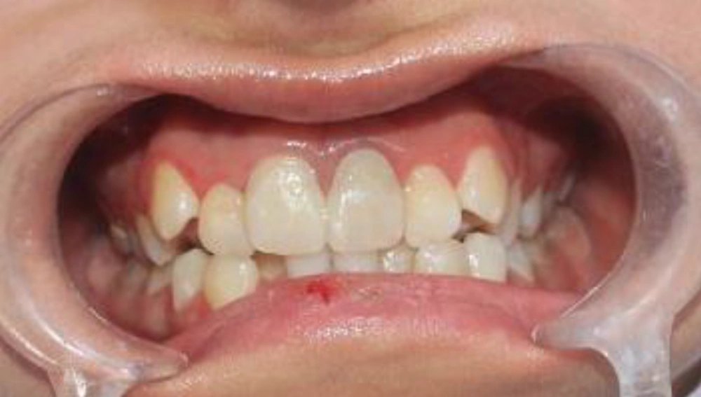
During the 3-year follow-up, the patient was satisfied with the outcome and did not tend to continue the treatment for the aesthetical or functional improvement (Figure 3). However, the teeth 11 and 21 were infra-positioned since there was an interference of ankylosis in the vertical growth of the alveolar process.
3. Discussion
A large number of factors, including the status of the avulsed tooth, stages of root development, period of extra-alveolar dryness, storage environment, treatment time, and modality, can affect the success of the replanted avulsed tooth (12). According to the International Association of Dental Traumatology (IADT) (2017), delayed replantation has a poor long-term prognosis in cases with a dry time of over 60 min (for avulsed closed apex tooth) or those with nonviable periodontal cells (1).
In our case, it was not possible to find suitable prosthetic solutions even if the replantation treatment plan was followed. As a child continues to grow, the reduced formation of alveolar bone in this area results in a narrow alveolar ridge and difficult restoration of bridging or implanting in the future. The loss of two anterior teeth in children can have detrimental effects on their physical attractiveness and self-confidence. Accordingly, the replantation of avulsed teeth was performed with the consent of the child’s parents.
In order to prevent infection-related resorption, the root surface of the tooth was cleaned with 0.5% sodium hypochlorite to separate the remaining dead PDL cells, which in turn slowed down the osseous replacement of the root surface (13). Later, the tooth was immersed in sodium fluoride (NaF) solution for 20 min. The application of NaF has been suggested for the pretreatment of the root surface since 1968 due to its beneficial effects. In other words, NaF delays the progress of root surface resorption after replantation.
Trope (14) mentioned that on some occasions, there is a need for slowing down the process of root replacement by the bone in order to keep the tooth in the mouth as long as possible. These occasions include the time when there is no way to avoid the serious additional damage and osseous replacement of the root. In this case, the root canal was cleaned before replantation. Moreover, the canal was filled with calcium hydroxide in order to avoid inflammatory external root resorption. However, there is no clear estimation of clinical time for the use of calcium hydroxide in avulsed teeth.
The obtained results of long-term clinical studies and case reports are indicative of the beneficial effects of calcium hydroxide on avulsed teeth (8). However, long-term calcium hydroxide treatment may have some disadvantages. This treatment requires multiple appointments extending over a long period of time, which needs highly cooperative patients. In other words, a patient’s lack of cooperation can result in the infection of the root canal, leading to the probable loss of the tooth (8).
Ankylosis of the tooth causes a delay in the replantation of avulsed incisors. The incidence of this condition is mostly due to the presence of osteoclasts sourcing from the surrounding alveolar bone, which are instantly followed by osteoblasts. Accordingly, osteoclasts reach the root surface after passing through the damaged PDL and pre-cementum. This process leads to the bony replacement of the root cementum and root dentin in older children. The implementation of replacement resorption through spontaneous or surgical procedures leads to the loss of dental crowns (15).
The comparison of the mature teeth in adults and children revealed that replantation can lead to extensive inflammatory root resorption. Regarding ankylosed teeth, decoronation is suggested because it maintains the contour of the alveolar ridge in case the infra-position of the tooth crown is more than 1 mm (16). Accordingly, the patients who are satisfied with replantation are not mostly interested in undergoing the procedures targeted toward the aesthetical or functional improvement of their teeth.
Delayed dental replantation can lead to an acceptable level of functionality and stability in a tooth despite the prolonged extra-alveolar dry storage time. Moreover, it is better to postpone the implantation in young patients who have not reached puberty until they have passed their growth spurt. In such cases, the patient can benefit from dental replantation for the maintenance of the surrounding bones.


