Cover image of the article: Our Experience With Regional Anesthesia in a Case of Pierre Robin Syndrome

Image Credit:Anesth Pain Med

Our Experience With Regional Anesthesia in a Case of Pierre Robin Syndrome

Authors

Ozkan Onal1,*, Muhammed Emin Zora1, Ali Saltali1, Serdal Bozdogan1, Jale Bengi Celik1
1Department of Anesthesiology and Reanimation, Selcuk University Medical Faculty, Konya, Turkey
*Corresponding Author: Corresponding author: Ozkan Onal, Department of Anesthesiology and Reanimation, Selcuk University Medical Faculty, Konya, Turkey. Tel: +90-5059052252, Fax: +90-3322245178, E-mail: Email: drozkanonal@selcuk.edu.tr

Anesthesiology and Pain Medicine:Vol. 6, issue 1; e33480
Published online:Jan 31, 2016
Article type:Letter
Received:Sep 29, 2015
Accepted:Dec 02, 2015
How to Cite:Onal O, Zora ME, Saltali A, Bozdogan S, Bengi Celik J. Our Experience With Regional Anesthesia in a Case of Pierre Robin Syndrome. Anesth Pain Med. 2016;6(1):e33480. doi: https://doi.org/10.5812/aapm.33480

Dear Editor,
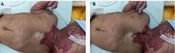
Pierre-Robin Syndrome (PRS) is characterized by micrognathia, glossoptosis, and a “u-shaped” cleft palate (1). The incidence of PRS varies from 1: 5000 to 1: 85000 (2). Neonates with PRS present with respiratory obstruction varying from mild to severe directly after birth or during the first week of life. A sufficiently severe obstruction can result in obstructive apnea, hypoxia, failure to thrive, and death (3). We report a case of a patient with PRS who could not be intubated previously and who had a history of tracheostomy; this patient was scheduled to undergo an operation for hip dysplasia. We present our anesthesia management and approach. An 8-year-old boy weighing 18 kg and diagnosed with PRS underwent preoperative evaluation that revealed restricted mouth opening, micrognathia, short extremities, mild airway obstruction, and respiratory distress (Figure 1).
Image of the Patient With Pierre Robin Syndrome
Figure 1.
Image of the Patient With Pierre Robin Syndrome
His Mallampati score was 3, his head-neck extension was comfortable, and his thyromental distance was within the normal range. Preoperative laboratory and imaging tests yielded normal results. This patient had a history of difficult intubation, and tracheostomy preparations were made to allow for a difficult intubation. The patient had no other known diseases and conditions that contraindicated spinal anesthesia (SA). Preoxygenation was performed under routine monitoring. The patient’s baseline heart rate was 131 beats min-1, his blood pressure was 112/64 mmHg, and his SpO2 was 94%. Premedication and amnesia treatment consisted of administration of 10 mg midazolam (Dormicum, Roche, Istanbul, Turkey) intranasal 10 minutes prior to the procedure. The patient was then administered 2 mg/kg propofol, 25 µg fentanyl (Talinat, Vem Ilac, Istanbul, Turkey), and 40 mg methylprednisolone (Prednol-l, Mustafa Nevzat, Istanbul, Turkey) intravenously. A no. 2.0 I-gel was placed while the patient was in the supine position, and the patient was then placed in a lateral decubitus position. The skin was prepared with antiseptic and a spinal block was carried out by administering 0.4 mg/kg hyperbaric bupivacaine to the L4-5 vertebral space, as described previously (4) (Figures 2 and 3).
Image of the Patient During Spinal Anesthesia
Figure 2.
Image of the Patient During Spinal Anesthesia
Image of the Patient After Extubation
Figure 3.
Image of the Patient After Extubation
After the procedure, the patient was placed in a supine position. Muscle relaxants were not used during surgery and anesthesia was maintained with 100 µg/kg/min continuous propofol infusion. The operation lasted for two hours. Spontaneous respiration was sufficient at the end of the operation and the patient was extubated and transferred to the recovery unit. In follow up, active extremity movements started and the patient was transferred to the orthopedics clinic. Micrognathia may be an isolated problem or may occur as part of a sequence or a syndrome such as PRS (5). Patients with PRS have a high risk of airway obstruction and a high probability of the development of malnutrition, failure to thrive, hypoxemia, cor-pulmonale, loss of strength, and cerebral anoxia. In these patients, respiratory problems lead to aggravation of the clinical picture. Airway obstruction is caused by the presence of glossoptosis, in addition to cleft palate and micrognathia with retrognati (6). The small size and backward position of the jaw in children with PRS create serious difficulties in using a mask and maintaining an open airway in the supine position. The airway obstruction risk will increase when the soft tissues are relaxed; therefore, the use of muscle relaxants should be avoided (7). No problems were encountered with mask use in our case, so an I-gel was placed under propofol anesthesia without using any muscle relaxants, and a spinal block was carried out for postoperative analgesia.
The intubation and extubation of PRS cases requires special care, and preoperative monitoring is warranted. For operations under the umbilicus, regional anesthesia can be a good alternative in children who are at high risk from general anesthesia (8). In fact, previous studies have demonstrated that regional anesthesia is a safe alternative in neonates (9). In our opinion, preparations should be made for difficult airway management in these cases, and spinal anesthesia may be reliably and safely used in lower extremity surgeries.
Footnotes

  • Authors’ Contribution:Study concept and design: Ozkan Onal; acquisition of data: Muhammed Emin Zora, Serdal Bozdogan, Ali Saltali; analysis and interpretation of data: Ozkan Onal, Muhammed Emin Zora; drafting of the manuscript: Ozkan Onal, Muhammed Emin Zora; critical revision of the manuscript for important intellectual content: Jale Bengi Celik.

References

  • 1.
    Portnoy JE, Tatum S. Retrograde nasal intubation via the cleft in Pierre-Robin Sequence neonates: a case series. Int J Pediatr Otorhinolaryngol. 2009;73(12):1828-32. [PubMed ID: 19836088]. https://doi.org/10.1016/j.ijporl.2009.09.030.
  • 2.
    Cladis F, Kumar A, Grunwaldt L, Otteson T, Ford M, Losee JE. Pierre Robin Sequence: a perioperative review. Anesth Analg. 2014;119(2):400-12. [PubMed ID: 25046788]. https://doi.org/10.1213/ANE.0000000000000301.
  • 3.
    Kucukyavuz Z, Ozkaynak O, Tuzuner AM, Kisnisci R. Difficulties in anesthetic management of patients with micrognathia: report of a patient with Stickler syndrome. Oral Surg Oral Med Oral Pathol Oral Radiol Endod. 2006;102(6):e33-6. [PubMed ID: 17138163]. https://doi.org/10.1016/j.tripleo.2006.06.002.
  • 4.
    Duman A, Apiliogullari S, Duman I. Effects of intrathecal fentanyl on quality of spinal anesthesia in children undergoing inguinal hernia repair. Paediatr Anaesth. 2010;20(6):530-6. [PubMed ID: 20456062]. https://doi.org/10.1111/j.1460-9592.2010.03315.x.
  • 5.
    Frawley G, Espenell A, Howe P, Shand J, Heggie A. Anesthetic implications of infants with mandibular hypoplasia treated with mandibular distraction osteogenesis. Paediatr Anaesth. 2013;23(4):342-8. [PubMed ID: 23043528]. https://doi.org/10.1111/pan.12049.
  • 6.
    Semjen F, Bordes M, Cros AM. Intubation of infants with Pierre Robin syndrome: the use of the paraglossal approach combined with a gum-elastic bougie in six consecutive cases. Anaesthesia. 2008;63(2):147-50. [PubMed ID: 18211445]. https://doi.org/10.1111/j.1365-2044.2007.05097.x.
  • 7.
    Patra P. Capnography guided awake nasal intubation in a 4 month infant with pierre robin syndrome for cleft lip repair-a better technique. Indian J Anaesth. 2009;53(6):692-5. [PubMed ID: 20640099].
  • 8.
    Onal O, Demirci A, Bayrak O. Caudal Anesthesia for Open Bladder Surgery in High-Risk Adolescent With Scoliosis and Neural Tube Defect. Anesth Pain Med. 2015;5(4). eee26878. [PubMed ID: 26478864]. https://doi.org/10.5812/aapm.26878.
  • 9.
    Imani F, Entezary SR, Alebouyeh MR, Parhizgar S. The maternal and neonatal effects of adding tramadol to 2% lidocaine in epidural anesthesia for cesarean section. Anesth Pain Med. 2011;1(1):25-9. [PubMed ID: 25729652]. https://doi.org/10.5812/kowsar.22287523.1271.

Copyright

Copyright © 2016, Iranian Society of Regional Anesthesia and Pain Medicine (ISRAPM). This is an open-access article distributed under the terms of the Creative Commons Attribution-NonCommercial 4.0 International License (http://creativecommons.org/licenses/by-nc/4.0/) which permits copy and redistribute the material just in noncommercial usages, provided the original work is properly cited.

Similar Articles

10
Nov
2013
The First Report of Robinow Syndrome in Iran and Literature Review

The First Report of Robinow Syndrome in Iran and Literature Review

Zohreh Kavehmanesh,
Ali Reza Shafiee

Kavehmanesh Z, Shafiee AR. The First Report of Robinow Syndrome in Iran and Literature Review. Thrita J Neu. 2013;2(4):84-6. doi: https://doi.org/10.5812/thrita.11928

16
Mar
2023
General Anesthesia in Schwartz-Jampel Syndrome: A Case Report

General Anesthesia in Schwartz-Jampel Syndrome: A Case Report

Pooya Derakhshan,
Azadeh Habibi,
Saied Amniati

Derakhshan P, Habibi A, Amniati S. General Anesthesia in Schwartz-Jampel Syndrome: A Case Report. Anesth Pain Med. 2023;13(3):e129305. doi: https://doi.org/10.5812/aapm-129305

11
Jan
2024
Facing PHACES Syndrome; Anesthesiologist’s Point of View

Facing PHACES Syndrome; Anesthesiologist’s Point of View

Dariush Abtahi,
Alireza Shakeri,
Ardeshir Tajbakhsh

Abtahi D, Shakeri A, Tajbakhsh A. Facing PHACES Syndrome; Anesthesiologist’s Point of View. Anesth Pain Med. 2023;13(6):e141896. doi: https://doi.org/10.5812/aapm-141896

5
Jan
2022
The Neurologic Complications Associated with Anesthesia in Pediatrics Treated with Radiotherapy Under Anesthesia

The Neurologic Complications Associated with Anesthesia in Pediatrics Treated with Radiotherapy Under Anesthesia

Shahryar Sane,
Behzad Sinaei,
Parang Golabi,
Hadi Talebi,
Nazila Rahmani,
Reza Foruhar
,et al.

Sane S, Sinaei B, Golabi P, Talebi H, Rahmani N, et al. The Neurologic Complications Associated with Anesthesia in Pediatrics Treated with Radiotherapy Under Anesthesia. Inn J Pediatr. 2022;32(1):e116822. doi: https://doi.org/10.5812/ijp.116822

29
Nov
2016
Anesthesia Management of a Patient with Papillon-Lefevre Syndrome: A Case Report

Anesthesia Management of a Patient with Papillon-Lefevre Syndrome: A Case Report

Afshin Iranpour,
Ata Mahmoodpoor

Iranpour A, Mahmoodpoor A. Anesthesia Management of a Patient with Papillon-Lefevre Syndrome: A Case Report. Anesth Pain Med. 2017;7(1):e39556. doi: https://doi.org/10.5812/aapm.39556

More by these authors

Ozkan OnalPubMedScholar
Muhammed Emin ZoraPubMedScholar
Ali SaltaliPubMedScholar
Serdal BozdoganPubMedScholar
Jale Bengi CelikPubMedScholar
Share
Cited by
Metrics