
1. Context
Chronic postsurgical pain (CPSP) is an important consequence of surgery and is characterized by pain that develops or persists after a surgical procedure beyond the expected period of tissue healing, typically for at least 3 months. It may occur within the surgical field or in the distribution of an affected nerve or dermatome and may include persistent pain, hyperalgesia, or allodynia. Current evidence indicates that CPSP is a multifactorial condition involving both peripheral and central sensitization. Persistent nociceptive input from injured tissues can promote changes in peripheral and central nociceptive pathways, while inflammatory and neuroimmune mechanisms may contribute to pain persistence (1).
CPSP is a broad clinical category and is not synonymous with persistent postsurgical neuropathic pain. Although neuropathic mechanisms may contribute to CPSP, not all persistent postsurgical pain is neuropathic. Persistent postsurgical neuropathic pain represents a more specific presentation in which pain is attributable to a lesion or disease affecting the somatosensory nervous system. Accordingly, findings from studies of neuropathic pain after nerve injury should not automatically be generalized to the full spectrum of CPSP.
The extent and nature of surgical tissue injury may influence the subsequent development of persistent pain. Following tissue injury, inflammatory mediators and changes in peripheral nerve excitability can enhance nociceptive signaling, while sustained peripheral input may contribute to central sensitization (1).
When a peripheral nerve is injured, the biological response extends beyond the initial structural damage. Interruption of axonal continuity initiates a coordinated sequence of degeneration, inflammation, cellular reprogramming, and regeneration (Table 1). Distal to the injury site, Wallerian degeneration is accompanied by axonal and myelin fragmentation, changes in Schwann-cell phenotype, disruption of the blood–nerve barrier, and recruitment of immune cells. These processes contribute to the removal of damaged tissue and establish the conditions required for subsequent axonal regeneration (2, 3).
Table 1.
Processes in Peripheral Nerve Injury and Regeneration: Adaptive and Maladaptive Outcomes a
| Process | Key Biological Events | Adaptive/Functional Trajectory | Maladaptive/Pain-Associated Trajectory |
|---|---|---|---|
| Surgical nerve injury | Direct or indirect nerve injury; disruption of axonal continuity; mechanical or ischemic damage | Preservation or restoration of neural continuity and initiation of repair | Persistent or severe nerve injury may provide the initial substrate for abnormal sensory signaling |
| Wallerian degeneration | Distal axonal and myelin fragmentation; blood–nerve barrier changes; immune-cell recruitment; debris clearance | Efficient removal of damaged tissue prepares the distal nerve for regeneration | Persistent inflammatory/neuroimmune signaling may influence neuronal excitability |
| Schwann-cell response | Schwann-cell phenotypic reprogramming; proliferation and migration; formation of bands of Büngner; production of trophic and signaling molecules | Creation of a permissive environment and guidance for regenerating axons | Dysregulated signaling may coexist with or contribute to abnormal nociceptive activity |
| Immune and regenerative microenvironment | Macrophage recruitment; cytokine/chemokine and growth-factor signaling; extracellular-matrix remodeling; transition toward a reparative state | Resolution of inflammation and coordinated tissue remodeling support regeneration | Persistent or dysregulated inflammation may sustain abnormal neuronal signaling |
| Axonal regeneration | Intrinsic neuronal regenerative response; axonal extension through the injured nerve | Directed axonal growth toward appropriate distal pathways | Disorganized or incomplete growth; aberrant sprouting or failure to reach an appropriate target |
| Target reinnervation | Regenerating axons establish connections with distal sensory, motor, or other target tissues | Appropriate target reinnervation and restoration of neural function | Misrouting, incomplete reinnervation, or absence of an appropriate target |
| Post-regeneration functional outcome | Remyelination, maturation of regenerated axons, restoration of sensory/motor function | Functional recovery with normalization or improvement of sensory function | Neuroma formation, ectopic activity, persistent abnormal sensory signaling; regenerative PNP remains an experimental concept |
| Long-term clinical trajectory | Interaction of peripheral repair with ongoing nociceptive processing and central mechanisms | Recovery with resolution or reduction of pain | Persistent neuropathic pain and possible contribution to CPSP through interacting peripheral and central mechanisms |
a The adaptive and maladaptive trajectories represent a conceptual framework rather than a proven causal sequence. The two trajectories are not mutually exclusive, and functional recovery may coexist with persistent pain. Abbreviations: CPSP, chronic postsurgical pain; PNP, peripheral neuropathic pain.
Schwann cells respond dynamically to nerve injury and interact with neurons and immune cells through growth factors, cytokines, chemokines, and other mediators. Thus, the regenerative microenvironment is not biologically isolated from the mechanisms involved in neuropathic pain (2, 4).
Do the quality and organization of peripheral nerve regeneration influence the clinical consequences of surgical nerve injury? Is axonal regrowth alone sufficient for functional recovery, or must regenerating axons navigate the injured nerve, reach appropriate distal pathways, and establish functional target reinnervation? (5)
This review examines the potential relationship between the quality of peripheral nerve regeneration and chronic postsurgical pain, with particular emphasis on Wallerian degeneration, Schwann cells, neuroimmune interactions, maladaptive regeneration, and emerging regenerative strategies. The available evidence is considered across experimental and clinical studies, while established mechanisms are distinguished from emerging hypotheses. This review does not assume that maladaptive regeneration is a direct or sufficient cause of CPSP; rather, it examines whether the organization and functional outcome of nerve regeneration may represent one component of the complex biological processes underlying chronic postsurgical pain.
2. Evidence Acquisition
A focused narrative literature review was conducted to examine the potential relationship between the quality and functional organization of peripheral nerve regeneration and the development or persistence of chronic postsurgical pain (CPSP).
The initial literature search was conducted on September 27, 2026, using Google and PubMed. Google was used for web-based searches, whereas PubMed was used to locate bibliographic records and abstracts. Publisher platforms, including Elsevier, were subsequently used to access available full-text publications and were not treated as separate bibliographic databases.
The initial search used combinations of the concepts “chronic pain,” “regenerative medicine,” and “surgery.” To clarify the search strategy, the principal concepts and related terms can be represented by the following reconstructed search combinations:
(“chronic pain” OR “chronic postsurgical pain” OR “chronic postoperative pain”) AND (“surgery” OR “surgical injury”).
(“peripheral nerve regeneration” OR “axonal regeneration” OR “target reinnervation” OR “Wallerian degeneration”) AND (“pain” OR “neuropathic pain”).
(“Schwann cells” OR “neuroimmune interactions” OR “neuroma”) AND (“peripheral nerve injury” OR “nerve regeneration”).
Additional topic-specific searches addressed regenerative peripheral nerve interfaces (RPNI), targeted muscle reinnervation (TMR), neuroma formation, mesenchymal stem cells and extracellular vesicles, and other regenerative approaches. Subsequent supplementary searches also addressed autologous conditioned serum (ACS), platelet-rich plasma (PRP), osteoarthritis-related literature, the transition from acute postoperative pain to chronic pain, and the ICD-11 definition of CPSP. These records were considered in relation to the review question but were not treated as direct evidence that regenerative interventions prevent or treat CPSP.
Across the initial and expanded search rounds, 20 records were identified. Of these, 18 full-text articles were available as PDFs and were examined in detail; full-text access was not obtained for two records. Subsequent topic-specific searches identified six additional records: four addressing autologous conditioned serum, platelet-rich plasma, or osteoarthritis (ACS/PRP/OA), and two addressing chronic postsurgical pain mechanisms or definition. These six records were identified during a later, separate search stage. Reference-based searching identified one additional relevant publication by Navarro. Thus, 27 records were identified across the initial and expanded searches, subsequent topic-specific searches, and reference-based identification. The original submitted manuscript cited 12 references selected to support its specific mechanistic and translational topics; this was not an exhaustive list of all records identified. Following further literature assessment and citation tracing during revision, the revised manuscript contains 29 references.
Literature was considered relevant to the review if it addressed one or more of the following domains: mechanisms of CPSP; peripheral nerve injury and Wallerian degeneration; Schwann-cell and neuroimmune responses; axonal regrowth, remyelination, and functional target reinnervation; neuroma formation and neuroma-related pain; experimental regenerative neuropathic pain; or regenerative interventions relevant to peripheral nerve repair. Studies were assessed narratively according to their relevance to the central review question, study design, experimental model or clinical population, outcomes evaluated, and the extent to which their findings could be related to postsurgical pain. Experimental nerve-injury studies and studies of postamputation or neuroma-related pain were considered indirect or context-specific evidence and were not assumed to represent the full spectrum of CPSP.
The original search documentation did not specify prespecified publication-date or language restrictions; therefore, no such restrictions are claimed retrospectively. The review was conducted as a focused narrative synthesis rather than a systematic review or meta-analysis. No prospective protocol registration, formal PRISMA screening process, independent duplicate study selection, quantitative pooling, or formal risk-of-bias assessment was undertaken. In addition, full-text publication availability was variable and may have influenced the depth of assessment of some records. The findings were therefore interpreted cautiously, with particular attention to distinguishing direct clinical evidence for CPSP from mechanistic and translational evidence derived from experimental nerve injury, neuroma-related pain, and other conditions.
2.1. Supplementary Literature Identification During Revision
Following peer review, the evidence base was expanded through an AI-assisted literature identification process using ChatGPT (GPT-6; OpenAI). Additional publications were identified primarily by examining the reference lists of articles included in the original review and following relevant citations. The supplementary identification process focused on primary experimental and clinical evidence concerning Wallerian degeneration, Schwann-cell and immune responses, axonal regeneration, functional target reinnervation, regeneration-associated pain, and regenerative interventions, including targeted muscle reinnervation (TMR), regenerative peripheral nerve interfaces (RPNI), and mesenchymal stem cell-derived extracellular vesicles.
The identified publications were reviewed by the author for relevance to the review question. Primary studies directly evaluating peripheral nerve repair, functional reinnervation, regeneration-associated pain, or regenerative interventions were selected for inclusion in Table 2. Other primary studies that addressed pain-related mechanisms without directly evaluating nerve regeneration or examined pain outcomes without assessing nerve repair were retained in the narrative where relevant but were not included in the table.
Table 2.
Selected Primary Experimental and Clinical Studies Addressing Peripheral Nerve Repair, Functional Reinnervation, and Regeneration-Related Pain a
| Study | Model/Population | Design/Intervention | Outcomes/Follow-Up | Main Findings | Limitations/Relevance to CPSP |
|---|---|---|---|---|---|
| Verdú & Navarro, 1997 (6) | 36 female mice; sciatic crush | Morphological and functional reinnervation assessment | Sensory responses, sweat-gland response, CMAP; up to 100 days | Histological reinnervation preceded recovery of some functional responses; morphology and function did not fully coincide. | Animal crush model; no chronic-pain endpoint. |
| Valero-Cabré & Navarro, 2002 (7) | 59 rats; 8-mm sciatic nerve resection | Autograft and tube-repair strategies; four repair groups included 42 rats within the 59-animal study | CMAP and walking-track SFI; 90 days | Axonal misdirection varied by repair method and was associated with poorer functional recovery. | Severe experimental lesion; no pain endpoint. |
| Boivin et al., 2007 (8)b | 208 adult male mice and 178 adult male rats across multiple experiments; sciatic crush | TLR2/TLR4/MyD88 signaling disruption | Myelin clearance, regeneration, motor recovery | Disrupted TLR signaling impaired debris clearance and delayed regeneration and functional recovery. | Animal study; no direct chronic-pain outcome; reported totals span multiple experiments. |
| Barrette et al., 2008 (9)b | 452 adult female mice across multiple experiments; peripheral nerve injury | Myeloid-cell depletion/requirement for myeloid cells | Debris clearance, axon regeneration, functional recovery | Myeloid cells were required for efficient debris clearance and axonal regeneration. | Animal study; no direct pain endpoint; exact cohort sizes vary by experiment. |
| Orita et al., 2013 (10) | Mouse; Schwann-cell-specific LRP1 deletion | Genetic disruption of Schwann-cell LRP1 | Remak bundles, axon–glia organization, mechanical sensitivity | LRP1 disruption altered Remak bundle organization and was associated with mechanical hypersensitivity and impaired repair. | Animal model; not CPSP; n varied by assay. |
| Su et al., 2019 (11) | Mouse sciatic crush | Schwann-cell P2X4 overexpression/BDNF signaling | Remyelination, motor and sensory function; multiple assay time points | P2X4/BDNF signaling supported remyelination and functional recovery; sensory testing assessed recovery after crush. | Animal study; mechanical sensitivity is not equivalent to a chronic neuropathic-pain endpoint; n varied by assay. |
| Xie et al., 2017 (12) | Rat SNL and SNI models | Assessment/manipulation of active regeneration and target reinnervation | Spontaneous activity, histology, pain-related behavior | Regenerating axons with failed target reinnervation were associated with persistent pain-related behavior; blocking regeneration reduced behavior in tested models. | Animal-only evidence; model-specific; no direct CPSP evidence. |
| Ren et al., 2016 (13) | Adult rats; sciatic nerve crush | Behavioral and histological follow-up of reinnervated skin | Mechanical/thermal sensitivity and histology to 8 weeks | Pain-related sensory abnormalities persisted in reinnervated skin despite progressive reinnervation. | Animal-only; stimulus-evoked outcomes; completeness/accuracy of regeneration not fully established. |
| Oliveira et al., 2018 (14) | 30 male rats; forelimb amputation | Time-course histology of traumatic neuroma formation | Histology at days 3, 7, 28, 60, and 90; n = 6 at each time point | Axonal sprouts were present early; disorganized axonal growth and histological neuroma features appeared from day 28 and became more prominent over time. | No pain behavior or electrophysiology; histology cannot distinguish painful from asymptomatic neuroma. |
| Dumanian et al., 2019 (15) | 28 human amputees; 30 residual limbs | Randomized trial: TMR vs standard neuroma treatment | Residual-limb and phantom pain; 1-year outcomes | TMR showed a favorable longitudinal signal for phantom-limb pain; primary intention-to-treat comparisons were not uniformly statistically significant. | Clinical evidence is specific to postamputation pain; trial stopped below target enrollment; pain change does not prove a regeneration-mediated mechanism. |
| Woo et al., 2016 (16) | 16 patients; 17 residual limbs | Retrospective RPNI pilot/case series; 46 RPNIs constructed | Neuroma and phantom pain; telephone-interview follow-up mean 7.5 ± 3.4 months (range 3–15) | Reported reductions in neuroma-related and phantom pain after RPNI. | No control group; postamputation setting; no direct measurement proving regeneration-mediated analgesia. |
| Senger et al., 2023 (17) | 36 Fischer rats; painful tibial neuroma model | Two experiments: (1) neuroma in situ vs excision (n = 6/group); (2) neuroma in situ vs TMR vs RPNI (n = 8/group) | Pain-related behavior (thigh tap and von Frey) and neuromuscular-junction reinnervation | TMR and RPNI reduced pain scores versus untreated neuroma controls in the second experiment. | Animal neuroma model; two experiments had different group sizes; findings are specific to this model and do not establish efficacy for general CPSP. |
| Bucan et al., 2019 (18) | 12 Wistar rats; sciatic nerve crush; in-vitro Schwann-cell and DRG-neuron assays | Rat adipose-derived MSC exosomes injected locally proximal and distal to the crush site | Footprint/SFI weekly to 21 days; neurofilament immunohistochemistry at day 21; in-vitro proliferation and neurite assays | Exosomes increased Schwann-cell proliferation in vitro; in vivo regenerated neurofilament-positive fibers were approximately 40% higher and SFI recovery improved. The in-vitro neurite-length increase was not statistically significant. | No pain-related behavior, neuropathic-pain, or CPSP outcome was assessed; short-term preclinical nerve-repair evidence only. |
| Chen et al., 2019 (19) | Rat sciatic nerve transection with a 10-mm nerve gap | Human adipose-derived stem-cell exosomes delivered in Matrigel within a silicone conduit | Schwann-cell proliferation, migration and myelination; axonal regeneration; muscle atrophy recovery | Exosomes improved Schwann-cell function, axonal regeneration, myelination, and recovery of denervation-related muscle atrophy. | No pain-related behavior or CPSP outcome was assessed; preclinical nerve-repair evidence only. |
| Zhao et al., 2020 (20) | Rat sciatic nerve crush; four exosome-dose groups (n = 6 per dose group) | Intramuscular BMSC-derived exosomes on days 0, 7, and 14 | SFI and thermal paw-withdrawal latency through day 28; regenerated-fiber number and diameter; DRG gene expression | Exosomes improved nerve regeneration and functional recovery; the 0.9 × 10¹⁰ particles/ml dose showed favorable results in several measures, but no clear linear dose–response relationship was established. | Short-term preclinical study; thermal withdrawal was an evoked sensory measure, not evidence of analgesia; no persistent neuropathic-pain or CPSP outcome was assessed. |
a This table presents selected primary experimental and clinical studies relevant to peripheral nerve repair, functional reinnervation, and regeneration-associated pain. It is not an exhaustive summary of all studies identified during the literature search. Most included evidence is experimental or relates to specific clinical conditions, particularly neuroma-related and postamputation pain, and should not be interpreted as direct evidence for the prevention or treatment of CPSP across general surgical populations. Abbreviations: BDNF, brain-derived neurotrophic factor; BMSC, bone marrow-derived mesenchymal stromal cell; CMAP, compound muscle action potential; CPSP, chronic postsurgical pain; DRG, dorsal root ganglion; LRP1, low-density lipoprotein receptor-related protein 1; MSC, mesenchymal stromal cell; RPNI, regenerative peripheral nerve interface; SFI, sciatic functional index; SNI, spared nerve injury; SNL, spinal nerve ligation; TLR, Toll-like receptor; TMR, targeted muscle reinnervation.
b Sample sizes varied across experimental groups and outcome assessments in some studies. Where a single representative sample size could not be accurately reported, it has not been specified. Reported totals across multiple experiments should not be interpreted as the sample size of a single experimental cohort.
3. Results
3.1. Peripheral Nerve Injury Following Surgery
Surgical procedures may expose peripheral nerves to direct trauma or indirect mechanical and ischemic stress. The likelihood and consequences of nerve injury depend on the anatomical site, procedural factors, and the nature and severity of the insult (2).
The response to peripheral nerve injury is not solely destructive; it also establishes the cellular and extracellular conditions required for subsequent repair. In addition, inflammatory and neuroimmune changes accompanying nerve injury may influence neuronal excitability and neuropathic pain (2-4).
3.2. Wallerian Degeneration: The Beginning of Regeneration
Following axonal disruption, the distal segment of the injured nerve undergoes Wallerian degeneration (WD), a tightly regulated sequence of cellular and molecular events that prepares the nerve for subsequent regeneration. Axonal and myelin fragmentation occurs alongside changes in Schwann-cell phenotype, disruption of the blood–nerve barrier, and recruitment of immune cells, including neutrophils and macrophages. Efficient clearance of axonal and myelin debris is an essential component of this process because myelin debris contains factors that can inhibit axonal growth (3).
A central event in WD is the transformation of Schwann cells from their mature myelinating state to a repair-supportive phenotype. After losing axonal contact, Schwann cells downregulate components associated with myelination and acquire functions that support regeneration. They proliferate, migrate within the injured nerve, and become organized longitudinally to form the bands of Büngner, which provide a structural pathway and a source of trophic support for regenerating axons (2, 3).
Macrophages also play an important role during WD. Their recruitment to the injured nerve is associated with changes in the blood–nerve barrier and signals generated by Schwann cells and other cells within the injured tissue. Macrophages contribute to the removal of degenerating myelin and axonal debris and release cytokines and neurotrophic mediators that can influence the regenerative environment (2, 3).
Successful regeneration ultimately requires more than clearance of damaged tissue. WD therefore represents not merely a degenerative phase following nerve injury but also a biological transition that prepares the injured nerve for regeneration (3).
3.3. Schwann Cells, Immune Response and the Regenerative Microenvironment
Peripheral nerve regeneration depends on coordinated interactions among Schwann cells, immune cells, and components of the extracellular environment. Experimental studies have demonstrated that immune-cell recruitment and signaling are important for clearing degenerating myelin and supporting subsequent axonal regeneration. For example, disruption of Toll-like receptor signaling or depletion of myeloid cells impairs debris clearance and functional recovery after peripheral nerve injury. These findings indicate that the inflammatory response is not merely a consequence of nerve damage but an active component of the regenerative process (8, 9).
Schwann cells play an active role in peripheral nerve repair through axonal support, myelination, and interactions with surrounding cells. Schwann-cell LRP1 has been identified as a pro-survival receptor (21), and its disruption alters Remak bundle organization and is associated with pain-related behavior in animal models (10). Schwann-cell P2X4/BDNF signaling may also support remyelination and functional recovery after nerve injury (11).
Early immune-cell activity supports debris clearance and nerve regeneration (8, 9). Macrophage accumulation in the dorsal root ganglia (DRG) has been observed after peripheral nerve injury (22), and DRG macrophages contribute to the initiation and persistence of neuropathic pain (23). Separately, NFκB–COX2 signaling in non-myelinating Schwann cells is required for neuropathic pain maintenance in vivo (24).
Schwann cells may also influence nociceptive signaling through interactions with sensory axons and expression of pain-related molecules. In a model of ethanol-induced neuropathy, Schwann-cell TRPA1 signaling was shown to contribute to mechanical allodynia. These findings suggest that Schwann cells may modulate pain-related responses through distinct mechanisms, although their relevance to chronic postsurgical pain (CPSP) remains uncertain (25).
Taken together, these findings indicate that the regenerative microenvironment is an active component of peripheral nerve repair rather than merely a passive substrate for axonal growth. Its cellular composition, molecular signaling, and structural organization may influence axonal regeneration, remyelination, and functional recovery. Some of these same mechanisms may also modulate nociceptive signaling. Nevertheless, whether specific characteristics of the regenerative microenvironment can predict the development of chronic postsurgical pain in humans remains unknown.
3.4. When Regeneration Becomes Maladaptive
Peripheral nerve regeneration does not necessarily result in complete functional recovery (Figure 1). Although axonal regrowth is essential for nerve repair, functional recovery also depends on the accurate guidance of regenerating axons toward appropriate distal pathways and target tissues. Regenerating axons may establish inappropriate connections, and morphological evidence of reinnervation does not always correspond to the degree of functional recovery. Thus, the quality of nerve regeneration should be assessed not only by axonal growth but also by the accuracy of target reinnervation and the restoration of sensory and motor function (6, 7).
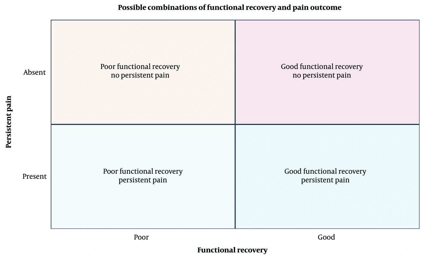
Figure 1.
Conceptual framework illustrating the relationship between peripheral nerve repair, functional recovery, and pain outcomes. Functional recovery and persistent pain are distinct dimensions that may coexist. The four combinations are possible clinical states, not sequential stages or probabilities. The framework does not imply that regeneration quality causes a particular pain outcome.
Following peripheral nerve transection, regenerating axons that fail to establish appropriate distal connections may form disorganized neuromatous tissue consisting of irregular axonal bundles embedded in fibrous connective tissue (14). Experimental nerve injury models have shown that actively regenerating axons can exhibit abnormal spontaneous activity and contribute to persistent pain-related behaviors, particularly when appropriate target reinnervation fails (12). However, neuroma formation does not invariably result in symptoms. An asymptomatic traumatic neuroma of the inferior alveolar nerve has been reported four years after mandibular sagittal split osteotomy (26).
Regenerative peripheral neuropathic pain (regenerative PNP) has been proposed based on rodent studies showing persistent pain-related behaviors during axonal regeneration. These findings suggest that axonal regrowth does not necessarily restore normal sensory function; however, their clinical relevance remains uncertain (12, 13, 27).
3.5. Regenerative Medicine: From Promoting Regeneration to Guiding Functional Regeneration
Regenerative strategies discussed here include target-directed procedures and cell-derived products, but their evidence bases and measured outcomes differ (Figure 2).
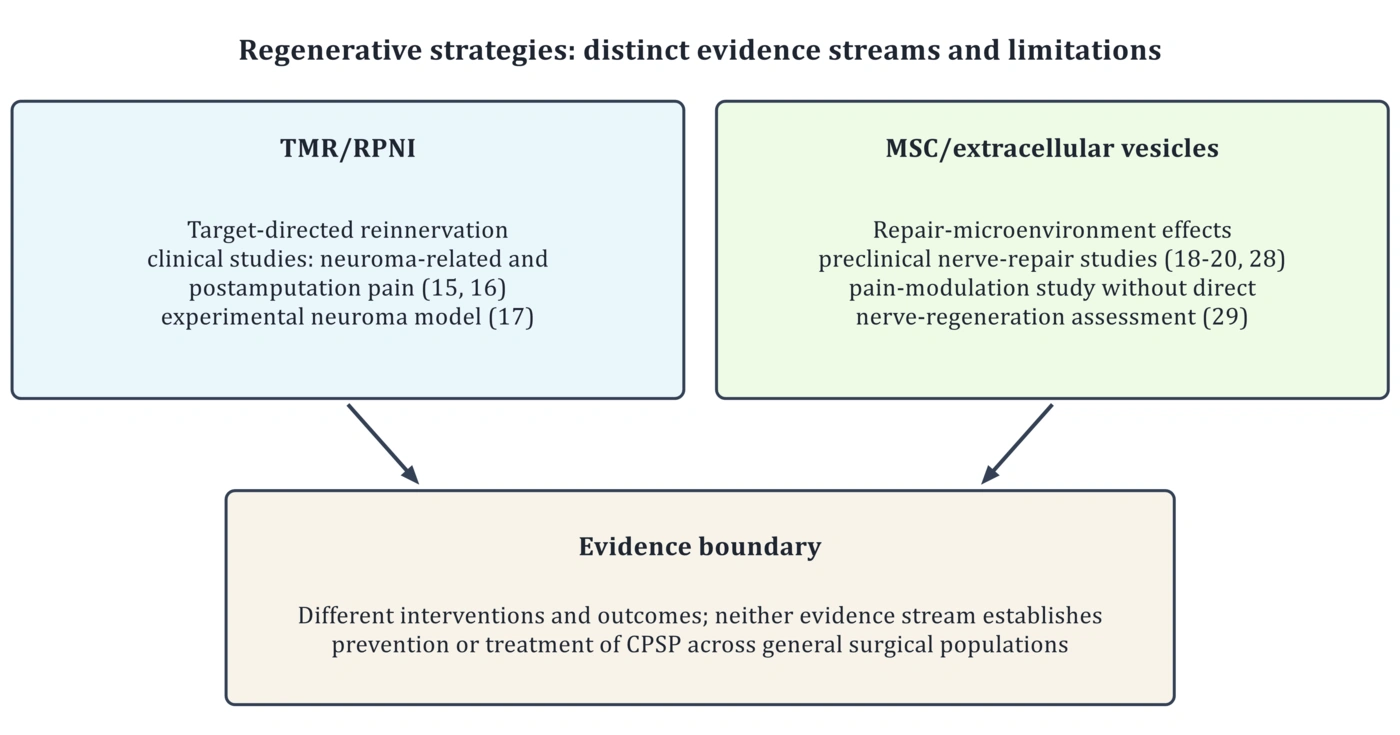
Figure 2.
Regenerative strategies discussed in this review. TMR and RPNI provide target-directed reinnervation and have been studied in clinical postamputation settings and experimental neuroma models (15-17). MSC/exosome approaches have shown nerve-repair effects mainly in preclinical models (18-20, 28). The pain-modulation study (29) did not directly assess nerve regeneration. Neither evidence stream establishes prevention of general CPSP.
Targeted muscle reinnervation (TMR) and regenerative peripheral nerve interfaces (RPNI) provide physiological targets for regenerating axons and have been investigated as approaches to reduce neuroma-related and postamputation pain. Although some clinical and experimental studies have reported pain improvement, the available evidence remains limited and does not establish regeneration-mediated analgesia or efficacy in other forms of CPSP. The relevant studies are summarized in Table 2 (15-17).
Mesenchymal stromal cell (MSC)-derived extracellular vesicles have shown potential to support peripheral nerve repair in preclinical models. Preclinical studies have reported improvements in selected measures of Schwann-cell responses, axonal regeneration, myelination, and functional recovery after peripheral nerve injury (18-20). These findings should be distinguished from those of studies primarily investigating pain modulation. For example, intrathecal administration of human umbilical cord-derived MSC exosomes reduced pain-related behaviors and altered inflammatory and glial responses in a spinal nerve ligation model, without directly assessing structural or functional nerve regeneration (29). Therefore, evidence of pain reduction does not necessarily demonstrate enhanced nerve regeneration, just as improved nerve repair does not establish prevention of CPSP.
4. Evidence Gaps and Future Directions
Despite advances in understanding peripheral nerve regeneration, its clinical relationship with CPSP remains poorly defined. Most evidence linking regeneration to persistent pain is derived from experimental nerve injury models, whereas CPSP is a heterogeneous condition involving multiple peripheral and central mechanisms (1, 12). The primary experimental and clinical evidence relevant to this review is summarized in Table 2.
A major challenge is identifying and characterizing nerve injuries in surgical patients that are comparable to those studied in experimental models, an essential step in evaluating the relevance of regenerative PNP to human postsurgical pain (12, 27).
A key research challenge is distinguishing structural axonal regrowth from successful target reinnervation and measurable sensory or motor recovery in clinical studies (5-7).
An important therapeutic challenge is promoting organized nerve regeneration without sustaining abnormal sensory activity. Because some cellular and molecular pathways involved in nerve repair may also modulate nociceptive signaling, regenerative interventions require evaluation of both functional recovery and pain-related outcomes (10, 12).
Clinical evidence for TMR/RPNI is concentrated in neuroma-related and postamputation pain (15, 16), whereas evidence for MSC/exosome nerve repair remains predominantly preclinical (18-20, 28). Neither establishes that these interventions prevent CPSP, and pain-modulation findings (29) do not demonstrate nerve regeneration.
Prospective clinical studies are needed to determine whether measurable characteristics of nerve regeneration are associated with long-term pain outcomes after surgery. Such studies should assess the extent and organization of axonal regrowth, target reinnervation, remyelination, sensory and motor recovery, and standardized pain outcomes over adequate follow-up periods.
5. Conclusions
Peripheral nerve injury initiates overlapping repair processes, and axonal regrowth does not necessarily ensure accurate target reinnervation or functional recovery (5-7). Maladaptive repair may contribute to persistent neuropathic pain but is not established as an independent cause of CPSP (1, 12).
Although TMR/RPNI have shown signals in clinical studies of patients after amputation and in experimental neuroma models (15-17), and MSC/exosome approaches have shown nerve-repair effects mainly in preclinical models (18-20, 28), their ability to prevent or treat CPSP remains unproven. Pain-modulation findings from MSC-derived exosomes should be interpreted separately from evidence of nerve regeneration (29). Prospective studies should relate objective measures of nerve repair to standardized long-term pain outcomes after surgery.