Cover image of the article: Facial Myositis: A Very Rare Syphilitic Symptom

Image Credit:Ann Mil Health Sci Res

Facial Myositis: A Very Rare Syphilitic Symptom

Authors

Mohamed Islam KedihaMohamed Islam Kediha ORCID1,*, Farida Mostefaoui1, Lamia Ali Pacha1
1Department of Neurology, Mustapha Bacha University Hospital, Benyoucef Benkhedda University Alger 1, Algeria
*Corresponding Author: Department of Neurology, Benyoucef Benkhedda University Alger 1, Algeria. Email: kediha.islam@gmail.com

Annals of Military and Health Sciences Research:Vol. 21, issue 2; e140861
Published online:Oct 16, 2023
Article type:Case Report
Received:Sep 10, 2023
Accepted:Oct 07, 2023
How to Cite:Kediha MI, Mostefaoui F, Ali Pacha L. Facial Myositis: A Very Rare Syphilitic Symptom. Ann Mil Health Sci Res. 2023;21(2):e140861. doi: https://doi.org/10.5812/amh-140861

Abstract

Introduction:

Myositis is an inflammation of striated muscle tissue. Among its causes, autoimmune diseases are the main ones. However, infectious causes are also incriminated, such as Coxsackie B virus epidemic myositis. Treponemal myositis or syphilitic myositis is, however, exceptional. Its localization or predominance on the facial muscles is even rarer.

Case Presentation:

We report a case of a facial form of syphilitic myositis with clinical and radiological study in a 25-year-old patient of Malian origin, presented to the neurology department with a painful facial diplegia that had been evolving for one month. Infectious myositis is a rare entity. Its symptomatology (myalgia/muscle weakness) may be diffuse or localized, as in the case of our patient. The infectious syndrome may be absent and limited to a flu-like syndrome. Furthermore, direct involvement of a skeletal muscle is rarely reported in the literature. The evolutionary profile is, therefore, little known.

Conclusions:

Syphilitic myositis can have complex clinical aspects and constitute a real diagnostic challenge, as reported in our patient's case.

Highlights

1. Introduction

Syphilis is a sexually transmitted disease caused by the bacterium Treponema pallidum. Syphilis can cause serious health effects without adequate treatment.
Some so-called "rare" clinical presentations in syphilis pose a real diagnostic challenge; sometimes, this diagnosis can be missed or delayed with delayed therapeutic management. Although most physicians think to include syphilis in the differential diagnosis of a wide range of symptoms, some presentations may surprise even the most experienced clinicians.
Myositis is an inflammation of striated muscle tissue. Among its causes, autoimmune conditions are the main ones. However, infectious causes are also incriminated, such as Coxsackie B virus epidemic myositis (1). Treponemal myositis or syphilitic myositis is, however, exceptional. Its localization or predominance on the facial muscles is even rarer. We report the case of a facial form of syphilitic myositis with a study of the clinical and radiological characteristics. To our knowledge, this myositis manifestation and its facial localization have never been reported.

2. Case Presentation

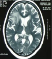
A patient aged 25 years of Malian origin presented to the Neurology Department of Mustapha Bacha University Hospital with a painful facial diplegia. The neurological picture of sub-acute onset was marked by a reduction or even abolition of facial mimicry, with the impossibility of whistling, blowing, or even smiling. The closure of the eyes was partial when falling asleep. This picture was completed after a few days of pain that felt like cramps, especially when palpating his face. He did not report any previous dermatological lesions or adenopathy. The neurological examination revealed facial diplegia with Charles Bell's sign on both sides, an inability to smile, and paralytic dysarthria. There were no swallowing or phonation disorders. Nausea and soft tissue reflexes were present. The rest of the neurological examination was normal (Figure 1).
Facial diplegia
Figure 1.
Facial diplegia
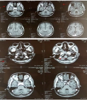
Brain and facial MRI showed diffuse thickening of the Temporalis muscles bilaterally, the internal and external pterygoid muscles, and the paraspinal and long neck muscles, with heterogeneous enhancement after injection of the contrast medium (Figure 2).
Facial and cerebral MRI
Figure 2.
Facial and cerebral MRI
The inflammatory panel was disturbed with positive CRP with a rate of 34 and accelerated sedimentation rate (35/80). Muscle enzymes were normal. A lumbar puncture showed medium cellularity (55 elements) with a predominance of leukocytes, and syphilitic serology was positive. HIV serology was requested, but no result was obtained. The patient was referred to the infectious diseases department for treatment.

3. Discussion

In syphilis, direct skeletal muscle involvement is uncommon, although several case reports have appeared in the literature (1). In the primary and secondary stages of the disease, generalized myalgia is a common symptom, but its presentation as an inflammatory myopathy is rare. The first case was reported in 1965 by Towpik and Mrozek (2) and involved the limb belts. Only 2 other cases were subsequently published, in 2010 and 2011, but only involved the musculature of the limbs, never the face (3, 4). Involvement of the facial muscles has never been reported, making our case the first reported case of inflammatory involvement of the facial muscles of syphilitic origin with suggestive imaging. Our patient's bilateral facial myopathy mimicked facial diplegia. It is, therefore, advisable to consider facial syphilitic myositis in any subject presenting with this symptomatology. Syphilis, in its myositic form, has rarely been evoked, and this patient bears witness to the fact that the resurgence of this condition can manifest itself in atypical disorders of neurological order other than those already identified in the literature.

3.1. Conclusions

Although myalgia is commonly seen in primary or secondary syphilis, inflammation of muscle tissue is not. Our "anecdotal" case confirms that syphilis should be considered when faced with myositis of uncertain etiology and that facial muscle involvement can be confusing as it is exceptional. However, imaging and infectious work-up can point to the correct diagnosis.


Footnotes

  • Authors' Contribution: Study concept and design, drafting of the manuscript, Kediha; acquisition of data, Mostefaoui; study supervision, Ali Pacha.

  • Conflict of Interests: The authors declare no conflict of interests.

  • Ethical Approval: This study is approved under the ethical approval of Mustapha Bacha University Hospital.

  • Funding/Support: No funding/support was received for this study.

  • Informed Consent: Informed consent was obtained from the patient.

References

Copyright

Copyright © 2023, Annals of Military and Health Sciences Research. This is an open-access article distributed under the terms of the Creative Commons Attribution-NonCommercial 4.0 International License (CC BY-NC 4.0) (https://creativecommons.org/licenses/by-nc/4.0/) which permits copy and redistribute the material just in noncommercial usages, provided the original work is properly cited.

Similar Articles

30
Nov
2011
Syphilitic Retinitis and Panuveitis in an Immunocompetent Patient

Syphilitic Retinitis and Panuveitis in an Immunocompetent Patient

Shirin Manshouri,
Zohreh Aminzadeh,
Shima Sayanjali

Manshouri S, Aminzadeh Z, Sayanjali S. Syphilitic Retinitis and Panuveitis in an Immunocompetent Patient. Arch Clin Infect Dis. 2011;6(Suppl):. doi:

25
Jul
2015
An Unusual Presentation of Tuberculous Meningitis

An Unusual Presentation of Tuberculous Meningitis

Shahnaz Sali,
Niloufar Valipour

Sali S, Valipour N. An Unusual Presentation of Tuberculous Meningitis. Arch Clin Infect Dis. 2015;10(3):e26083. doi: https://doi.org/10.5812/archcid.26083

30
Sep
2014
A Case of Myopericarditis: Very Rare Complication of Brucellosis

A Case of Myopericarditis: Very Rare Complication of Brucellosis

Ali Akbar Khorasani,
Mehrdad Farrokhnia

Khorasani AA, Farrokhnia M. A Case of Myopericarditis: Very Rare Complication of Brucellosis. Arch Clin Infect Dis. 2014;9(4):e18256. doi: https://doi.org/10.5812/archcid.18256

13
Jan
2023
A Case-report of Concurrent Pulmonary and Cerebral Lesions in a Patient with Polymyositis: Invasive Aspergillosis or Astrocytoma?

A Case-report of Concurrent Pulmonary and Cerebral Lesions in a Patient with Polymyositis: Invasive Aspergillosis or Astrocytoma?

Shabnam Tehrani,
Davood Yadegarynia,
Amirreza Keyvanfar

Tehrani S, Yadegarynia D, Keyvanfar A. A Case-report of Concurrent Pulmonary and Cerebral Lesions in a Patient with Polymyositis: Invasive Aspergillosis or Astrocytoma?. Jundishapur J Microbiol. 2022;15(11):e132821. doi: https://doi.org/10.5812/jjm-132821

27
Jun
2015
Idiopathic Orbital Myositis in a 9-Year-Old Girl: A Case Report

Idiopathic Orbital Myositis in a 9-Year-Old Girl: A Case Report

Titap Yazicioglu,
Isil Kutluturk

Yazicioglu T, Kutluturk I. Idiopathic Orbital Myositis in a 9-Year-Old Girl: A Case Report. Inn J Pediatr. 2015;25(3):-. doi: https://doi.org/10.5812/ijp.25(3)2015.371

More by these authors

Mohamed Islam KedihaPubMedScholar
Farida MostefaouiPubMedScholar
Lamia Ali PachaPubMedScholar
Share
Cited by
Metrics
Indexed in