
1. Introduction
Physical inactivity is recognized as the fourth leading risk factor for global mortality and is a major contributor to the development of non-communicable diseases worldwide (1). A growing body of evidence highlights the beneficial effects of physical exercise on overall health, including cognitive and neurological function. In the general population, regular physical activity has been shown to enhance cognitive processes such as attention, memory, processing speed, and executive function. In addition, numerous studies have demonstrated that various forms of exercise promote brain health by improving cerebrovascular regulation, enhancing neural plasticity, reducing inflammation and neuronal apoptosis, and mitigating the progression of neurodegenerative diseases (2).
One of the earliest physiological responses to exercise is an elevation in circulating lactate levels, a metabolite historically regarded as a mere byproduct of glycolysis. However, contemporary research has fundamentally reshaped this view, revealing lactate’s integral role in brain metabolism and function (3, 4). Since its initial identification in 1780, the scientific understanding of lactate has evolved considerably. Once believed to be a waste product responsible for muscle fatigue, lactate is now recognized as a critical energy substrate and signaling molecule with diverse biological functions (5). For instance, the astrocyte-neuron lactate shuttle is essential for long-term memory formation. Moreover, lactate has been implicated in the amelioration of depressive symptoms, the promotion of angiogenesis and neurogenesis, and the modulation of exercise-induced cerebrovascular adaptations and brain-derived neurotrophic factor (BDNF) expression (5).
BDNF is a neurotrophin that plays a pivotal role in the development and maintenance of the central nervous system. It supports the growth and survival of neurons and glial cells, facilitates synaptic plasticity, and confers neuroprotection (6). Exercise-induced elevations in BDNF, particularly within the hippocampus, are believed to underlie many of the cognitive and neuroprotective benefits associated with physical activity. Despite these associations, the precise molecular mechanisms by which exercise and lactate stimulate BDNF expression remain incompletely understood (7). Identifying key mediators of exercise-induced neuroplasticity may enable the development of novel therapeutic strategies to enhance cognitive function and mitigate neurological disorders (7).
A substantial body of research indicates that physical exercise enhances neuroplasticity, the nervous system’s ability to reorganize and adapt in response to internal and external stimuli, thereby contributing to improved cognitive and vascular function. Several studies suggest that exercise facilitates neuroplasticity by augmenting the downstream signaling of neurotrophic factors such as BDNF (8, 9). Neuroplasticity enables the brain to compensate for injury, adapt to environmental changes, and optimize functional capacity, underscoring its importance in maintaining neurological health. Notably, exercise appears to confer dual benefits by simultaneously promoting neuroplastic and cerebrovascular adaptations.
Given the breadth of evidence supporting the neurobiological effects of physical activity, this review aims to elucidate the influence of exercise intensity on brain neuroplasticity. We begin by examining recent findings on the impact of different exercise modalities and intensities on cognitive and vascular function, with a particular focus on the neuroprotective properties of lactate. Subsequently, we explore the role of BDNF as a potential mediator of exercise-induced neuroplasticity and its implications for brain injury rehabilitation and cognitive enhancement.
2. Methods
In the present systematic review, studies published between 2004 and 2024 that investigated the relationship between physical exercise and neuroplasticity were examined through a systematic search of the Web of Science Core Collection, Scopus, PubMed/MEDLINE, Embase, and ScienceDirect databases. Keywords included exercise, neuroplasticity, BDNF, and lactate, in accordance with the PRISMA guidelines. The inclusion criteria were: 1) original research articles, 2) human or animal studies, 3) defined exercise-intensity interventions, 4) reporting of at least one neuroplasticity marker, and 5) the presence of a control or comparison group. The exclusion criteria were reviews, studies without defined exercise intensity or neuroplasticity outcomes, non-English publications, letters, conference abstracts, and uncontrolled studies.
After removing 25 duplicates, 95 articles underwent title and abstract screening, and 87 were excluded because they did not meet the criteria. During this stage, studies were preliminarily grouped and evaluated based on two key criteria: (i) clearly defined and quantifiable exercise-intensity parameters (e.g., %HRmax, VO2max, lactate threshold, or %1RM), and (ii) objective neuroplasticity-related outcomes, including molecular biomarkers (e.g., BDNF, lactate, IGF-1, VEGF) and/or neurophysiological or neuroimaging measures. Seventeen articles were assessed in full text, of which nine were excluded for not meeting the inclusion criteria, resulting in eight studies included in the final analysis.
All screening stages (title/abstract and full-text) were performed independently by two authors. Disagreements were resolved through discussion or consultation with a third reviewer. The initial inter-rater agreement was 92%. The PRISMA flow diagram illustrating each step of the selection and inclusion process is presented in Figure 1.
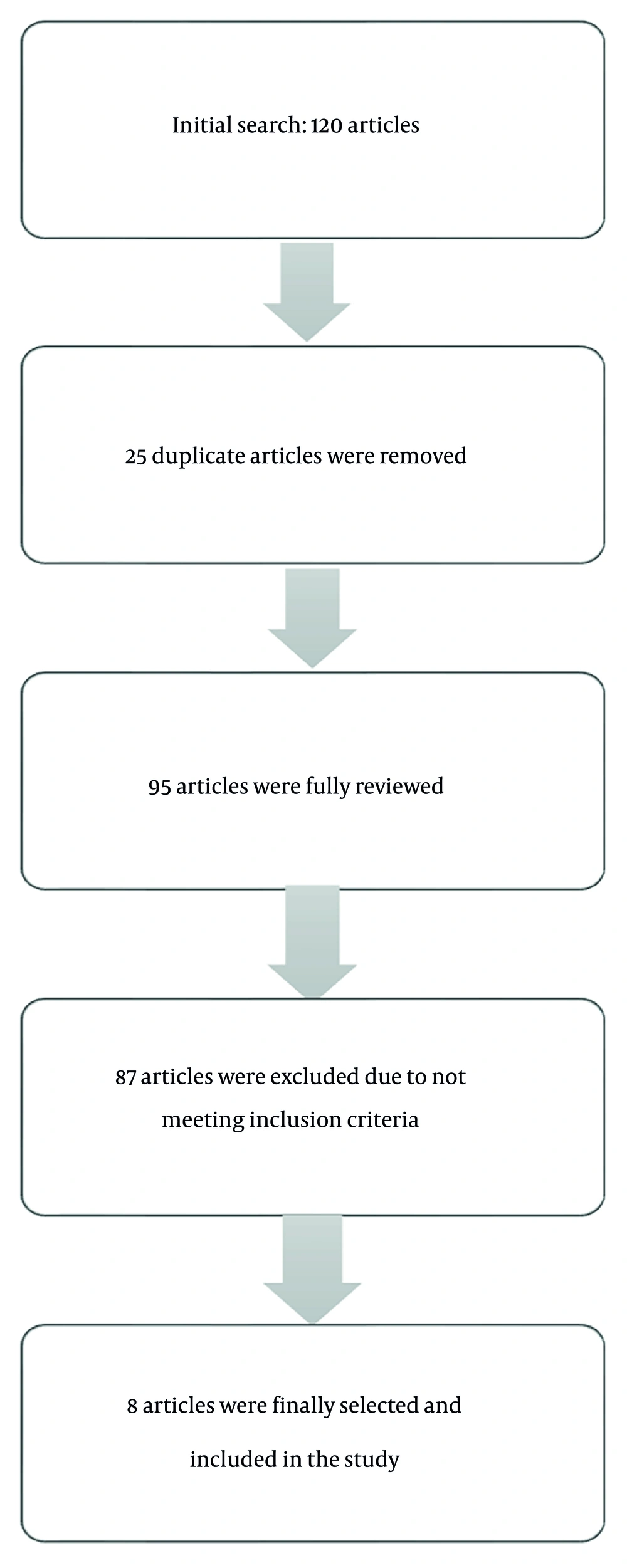
Figure 1.
Chart of selection of articles in the present study
2.1. Scope and Rationale for Limited Included Studies
This review applied strict inclusion criteria to address a highly focused research question: the direct, quantitative relationship between prescribed exercise intensity and objective measures of neuroplasticity (e.g., biomarkers such as BDNF and lactate or neuroimaging outcomes). This specificity necessarily excluded a large body of relevant literature that: 1) examined exercise and brain health without precisely quantifying or reporting intensity parameters; 2) focused solely on cognitive outcomes without concomitant neuroplasticity biomarkers; or 3) used multimodal exercise interventions in which the isolated effect of intensity could not be discerned. Consequently, although the final pool of eight studies is limited in breadth, it represents a homogeneous and methodologically rigorous subset of the literature that enables a meaningful, albeit preliminary, synthesis of intensity-specific effects. This focused approach prioritizes depth of mechanistic insight over breadth, with the aim of clarifying dose-response relationships that are often obscured in broader reviews.
3. Results
The results presented below are based exclusively on studies that met the predefined inclusion criteria and were selected through the PRISMA-guided screening process described in the Materials and Methods section. A summary of all selected studies is presented in Table 1.
Table 1.
Summary of Studies on the Effects of Different Types of Physical Exercise on Brain Neuroplasticity
| Authors (y) | Title of Studies | Subjects | Type & Duration of Exercise | Findings |
|---|---|---|---|---|
| Hill et al. (2023) (16) | Moderate-intensity aerobic exercise may enhance neuroplasticity of the contralesional hemisphere after stroke: A randomized controlled trial | 33 stroke patients | Moderate-intensity cycling for 20 min (70 - 80% of max HR); groups: moderate exercise, motor evoked potentials (MEPs), and intermittent theta burst stimulation (iTBS) | Moderate cycling may enhance neuroplasticity in individuals after stroke. |
| Donell et al. (2013) (17) | A single session of aerobic exercise increases motor cortex neuroplasticity | 25 healthy adults | Three intensities: low (55% HR), moderate (65% HR), high (75% HR) | A single aerobic session improves brain neuroplasticity; low and moderate intensities have better effects. |
| Cassilhas et al. (2012) (18) | Spatial memory improves through aerobic and resistance training via different molecular mechanisms | Adult male Wistar rats | 8 weeks of either treadmill aerobic training (Group AERO) or vertical ladder resistance training (Group RES) | Aerobic and resistance exercise positively impact spatial learning and memory via different mechanisms. |
| Sleiman et al. (2016) (19) | Exercise increases brain-derived neurotrophic factor (BDNF) expression via beta-hydroxybutyrate | 20 male mice, control and running groups | 4 weeks of voluntary wheel running | BDNF levels increased in the hippocampus after 30 days of voluntary wheel access. |
| Hakansson et al. (2017) (20) | BDNF responses to 35-minute physical, cognitive, and mindfulness exercises in healthy older adults: Association with working memory performance | 19 healthy elderly individuals | Moderate-intensity physical exercise (Borg RPE 11 - 13), cognitive training, and mindfulness | A single session of physical exercise significantly increased serum BDNF compared to cognitive or mindfulness training. |
| Vints et al. (2024) (23) | Effects of resistance training on hippocampal subfields, neuroplasticity, and neuroinflammation in older adults at low/high risk for mild cognitive impairment: RCT | 70 healthy men/women aged 60 - 85 | 12 weeks of resistance training at 70 - 85% of 1RM | Improved brain neuroplasticity and prevention of age-related cognitive decline. |
| Pin-Barre et al. (2021) (37) | Effects of different HIIT protocols on endurance and neuroplasticity after cerebral ischemia | 42 adult male rats | HIIT1 (short intervals: 1 min) or HIIT4 (long intervals: 4 min) after transient middle cerebral artery occlusion for 2 weeks | Neuroplasticity markers increased in the contralateral cortex and hippocampus. |
| Lourenco et al. (2019) (47) | The novel exercise-induced hormone irisin protects against neural damage via activation of Akt and ERK1/2 signaling, aiding neuroprotection in cerebral ischemia | Male mice aged 8 - 12 weeks | Treadmill running: 10 m/min for 90 min over 2 weeks | Blocking Irisin/FNDC5 impairs the neuroprotective effects of exercise on synaptic plasticity and memory deficits in animal models of AD. |
Table 2.
Methodological Quality Assessment of Included Studies a
| Study (y) | Adequate Sample Size? | Controlled Confounders? | Blinded Assessment? | Appropriate Stats? | Overall Quality |
|---|---|---|---|---|---|
| Hill et al. (2023) | ✓ (RCT) | ✓ | ✓ (Assessor) | ✓ | High |
| Donell et al. (2013) | △ (n = 25) | ✓ | △ (Single-blind) | ✓ | Moderate |
| Cassilhas et al. (2012) | ✓ (Animal) | ✓ | N/A (Animal) | ✓ | High |
| Sleiman et al. (2016) | ✓ (Animal) | ✓ | N/A (Animal) | ✓ | High |
| Hakansson et al. (2017) | △ (n = 19) | ✓ | ✓ | ✓ | Moderate |
| Vints et al. (2024) | ✓ (RCT, n = 70) | ✓ | ✓ | ✓ | High |
| Pin-Barre et al. (2021) | ✓ (Animal) | ✓ | N/A (Animal) | ✓ | High |
| Lourenco et al. (2019) | ✓ (Animal) | ✓ | N/A (Animal) | ✓ | High |
a Legend: ✓ = Yes/Clearly reported; △ = Partially/Unclear; ✗ = No/Not reported; N/A = Not Applicable.
3.1. Aerobic Exercise
Aerobic exercise is widely recognized as a potent stimulus for enhancing neuroplasticity (10). Activities such as cycling, running, and swimming involve rhythmic, sustained movements that promote cardiovascular efficiency and facilitate increased oxygen delivery to the brain. Numerous studies have shown that aerobic exercise upregulates the expression of key neuroplasticity-related biomarkers, including brain-derived neurotrophic factor (BDNF), insulin-like growth factor 1 (IGF-1), and vascular endothelial growth factor (VEGF). These molecular mediators support critical neurobiological processes such as synaptogenesis, neurogenesis, and angiogenesis, while also contributing to improved cerebral blood flow (10-15).
Hill et al. (2023) examined the effects of moderate-intensity aerobic exercise in individuals with stroke and reported that 20 minutes of aerobic activity performed at 70 - 80% of maximum heart rate significantly enhanced markers of neural plasticity and functional recovery (16). Similarly, Donell et al. (2013) investigated the effects of varying aerobic exercise intensities—low (55% of maximum heart rate), moderate (65%), and high (75%)—on neuroplastic outcomes in healthy older adults. Their findings indicated that a single aerobic exercise session improved neuroplasticity, with low- and moderate-intensity sessions producing more favorable effects on brain function than high-intensity exercise (17).
Animal studies have provided additional insights into the molecular mechanisms underlying exercise-induced neuroplasticity. For instance, de Mello et al. demonstrated that aerobic exercise in rodents increased hippocampal levels of IGF-1, BDNF, tropomyosin receptor kinase B (TrkB), and calcium/calmodulin-dependent protein kinase II (β-CaMKII). Furthermore, resistance training was found to enhance both peripheral and hippocampal IGF-1 expression, accompanied by activation of the IGF-1 receptor (IGF-1R) and downstream AKT signaling pathways in the hippocampus, ultimately improving spatial learning and memory (18).
Aerobic exercise is particularly associated with the upregulation of neurotrophic factors in the brain. For example, Sleiman et al. observed significantly elevated BDNF levels in the hippocampus of rodents following 30 days of voluntary wheel running. The authors proposed that this increase may be mediated by endogenous production mechanisms, possibly involving the interaction of β-hydroxybutyrate with BDNF gene promoter regions (19). Consistent with these findings, Hakansson et al. (2016) reported increased serum BDNF levels in older adults after 35 sessions of moderate-intensity exercise, defined as 11 - 13 on the Borg Rating of Perceived Exertion scale (20). Similarly, a 20-minute session of moderate-intensity cycling (60 - 70% of maximum heart rate) was shown to elevate circulating levels of BDNF and IGF-1 in both male and female participants (13).
3.2. Resistance Exercise
Resistance exercise, a widely accessible and effective form of physical activity, involves repeated muscle contractions against external resistance. This includes bodyweight exercises (e.g., push-ups) as well as the use of resistance bands, free weights, or machines (10). Resistance training elicits neurophysiological adaptations through mechanisms distinct from those induced by aerobic exercise. Although both modalities contribute to neuroplasticity through increased cerebral blood flow and elevated expression of insulin-like growth factor 1 (IGF-1), aerobic training has been shown to exert a more substantial influence on brain-derived neurotrophic factor (BDNF) levels. In contrast, resistance training produces more pronounced effects on IGF-1 expression and reductions in pro-inflammatory cytokines such as interleukin-6 (IL-6) (21, 22). Notably, resistance exercise has been shown to increase IGF-1 levels both peripherally and within the hippocampus, along with activation of IGF-1 receptors and downstream signaling pathways, including protein kinase B (AKT) (18). In a recent clinical trial, Vints et al. (2024) demonstrated that a 12-week resistance training program performed at 70 - 85% of one-repetition maximum (1RM) significantly enhanced neuroplasticity and attenuated age-related cognitive decline (23).
Structural brain changes associated with resistance training are thought to arise, at least in part, from the modulation of molecular and cellular pathways critical to neuroplasticity. These changes are likely mediated through the regulation of neurotrophic and neuroprotective factors such as IGF-1, BDNF, and homocysteine. BDNF, in particular, plays a key role in mediating structural adaptations in response to physical training. Elevated serum BDNF levels following resistance exercise have been correlated with improvements in executive function and increases in hippocampal volume. However, the precise mechanisms linking resistance training to BDNF-mediated structural and functional brain changes remain incompletely understood, and further research is warranted to elucidate these associations.
IGF-1, produced in the liver, skeletal muscle, and brain, can cross the blood–brain barrier and exert effects on the central nervous system. It plays a vital role in synaptogenesis, axonal growth, neuronal survival, and overall neuroplasticity. Higher circulating IGF-1 levels have been associated with increased brain volume and improved cognitive performance, particularly in older adults. Nevertheless, the specific contributions of IGF-1 to central nervous system function and its precise role in mediating exercise-induced cognitive benefits remain areas for further investigation.
Additionally, resistance training has been shown to reduce plasma and serum levels of homocysteine, a sulfur-containing amino acid associated with neurotoxicity and neurodegenerative processes. Elevated homocysteine levels have been linked to white matter atrophy, neuronal damage, and impaired cognitive function. By lowering homocysteine concentrations, resistance training may contribute to improved brain structure and function. However, despite these promising findings, the mechanistic relationships among resistance exercise, homocysteine reduction, and neurocognitive outcomes are not yet fully established and warrant further exploration (24-28).
3.3. High-Intensity Interval Training
High-intensity interval training (HIIT) is characterized by repeated short bouts of vigorous physical activity performed near maximal aerobic capacity, interspersed with periods of low-intensity activity or rest. Although often perceived as exhaustive or overly strenuous, HIIT has been shown—both in preclinical and clinical research—to elicit physiological adaptations comparable to, or exceeding, those of traditional moderate-intensity continuous training (MICT). Notably, emerging evidence suggests that HIIT may induce more robust neuroplastic responses than MICT (29-31).
Several studies have demonstrated that HIIT enhances cognitive function and promotes neuroplasticity, particularly in post-stroke populations (32-34). A recent meta-analysis reported that HIIT facilitates neuroplasticity by increasing levels of key molecular mediators such as brain-derived neurotrophic factor (BDNF), vascular endothelial growth factor (VEGF), and lactate (35). However, findings regarding BDNF regulation during HIIT are not entirely consistent; for example, some studies using high-intensity cycling protocols have reported no significant changes in circulating BDNF levels (36). In contrast, Pin-Barre et al. found that various HIIT regimens in a rodent model of cerebral ischemia significantly upregulated neuroplasticity markers in both the contralateral cortex and hippocampus (37).
Mechanistically, HIIT appears to modulate several key molecular pathways involved in neuroplasticity. Notably, the upregulation of phosphorylated tropomyosin receptor kinase B (pTrkB), a central component of the BDNF signaling cascade, along with fibronectin type III domain-containing protein 5 (FNDC5), has been associated with improvements in neuronal survival, hippocampal neurogenesis, synaptic plasticity, and functional recovery. FNDC5, a myokine released from skeletal muscle during physical activity, has been shown to activate the TrkB/BDNF pathway in the brain. These molecular responses are further enhanced by HIIT-induced increases in cytochrome c (CytC) expression and VEGF, which are critical for mitochondrial function and angiogenesis, respectively (37-39).
3.4. Lactate and Neuroplasticity
Historically, lactate was regarded as a mere byproduct of anaerobic metabolism. However, contemporary research has redefined its role, recognizing lactate as a critical metabolic intermediate and signaling molecule involved in numerous physiological processes, particularly in the context of exercise (3). Energy production during physical activity primarily relies on three metabolic pathways: 1) the ATP–creatine phosphate system, 2) glycolysis, and 3) oxidative phosphorylation. When oxygen availability is limited, glycolysis predominates, resulting in lactate production, which helps buffer acidosis. Lactate can be transported to the liver for conversion into glucose via gluconeogenesis or used directly as an energy substrate by skeletal muscle, the heart, and the brain (40). The extent of lactate accumulation during acute exercise depends on the intensity and duration of the activity. The lactate threshold, also referred to as the anaerobic threshold, represents the highest exercise intensity at which lactate does not accumulate significantly in the blood and serves as a reliable indicator of cardiovascular fitness. Regular exercise elevates this threshold, thereby improving physical performance (41).
Lactate crosses the blood–brain barrier via monocarboxylate transporters (MCTs), with MCT2 predominantly expressed in neurons and MCT4 in astrocytes (42). Astrocytes, through complex interactions with neurons, regulate key aspects of brain homeostasis, including energy metabolism, ionic balance, and cell volume. By maintaining a glucose gradient—higher near the blood–brain barrier and lower near neurons—astrocytes facilitate efficient glucose transfer to neurons and also serve as glycogen reservoirs to support neuronal energy demands (43). Lactate generated by astrocytes is shuttled to neurons via MCTs, where it is converted into pyruvate and enters the tricarboxylic acid (TCA) cycle for ATP production. This neuronal lactate may originate from either astrocytic metabolism or peripheral sources such as skeletal muscle. Notably, in vitro studies have demonstrated that neurons preferentially use lactate over glucose as an energy substrate under certain conditions (42). Current evidence supports a critical role of the astrocyte–neuron lactate shuttle in memory consolidation, establishing a mechanistic link between physical activity and neuroplasticity. Pharmacological inhibition of MCTs impairs long-term memory formation, underscoring the importance of lactate transport in cognitive processes. Moreover, cerebral lactate uptake increases from approximately 8% at rest to 20% during exercise, with higher lactate metabolism observed in trained individuals than in sedentary controls (44).
Exercise is known to upregulate several neurotrophic and growth factors, including brain-derived neurotrophic factor (BDNF), insulin-like growth factor 1 (IGF-1), and vascular endothelial growth factor (VEGF). Although the interaction between lactate and BDNF remains incompletely understood, several mechanisms have been proposed. These include: 1) enhanced activation of N-methyl-D-aspartate (NMDA) receptors by lactate, leading to elevated intracellular calcium levels; 2) activation of G-protein-coupled receptor (GPCR)-mediated signaling pathways via lactate binding; and 3) stimulation of the SIRT1/PGC-1α/FNDC5/BDNF signaling cascade (42). Most of the current literature suggests that the neuroplastic effects of exercise are primarily mediated by increased expression of, or enhanced downstream signaling through, neurotrophic factors such as BDNF, IGF-1, and VEGF. In rodent models, inhibition of hippocampal BDNF signaling significantly impairs exercise-induced neuroplasticity. Similarly, blocking the entry of circulating IGF-1 or VEGF into the brain attenuates exercise-induced hippocampal neurogenesis. IGF-1, a pleiotropic growth factor with angiogenic and neurogenic properties, is essential for cerebral vascular adaptations to exercise. Systemic administration of IGF-1 has been shown to stimulate angiogenesis, whereas its inhibition reduces exercise-induced increases in cerebral capillary density (45).
Of particular interest is fibronectin type III domain-containing protein 5 (FNDC5), a muscle-derived, hormone-like myokine that mediates many of the neuroprotective effects of physical activity. Irisin, the cleaved and secreted form of FNDC5, is regulated by peroxisome proliferator-activated receptor gamma coactivator 1-alpha (PGC-1α). Physical exercise stimulates FNDC5 gene expression in skeletal muscle, leading to increased systemic irisin levels and downstream neurobiological benefits. The PGC-1α/FNDC5/BDNF axis has recently emerged as a key regulatory pathway underlying exercise-induced neuroprotection. In 2012, Boström et al. identified FNDC5 as a PGC-1α-dependent myokine released from contracting muscles, capable of improving systemic metabolism and glucose homeostasis (46). FNDC5/irisin is widely expressed across various brain regions, including the hippocampus, cerebellar Purkinje cells, and the hypothalamus, where it plays essential roles in neurodevelopment and cognitive function. For example, in mouse embryonic stem cells, FNDC5 is crucial for neuronal differentiation, and its deletion significantly reduces the expression of mature neuronal markers. Similarly, Forouzandeh et al. (2015) reported high FNDC5 expression during the differentiation of human embryonic stem cell-derived neural precursors into neurons. Other studies have demonstrated that exercise-induced irisin production in the hippocampus upregulates BDNF expression. Peripheral delivery of FNDC5 has also been shown to increase circulating irisin levels, subsequently enhancing hippocampal expression of BDNF and other neuroprotective genes.
Emerging evidence supports irisin as a key mediator of exercise-induced neuroprotection. Lourenco et al. reported significantly reduced hippocampal irisin levels in patients with Alzheimer’s disease (AD). In rodent models of AD, inhibition of FNDC5/irisin signaling attenuated the cognitive and synaptic benefits of exercise, whereas exogenous upregulation of FNDC5 rescued memory and synaptic deficits. Likewise, in a mouse model of cerebral ischemia, blockade of FNDC5/irisin abolished the neuroprotective effects of exercise on ischemia-induced neurological impairments compared with untreated controls (47, 48).
3.5. Exercise, BDNF, and Neuroplasticity
Exercise confers widespread neuroprotective benefits by activating diverse physiological pathways, notably those involved in neurotrophic and angiogenic signaling. Among these, the brain-derived neurotrophic factor (BDNF) signaling pathway is considered a key mediator of exercise-induced neuroplasticity. Preclinical studies have demonstrated that physical activity increases BDNF expression in the hippocampus, a brain region critical for learning and memory. For example, middle-aged mice subjected to daily treadmill training exhibit elevated BDNF protein and mRNA levels in the hippocampus, accompanied by improvements in spatial memory and object recognition (49, 50). In humans, aerobic exercise similarly enhances circulating BDNF levels, which are associated with increased hippocampal volume. These findings suggest that exercise-induced BDNF expression may counteract hippocampal atrophy and enhance cognitive performance, particularly spatial memory (49, 51).
The mechanisms underlying exercise-induced neuroplasticity are multifactorial, encompassing increased production and signaling of neurotrophic factors (e.g., BDNF, IGF-1, VEGF), attenuation of neuroinflammation, reduced psychological stress, and improvements in cardiovascular and metabolic parameters, such as arterial elasticity, blood pressure, and insulin sensitivity. BDNF has emerged as a central regulator within this framework. Experimental evidence from rodent models indicates that pharmacological blockade of BDNF signaling within the hippocampus significantly diminishes the neuroplastic effects of exercise. Additionally, BDNF produced in peripheral tissues in response to skeletal muscle contraction may cross the blood–brain barrier and influence central nervous system function (52).
Exercise also improves brain function and mitigates symptoms of various neurological disorders through enhanced neuroplasticity (53, 54). For instance, mice with access to voluntary running wheels demonstrate increased hippocampal neurogenesis and enhanced spatial and temporal memory. Neuroimaging studies have shown that both voluntary and sustained physical activity increase hippocampal volume. Similar effects have been reported in human populations: adults engaging in 12 weeks of moderate-intensity aerobic training exhibit volumetric increases in multiple brain regions, particularly the hippocampus. These structural adaptations are strongly correlated with improvements in cognitive function (55).
The beneficial effects of exercise are mediated by systemic physiological interactions involving the liver, muscles, and bones. These tissues release a range of signaling molecules, some of which remain unidentified, that circulate to the brain and facilitate learning, memory, and neuroplasticity (56, 57). Among the known exercise-induced factors, many converge on shared pathways, with BDNF signaling serving as a central integrative hub. The induction of BDNF expression in the hippocampus appears to be a principal mechanism by which these peripheral signals exert neuroprotective effects (19, 58, 59). Consequently, hippocampal BDNF expression is increasingly viewed as a potential biomarker for identifying novel exercise-responsive molecules that may inform therapeutic strategies for central nervous system disorders, such as depression (60).
BDNF promotes neuroplasticity primarily by binding to and activating its high-affinity receptor, tropomyosin receptor kinase B (TrkB). Experimental models have shown that exercise fails to induce neurogenesis or improve neural plasticity in TrkB-deficient mice. In humans, various forms of physical activity increase serum BDNF concentrations, which correlate with hippocampal growth and cognitive enhancement. However, genetic factors such as the Val66Met single nucleotide polymorphism (SNP) in the BDNF gene can impair exercise-induced BDNF signaling. Mice carrying the Met allele exhibit reduced synaptic plasticity and diminished gains in spatial learning following physical activity. Similarly, inhibition of BDNF signaling prevents the exercise-induced upregulation of key synaptic proteins and attenuates cognitive improvements. Collectively, these findings underscore that the beneficial effects of exercise on the brain arise from a complex, system-wide response, with BDNF signaling serving as a central and indispensable mediator (60).
4. Discussion
Neuroplasticity refers to the intrinsic capacity of the nervous system to strengthen synaptic connections and reorganize neural networks in response to internal or external stimuli (1). Among the various factors influencing neuroplasticity, physical exercise has emerged as a particularly potent modulator. Although the optimal exercise intensity for maximizing neurocognitive benefits remains under investigation, current evidence suggests that a combination of moderate- and high-intensity physical activity is most effective for promoting health and well-being in older adults (61). Extensive research has shown that regular physical activity not only improves cardiovascular and musculoskeletal function but also exerts profound effects on cognitive performance and brain structure (5). These cognitive benefits are mediated by multiple physiological and molecular mechanisms, which vary by exercise type, muscle recruitment patterns, and intensity (6, 9). For example, Sleiman and colleagues (19) demonstrated that both aerobic and resistance training enhance neuroplasticity across species by stimulating the production of neurotrophic factors, promoting intracellular signaling, and supporting neuronal growth and maturation.
A recent scoping review of physical activity guidelines highlights the importance of gradually progressing exercise intensity, particularly in older populations, to minimize the risk of injury or adverse events (62). Strength training at intensities between 60% and 85% of one-repetition maximum (1RM) is effective for increasing muscle mass, whereas higher intensities are better suited for enhancing the rate of force development (63). Accordingly, guidelines recommend a multimodal approach that combines moderate- and high-intensity aerobic activity with resistance training to promote physical and cognitive health in aging populations. Beyond physiological factors, successful aging is also influenced by demographic characteristics, health behaviors, family support, and social engagement (61). Nevertheless, increasing evidence underscores the central role of physical activity in healthy aging, as it is associated with reductions in all-cause mortality, risk of chronic disease, functional decline, anxiety, depression, and cognitive impairment (64).
The cognitive and health-related benefits of exercise in older adults appear to be at least partially dependent on exercise intensity. Although some studies suggest that high-intensity exercise may yield superior improvements in aerobic capacity and cardiovascular health compared with moderate-intensity activity, particularly in older individuals (61), others argue that moderate-intensity exercise provides comparable benefits with greater safety and adherence (65). Notably, high-intensity training has been linked to enhanced memory function, whereas moderate-intensity interventions have shown mixed results for cognitive outcomes (61). By contrast, low- to moderate-intensity activity has demonstrated protective effects against physical and mental decline, raising questions about whether higher intensity confers additive cognitive benefits (65). A systematic review and meta-analysis by Saunders et al. (2019) provides further nuance, showing that, in older adults with cognitive impairments, shorter but more frequent exercise sessions (e.g., five sessions per week) were more effective in improving cognitive outcomes than fewer, longer sessions (66). These findings highlight the importance of tailoring exercise interventions not only by type and intensity but also by frequency and duration.
The cognitive effects of exercise are influenced by several dosage parameters, including program duration (number of weeks), session duration (minutes per session including warm-up and cool-down), frequency (sessions per week), and intensity (typically measured as a percentage of maximal oxygen uptake, VO2max) (63). Meta-analytical evidence suggests that higher values across these parameters are associated with greater improvements in physical fitness, including increased muscle strength and aerobic capacity, in older adults. In individuals with Alzheimer’s disease, longer-duration programs are positively correlated with gains in endurance, lower-limb strength, balance, and the ability to perform daily living activities (67). Improvements in VO2max and muscular fitness are also associated with reduced all-cause mortality in healthy older populations (63). These enhancements in physical fitness may facilitate neuroplastic adaptations in the brain, thereby improving cognitive performance through increased neural activation and efficiency (68).
Taken together, these findings suggest a robust relationship between exercise dosage parameters and cognitive function. Higher-intensity, longer-duration, and more frequent exercise regimens are consistently associated with improved executive functions—including processing speed, attention, and inhibitory control—in both young and older adults (66). This dose-response relationship between physical activity and cognitive outcomes underscores the need for precision in exercise prescription, particularly in aging populations at risk of cognitive decline.
4.1. Quality Assessment and Consistency of Evidence
The eight included studies were assessed for methodological quality with respect to sample size, control of confounders, blinding of outcome assessors, and appropriateness of statistical analysis, as shown in Table 2. Overall, the studies demonstrated moderate to high quality, with clear experimental designs and well-defined intensity protocols. However, several limitations were noted: most human studies had modest sample sizes (n < 40), and only two were conducted in clinical populations (stroke and risk of MCI). All animal studies provided robust mechanistic data but inherently limit direct translation to humans. Findings regarding aerobic exercise were largely consistent, indicating a positive dose-response relationship between moderate intensity and neuroplasticity markers, particularly BDNF. The single study directly comparing intensities (17) provided critical evidence that low-to-moderate intensity may be more favorable than high intensity in older adults, aligning with the broader literature on intensity tolerance in aging. For resistance training, the evidence base is smaller but consistent in reporting increased IGF-1 and structural brain benefits. The primary contradiction in the broader literature—whether resistance training reliably increases circulating BDNF—was reflected in the included studies. The findings of this review suggest that resistance exercise may influence neuroplasticity more through IGF-1 and homocysteine pathways than through acute BDNF surges, distinguishing it from aerobic exercise. The most apparent inconsistency emerged in the HIIT literature. Although several studies and a meta-analysis (35) reported significant increases in BDNF and lactate, other high-quality trials (36) found no change in BDNF following HIIT. This discrepancy may be explained by critical protocol variables, including the nature of work/rest intervals, total session duration, and participant fitness level. Studies showing positive effects often employed protocols with longer work intervals (e.g., 4-minute bouts) or included clinical populations (post-stroke), in which relative physiological stress and response may differ from those of healthy subjects undergoing short Tabata-style HIIT. These inconsistencies do not necessarily weaken the data; rather, they highlight key gaps, including the lack of standardized HIIT protocols for brain outcomes and the need for studies that directly measure central versus peripheral lactate and BDNF kinetics in relation to specific intensity landmarks (e.g., lactate threshold).
4.2. Limitations and Future Research Directions
The primary limitation of this review is the limited number of studies (n = 8) meeting the stringent inclusion criteria, which precludes broad generalizations and definitive conclusions. This scarcity in the literature is itself informative, highlighting a critical gap: few exercise studies are designed with the primary aim of isolating and comparing the neuroplastic effects of different intensity regimens using direct biological endpoints. The findings presented here should therefore be interpreted as a proof-of-concept synthesis demonstrating that exercise intensity is a crucial variable, yet one that remains inadequately mapped. To build on this foundation, future research must expand both the quantity and quality of investigations in this domain. Priority should be given to large-scale, well-controlled randomized trials that directly compare multiple intensity levels (e.g., low, moderate, high, HIIT) within the same study protocol, using standardized intensity prescriptions and a combination of peripheral biomarkers (e.g., BDNF, lactate) and central nervous system measures (e.g., fMRI, MRS, TMS). Such work is essential for developing evidence-based, personalized exercise prescriptions for brain health.
4.3. Conclusion
Current evidence indicates that physical exercise, irrespective of modality, can enhance brain neuroplasticity through diverse molecular pathways, including the upregulation of BDNF, VEGF, IGF-1, and lactate. However, the neurobiological impacts of exercise are substantially modulated by its type and intensity. Although both low-to-moderate intensity aerobic exercise and high-intensity interval training (HIIT) have been shown to influence markers of neuroplasticity, existing literature remains insufficient to establish a single optimal intensity prescription for promoting brain health across varied populations, such as older adults, neurological patients, or younger individuals.