
1. Introduction
Aspergillus is a fungus found in the environment, such as in the air, water, soil, and decomposing organic debris. In an immunocompetent person, inhalation of Aspergillus spores may cause localized infection in the lungs, sinuses, or other sites. In immunocompromised patients, these fungi can cause life-threatening invasive infections of the lungs or sinuses, with dissemination to other organs (1).
Invasive aspergillosis has a poor prognosis, particularly in critically ill patients with cerebral involvement. The mortality rate of cerebral aspergillosis is 63% (2). We report a case of cerebral aspergillosis in an immunocompetent patient without involvement of the sinuses or lungs. There are several interesting aspects to this case. First, cerebral aspergillosis has been reported in only about 10% of all cases of aspergillosis (3). In addition, in most patients, invasive aspergillosis develops in the sinuses and lungs, and secondarily spreads to the brain hematogenously (4). Third, aspergillosis is unusual in immunocompetent patients.
2. Case Presentation
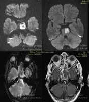
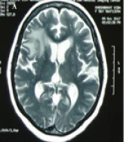
A 29-year-old woman was admitted to Imam Khomeini hospital due to seizures and headache. The patient had been well until four months before admission, when she developed seizures, abnormal mouth movement, lateral gaze, and tonic movement of the right hand. She went to a clinic, received phenytoin and carbamazepine, and returned home. The headaches and seizure attacks continued, and the patient was admitted to Imam Khomeini Hospital. On physical examination, she was oriented and her vital signs were normal. The neurologic examination was unremarkable, and she had no signs of meningeal irritation. She also had no signs of infection in the middle auditory canals, oral cavity, or nasal cavity. Magnetic resonance imaging (MRI) of the brain showed two masses, one with a diameter of 30 × 25 mm in the left frontal lobe and the other measuring 27 × 21 mm, in the left parietal lobe (Figure 1).

Figure 1.
Brain MRI
Abdominopelvic ultrasonography, whole-body bone scan, and echocardiography were normal. The patient’s laboratory data showed a WBC count of 9.8 × 1000/μL, hemoglobin of 11.8 g/dL, platelet count of 324 × 1000/μL, ESR of 2 mm per hour, normal liver function tests and electrolytes, and negative blood cultures. To reach a definitive diagnosis, excisional biopsies of both brain masses were performed. The operation notes revealed that there was discharge of purulent material during the surgery. Therefore, ceftriaxone (2 gr IV bd) and metronidazole (500 mg IV tds) were prescribed. Eight days after the surgery, the patient developed a severe headache. Brain CT revealed left hemisphere vasogenic edema with compression effect on the lateral ventricle and Mildline shift. Based on the CT scan findings, she was treated with mannitol and dexamethasone. Despite the treatment, her headache continued after the surgery.
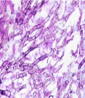
Microscopic examination of the biopsied material revealed granulomatous reactions, mixed inflammatory infiltration, fibrosis, and necropurulent material admixed with fungal elements. The hyphae of the fungus showed acute-angle branching and septation, compatible with aspergillosis (Figure 2).

Figure 2.
Fungal Hyphae With Septation and Acute-Angle Branching Is Seen on a Necrotic Background. Hand E Stain 40 × Magnification
Amphotericin B deoxycholate (1 mg/kg IV daily) was begun, and administration of ceftriaxone and metronidazole was stopped. The results of testing for human immunodeficiency virus (HIV) and nitroblue tetrazolin (NBT) for chronic granulomatous disease (CGD) were negative. However, the patient had a history of taking steroids (5 mg per day) for short periods. On follow-up MRI of the brain, she had a new abscess in the same location, but because the size of abscess was small, an operation was not performed.
Five months later, the size of the abscess had progressed, so a second surgery was performed. Amphotericin B deoxycholate continued for five months, and the patient was discharged with itraconazole (600 mg/day for three days, then 400 mg/day).
After three months, she had a seizure, and MRI of the brain showed two new lesions in the same locations, so surgery was performed and voriconazole (4 mg/kg IV q 12 hour, then 200 mg p. o. q 12 hour) was prescribed. After three months of follow-up, she did not have any lesions. She could not buy the voriconazole due to her poor socioeconomic condition; therefore, itraconazole (600 mg/day for three days, then 400 mg/day) was prescribed. After six months of therapy with itraconazole, she had no symptoms, and MRI of the brain showed no lesions. Presently, after two years of follow-up, she has no symptoms and her MRI is normal.
3. Discussion
Most cases of invasive aspergillosis show that this organism is pathogenic in immunocompromised patients; however, some case reports show that invasive aspergillosis may not be so rare in immunocompetent patients. In these patients, virulent and drug-resistant forms of aspergillus may be responsible for the disease, and treatment with antifungal agents is often ineffective, so that surgical excision is required (4). In an immunocompromised host, aspergillosis invades the brain hematogenously from the pulmonary organs or spreads from the paranasal sinuses, but in immunocompetent patients, this fungus spreads from sites near the brain, such as the sinuses. The clinical presentation of cerebral aspergillosis is nonspecific, and includes focal neurologic signs, altered mental status, and headaches; however, fever may be absent (4).
The risk factors for invasive aspergillosis include hematologic and lymphoreticular cancers (especially acute leukemia), hematopoietic stem cell transplantation, solid organ transplantation, the use of high-dose corticosteroids, and neutrophil deficiencies or dysfunction, such as CGD in children (1).
CT scan findings in invasive aspergillosis are similar to those of other infections that cause brain abscesses. MRI may show additional lesions, but the findings are nonspecific. The lesions are often located in the basal ganglia, thalami, corpus callosum, and other perforator artery territories (2). To confirm the diagnosis, biopsy may be helpful (5), showing hyaline-septated hyphae measuring 3-6 µm in width, with acute-angle branching. The hyphal elements occlude the intracerebral blood vessels, resulting in infarction. The fungi then penetrate into the ischemic brain parenchyma, leading to a mixed inflammatory reaction and necrosis (2). Surgical treatment can be effective in patients with focal CNS aspergillosis, and can reduce mortality from 64% to 39% (6).
Recently, voriconazole has been the therapy of choice and is recommended as a primary therapy for most patients with invasive aspergillosis. The primary use of combination therapy is not recommended because of the lack of prospective clinical trial data, but the addition of another antifungal drug, or a switch to another class in a salvage setting, may be considered due to the poor outcomes with single agents in progressive infections. Although the optimal duration of antifungal therapy is not known, improvement in the underlying host defenses is crucial to successful therapy (6).