Arch Pediatr Infect Dis

Image Credit:Arch Pediatr Infect Dis

Inflammatory Myofibroblastic Tumor of the Right Upper Lobe in an 11-Year-Old Boy Presenting with Dyspnea: A Case Report

Author(s):
Javad GhoroubiJavad Ghoroubi1, Shahed FarajiShahed Faraji2, Leila AzimiLeila AzimiLeila Azimi ORCID3,*
1Pediatric Surgery Research Center, Research Institute for Children’s Health, Shahid Beheshti University of Medical Sciences, Tehran, Iran
2Department of Pediatric Surgery, Mofid Children Hospital, Shahid Beheshti University of Medical Sciences, Tehran, Iran
3Pediatric Infections Research Center, Research Institute for Children’s Health, Shahid Beheshti University of Medical Sciences, Tehran, Iran

Archives of Pediatric Infectious Diseases:Vol. In Press, issue In Press; e168817
Published online:May 19, 2026
Article type:Case Report
Received:Dec 06, 2025
Accepted:Feb 21, 2026
How to Cite:Ghoroubi J, Faraji S, Azimi L. Inflammatory Myofibroblastic Tumor of the Right Upper Lobe in an 11-Year-Old Boy Presenting with Dyspnea: A Case Report. Arch Pediatr Infect Dis. 2026;In Press(In Press):e168817. doi: https://doi.org/10.5812/apid-168817

Abstract

Introduction:

Inflammatory myofibroblastic tumor (IMT) is a rare mesenchymal neoplasm, particularly in the pediatric lung. It often mimics malignancy clinically and radiologically, which can pose diagnostic challenges.

Case Presentation:

An 11-year-old boy presented with progressive dyspnea. Physical examination revealed decreased breath sounds over the right upper lung field. Chest computed tomography showed a 6 × 5.4 × 5.3 cm enhancing mass with central necrosis at the apex of the right lung, causing complete obstruction of the right upper lobe bronchus and encasement of the associated artery. Right hilar lymphadenopathy and adjacent pleural involvement were also noted. Bronchoscopy findings were unremarkable. The patient underwent surgical resection of the mass. Histopathological and immunohistochemical analyses confirmed the diagnosis of IMT. Postoperative recovery was uneventful, and the patient remained symptom-free during follow-up without the need for adjuvant therapy.

Conclusions:

IMT should be included in the differential diagnosis of pediatric pulmonary masses causing bronchial obstruction. Complete surgical excision is associated with favorable outcomes, even in cases with aggressive radiologic features.

1. Introduction

Inflammatory myofibroblastic tumor (IMT) is a rare mesenchymal neoplasm characterized by the proliferation of spindle-shaped myofibroblastic cells admixed with inflammatory infiltrates, primarily composed of plasma cells and lymphocytes (1). Historically, it has been described using various terms, including inflammatory pseudotumor, plasma cell granuloma, and fibrous histiocytoma (2). In 2013, the World Health Organization reclassified IMT as a soft tissue tumor of intermediate biological potential because of its capacity for local recurrence and rare metastasis (3).
Although IMT can occur in various organs, including the mesentery, retroperitoneum, liver, and bladder, the lung remains one of the most common sites in children (4). Pulmonary IMT is the most common benign lung tumor in children, accounting for more than 50% of benign pulmonary masses in previous pediatric case series and reviews (5, 6). However, IMT constitutes less than 1% of all primary lung tumors in the general population (7). Its overall incidence is low, with approximately 150 - 200 new cases reported annually in the United States (8).
Clinically and radiologically, pulmonary IMTs often mimic malignancy. Symptoms may include cough, hemoptysis, chest pain, or dyspnea; however, many cases are discovered incidentally on imaging (9). Computed tomography (CT) scans usually demonstrate a solitary, well-defined mass with heterogeneous enhancement, central necrosis, or adjacent pleural involvement (10). Bronchoscopy may be nondiagnostic, particularly when the tumor is peripherally located (11).
The diagnosis is established by histopathologic examination, with immunohistochemical staining often demonstrating positivity for smooth muscle actin (SMA) and, in some cases, anaplastic lymphoma kinase (ALK). Approximately 50% of IMTs harbor ALK gene rearrangements, which may guide targeted therapy in inoperable or recurrent cases (8). Complete surgical excision remains the standard of care and is typically curative, although close follow-up is recommended because of the risk of local recurrence (12).

1.1. Case Presentation

An 11-year-old boy with no significant medical, surgical, or perinatal history presented with progressive exertional dyspnea over several weeks. He denied fever, cough, chest pain, hemoptysis, or weight loss. There was no known exposure to tuberculosis or environmental toxins.
On physical examination, vital signs were within normal limits. Chest auscultation revealed markedly decreased breath sounds over the right upper lung field. The remainder of the physical examination was unremarkable.
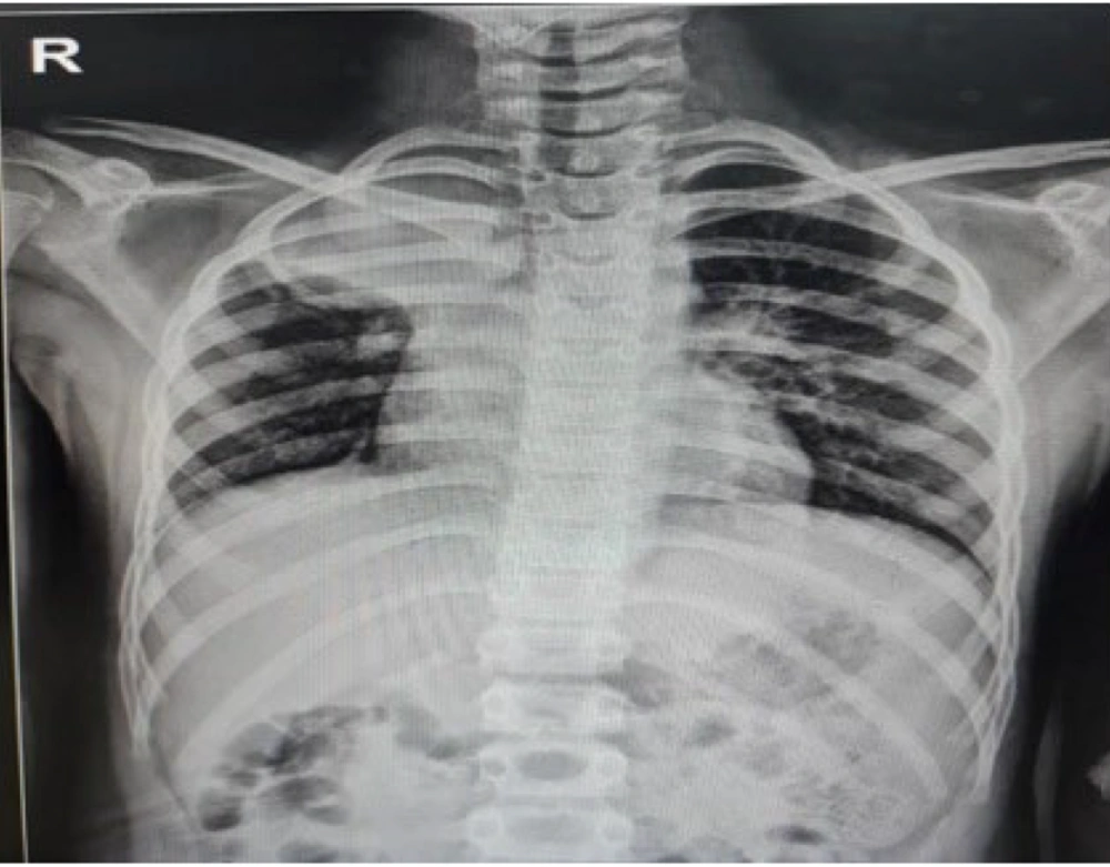
A chest radiograph revealed a dense opacity in the right upper lung zone, suggestive of a space-occupying lesion (Figure 1).
Chest X-ray, posteroanterior view, demonstrating a well-circumscribed mass lesion in the right upper lung zone, consistent with a parenchymal tumor. The lesion was later confirmed as an inflammatory myofibroblastic tumor following surgical resection and histopathological examination.
Figure 1.

Chest X-ray, posteroanterior view, demonstrating a well-circumscribed mass lesion in the right upper lung zone, consistent with a parenchymal tumor. The lesion was later confirmed as an inflammatory myofibroblastic tumor following surgical resection and histopathological examination.

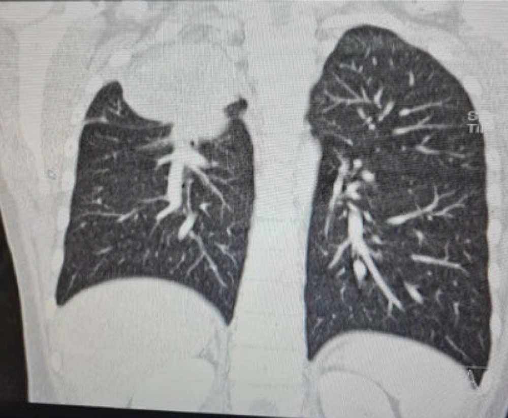
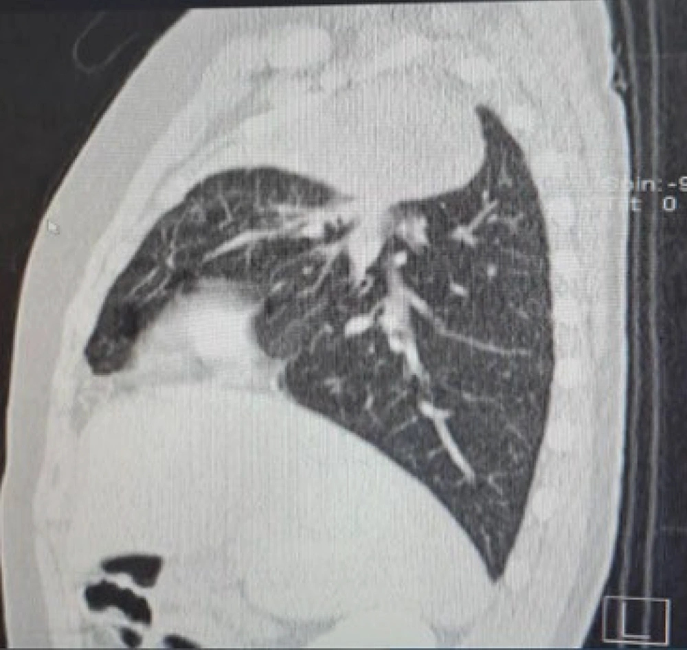
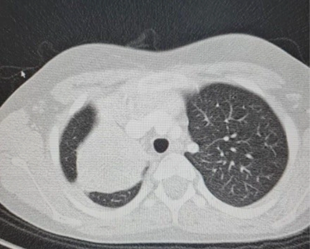
Contrast-enhanced chest CT demonstrated a 60 × 54 × 53 mm enhancing mass with central necrosis located at the apex of the right lung. The mass caused complete obstruction of the right upper lobe bronchus and extended medially, resulting in narrowing of the proximal bronchus intermedius. Encasement of the artery supplying the right upper lobe was evident, along with adjacent pleural involvement and right hilar lymphadenopathy, with a maximum short-axis diameter of 9 mm (Figures 2 - 4). Flexible bronchoscopy was performed and revealed no endobronchial lesion or mucosal abnormality.
The patient underwent a right posterolateral thoracotomy with complete surgical resection of the mass. Gross examination showed a firm, tan-gray mass with areas of necrosis. Histopathological analysis revealed lung parenchyma infiltrated by relatively uniform, hypocellular spindle cells within a collagenous stroma, accompanied by prominent lymphoplasmacytic infiltration, multifocal hyalinization, and lymphoid follicle formation. Special stains, including periodic acid-Schiff, periodic acid-Schiff with diastase, and Ziehl-Neelsen stains, were negative for fungal and acid-fast organisms.
Immunohistochemical staining showed diffuse positivity for SMA, focal positivity for desmin, and negativity for ALK, S100, and myogenin, confirming the diagnosis of IMT.
The postoperative course was uneventful. The patient was discharged on postoperative day 5 in good condition. During follow-up, he remained asymptomatic, with stable vital signs and no evidence of recurrence on imaging. A pediatric oncology evaluation concluded that no adjuvant therapy was necessary.

2. Discussion

Inflammatory myofibroblastic tumor is a rare pulmonary neoplasm, particularly in children, in whom it represents approximately 52% - 70% of benign lung tumors (5, 6), yet comprises less than 1% of all primary lung tumors in the general population (7). Despite being histologically benign, IMT is considered a neoplasm of intermediate biological potential because of its capacity for local recurrence and rare metastasis (3).
The clinical presentation of IMT varies widely and may include cough, dyspnea, chest pain, or hemoptysis; alternatively, the tumor may be entirely asymptomatic (9). In the present case, the patient presented with progressive exertional dyspnea. Imaging typically shows a well-defined solitary mass, often with heterogeneous enhancement, central necrosis, or adjacent pleural involvement (10). Bronchial obstruction and encasement of nearby vessels, as seen in this patient, can mimic aggressive malignancies such as sarcoma or lymphoma (9, 10).
The diagnosis of IMT relies on histopathologic evaluation and immunohistochemistry. The characteristic histologic pattern includes spindle-shaped myofibroblastic cells with an inflammatory background rich in lymphocytes and plasma cells (1). Bronchoscopy may be nondiagnostic, particularly when the tumor is peripherally located or lacks an endobronchial component. This limitation can delay diagnosis and necessitate further imaging or surgical biopsy to establish a definitive diagnosis (11). Approximately 50% of IMTs harbor ALK gene rearrangements, and ALK positivity supports the diagnosis (8).
Complete surgical resection remains the mainstay of treatment and is associated with an excellent prognosis (12, 13). Studies have shown that recurrence rates are very low after R0 resection, especially for intrapulmonary lesions (13). The patient in this case underwent complete resection via thoracotomy and had an uneventful postoperative course, with no recurrence during follow-up.
In cases in which the tumor is inoperable, recurrent, or metastatic, particularly in ALK-positive tumors, targeted therapy with ALK inhibitors, such as crizotinib, has shown promising results (12). However, in ALK-negative patients, such options are limited, and the role of systemic therapy remains unclear (8, 12).
We report a rare case of ALK-negative pulmonary IMT in an 11-year-old boy who presented with features mimicking malignancy. The diagnosis was confirmed histologically, and surgical resection led to complete resolution. The patient remained asymptomatic without the need for adjuvant therapy. This case highlights the importance of considering IMT in the differential diagnosis of pediatric lung masses and the excellent prognosis associated with complete excision.
Pulmonary IMT is a rare entity in pediatric patients that can clinically and radiologically mimic malignant lung neoplasms. Accurate diagnosis relies on histopathologic and immunohistochemical evaluation. Complete surgical resection remains the treatment of choice and is associated with an excellent prognosis. Long-term follow-up is essential because of the potential for recurrence, particularly in ALK-negative cases. Awareness of this tumor is crucial for timely diagnosis and appropriate management.
Coronal CT scan of the chest demonstrating a well-defined soft tissue mass occupying the apex of the right lung. The lesion causes complete obstruction of the right upper lobe bronchus, with associated collapse of the upper lobe parenchyma. Mild mediastinal shift and compression of adjacent structures are present.
Figure 2.

Coronal CT scan of the chest demonstrating a well-defined soft tissue mass occupying the apex of the right lung. The lesion causes complete obstruction of the right upper lobe bronchus, with associated collapse of the upper lobe parenchyma. Mild mediastinal shift and compression of adjacent structures are present.

Sagittal CT image showing the same mass extending posteriorly and superiorly, causing architectural distortion and narrowing of the proximal bronchial tree. The lesion demonstrates heterogeneous density, consistent with areas of central necrosis.
Figure 3.

Sagittal CT image showing the same mass extending posteriorly and superiorly, causing architectural distortion and narrowing of the proximal bronchial tree. The lesion demonstrates heterogeneous density, consistent with areas of central necrosis.

Axial CT image demonstrating a heterogeneous right upper lobe mass with central hypodensity and mild tracheal deviation, consistent with an inflammatory myofibroblastic tumor.
Figure 4.

Axial CT image demonstrating a heterogeneous right upper lobe mass with central hypodensity and mild tracheal deviation, consistent with an inflammatory myofibroblastic tumor.

Footnotes

References

  • 1.
    Coffin CM, Watterson J, Priest JR, Dehner LP. Extrapulmonary inflammatory myofibroblastic tumor: A clinicopathologic study of 84 cases. Am J Surg Pathol. 1995;19(8):859-872.
  • 2.
    Gleason BC, Hornick JL. Inflammatory myofibroblastic tumors: Where are we now? J Clin Pathol. 2008;61(4):428-437. [PubMed ID: 17938159]. https://doi.org/10.1136/jcp.2007.049387.
  • 3.
    Fletcher CDM, Bridge JA, Hogendoorn PCW, Mertens F. WHO Classification of Tumours of Soft Tissue and Bone. Mertens F. WHO Classification of Tumours of Soft Tissue and Bone. 4th ed. IARC; 2013.
  • 4.
    Soyer T, Karnak I, Ekinci S, et al. Surgical treatment of inflammatory myofibroblastic tumors in children. Eur J Pediatr Surg. 2016;26(3):250-254.
  • 5.
    Fabre A, Fadel E, Singhal S, de Montpreville VT. Pediatric inflammatory myofibroblastic tumor of the lung: A clinicopathologic study of 18 cases. Ann Thorac Surg. 2009;88(3):1024-1029.
  • 6.
    Camela F, Gallucci M, Di Palmo E, et al. Pulmonary inflammatory myofibroblastic tumor in children: A case report and literature review. Front Pediatr. 2018;6:35.
  • 7.
    Braham Y, Migaou A, Njima M, Achour A, Ben Saad A, Cheikh Mhamed S, et al. Inflammatory myofibroblastic tumor of the lung: A rare entity. Respir Med Case Rep. 2020;31. 101287. [PubMed ID: 33251105]. [PubMed Central ID: PMC7683262]. https://doi.org/10.1016/j.rmcr.2020.101287.
  • 8.
    Lovly CM, Gupta A, Lipson D, Otto G, Brennan T, Chung CT, et al. Inflammatory myofibroblastic tumors harbor multiple potentially actionable kinase fusions. Cancer Discov. 2014;4(8):889-895. [PubMed ID: 24875859]. [PubMed Central ID: PMC4125481]. https://doi.org/10.1158/2159-8290.CD-14-0289.
  • 9.
    Zhang N, Zeng Q, Chen C, Yu J, Yan D, Xu C, et al. Clinical characteristics and prognosis of pulmonary inflammatory myofibroblastic tumor: A 10-year retrospective analysis. Pediatr Investig. 2020;4(4):192-197. [PubMed ID: 33150313]. [PubMed Central ID: PMC7520109]. https://doi.org/10.1002/ped4.12218.
  • 10.
    Lichtenberger JP, Biko DM, Carter BW, et al. Primary lung tumors in children: Radiologic-pathologic correlation. Radiographics. 2018;38(7):2151-2172.
  • 11.
    Vounckx M, Jansen YJL, Fadaei S, Geers C, De Pauw V, Smets D. Unraveling the spectrum of inflammatory myofibroblastic tumors in the lung: A comprehensive case series highlighting endobronchial, pleural, and lung parenchymal tumors. JTCVS Open. 2024;17:297-305. [PubMed ID: 38420532]. [PubMed Central ID: PMC10897672]. https://doi.org/10.1016/j.xjon.2023.12.006.
  • 12.
    Kovach SJ, Fischer AC, Katzman PJ, Salloum RM, Ettinghausen SE, Madeb R, et al. Inflammatory myofibroblastic tumors. J Surg Oncol. 2006;94(5):385-391. [PubMed ID: 16967468]. https://doi.org/10.1002/jso.20516.
  • 13.
    Karnak I, Senocak ME, Ciftci AO, Tanyel FC, Büyükpamukçu N. Inflammatory myofibroblastic tumor in children: Diagnosis and treatment. J Pediatr Surg. 2001;36(6):908-912. https://doi.org/10.1053/jpsu.2001.23942.

Crossmark
Crossmark
Checking
Share on
Cited by
Metrics

Ordering Reprints

Articles are published under the Creative Commons license stated on each article. No permission or royalty fee is required for uses permitted by that license. CCC handles optional bulk and customized reprint orders. Any quotation covers production and delivery services only, not copyright permission. > Request Reprints from CCC 

Search Relations

Author(s):

Related Articles