4.3. Biofilms by SEM
The disks were exposed to three different VCZ concentrations (1, 4, and 16 µg/mL) and compared to the controls.
Figures 1 and
2 show SEM pictures of biofilms formed by
F. solani and
A. flavus, respectively. The biofilm architecture of the
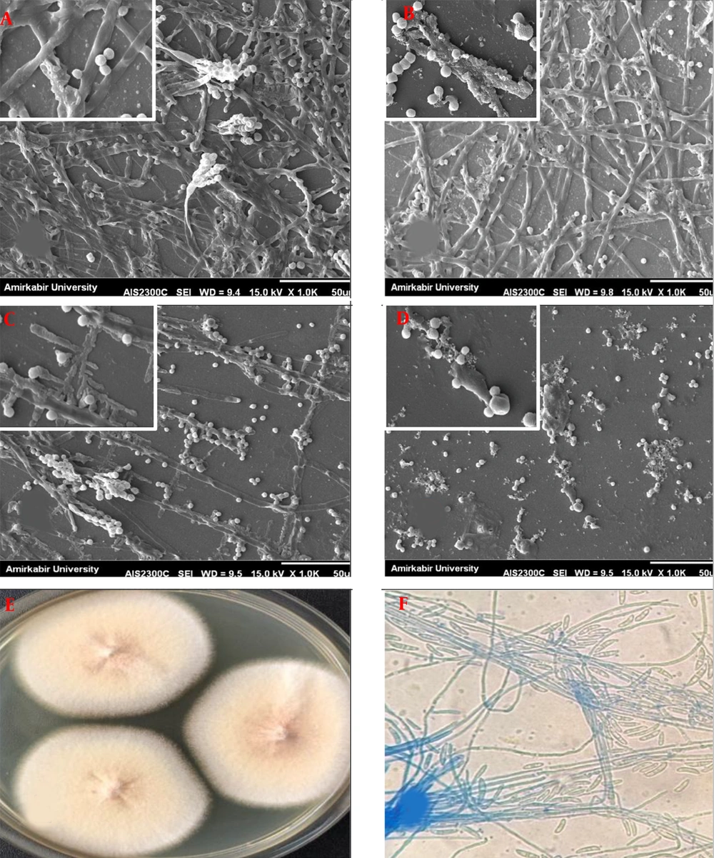
F. solani control (untreated) was highly mixed, comprised of a compact layer of yeasts, pseudo hyphae, and hyphal forms. A few typical microconidia, fusion of abundant hyphae with a well-defined septum, and macroconidia containing new conidia aggregated and associated with apical growth are depicted in
Figure 1A. After exposure to VCZ, biofilms of
F. solani showed fewer conidia, most of the hyphae appeared to be thinner after the 1µg/mL treatment (
Figure 1B). Lesser extent hyphae were also observed after the 4µg/mL treatment (
Figure 1C). Biofilms exposed to 16 µg/mL VCZ contained substantially fewer conidia (i.e., they were smaller than those observed at 1 and 4 µg/mL) than untreated biofilms; however, seemed completely affected by VCZ treatment (
Figure 1D).
Scanning electron micrographs of preformed Fusarium solani biofilms in the presence of different voriconazole concentrations. (A) F. solani biofilm control (without drug); (B through D) F. solani biofilms after exposure to voriconazole at 1 µg/mL, 4 µg/mL, and 16 µg/mL, respectively (1000 and 5000×magnification). (E) Macroscopic and (F) Microscopic observation.
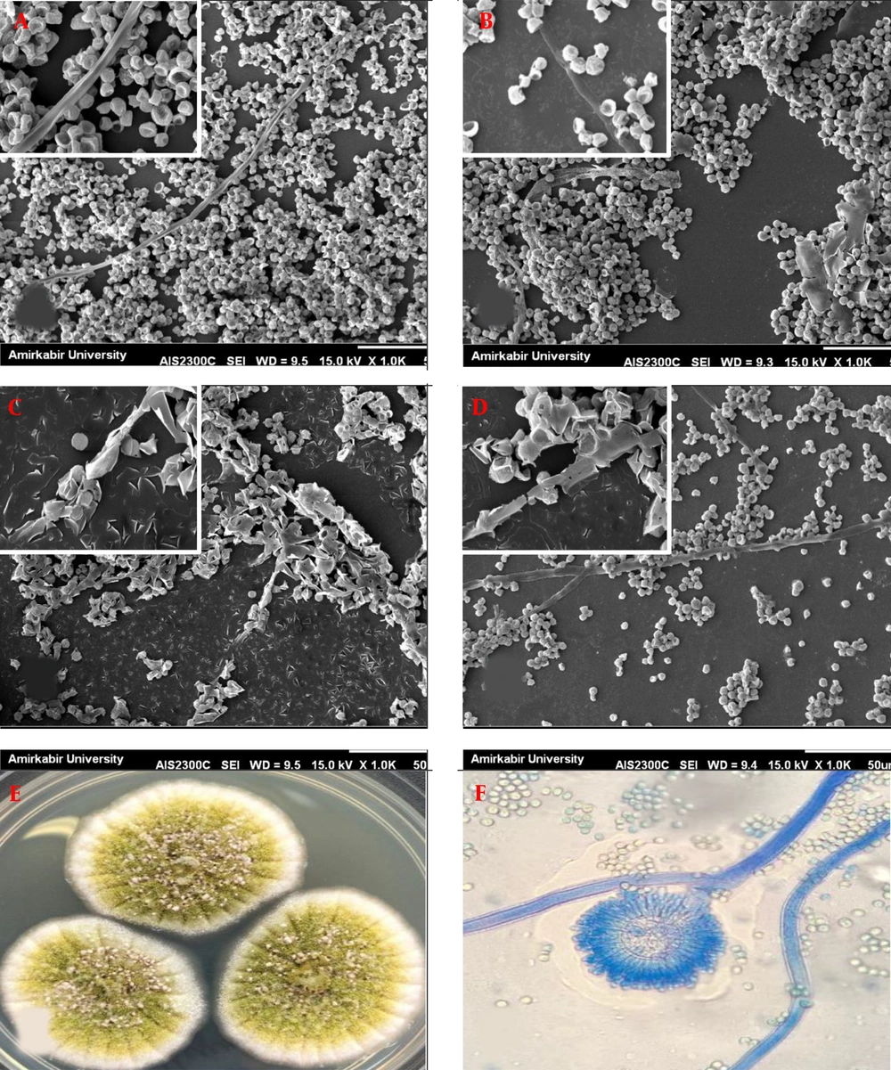
Scanning electron micrographs of preformed Aspergillus flavus biofilms in the presence of different voriconazole concentrations. (A) A. flavus biofilm control (without drug); (B through D) A. flavus biofilms after exposure to voriconazole at 1 µg/mL, 4 µg/mL, and 16 µg/mL, respectively (1000 and 5000×magnification). (E) Macroscopic and (F) Microscopic observation.
Unlike
F. solani,
A. flavus exhibited architecture containing abundant conidia (irregular groupings of microconidia) and few filamentous forms in control (
Figure 2A). No significant changes in morphology were observed in the presence of 1 µg/mL VCZ (
Figure 2B). All of the conidia revealed collapsed cell walls after exposure with 4 µg/mL VCZ, and degenerated hyphae were completely observed (
Figure 2C). There was less outcome on conidial and hyphal morphology at 16 µg/mL VCZ and biofilms appeared morphologically highly similar to untreated biofilms; nevertheless, density and accumulation of conidia and hyphae became much lower (
Figure 2D).
The growth of
Aspergillus niger on polyester mesh squares described biofilm formation by filamentous fungi (
14), and the growth of
Aspergillus fumigatus was revealed by scanning electron microscopy and laser scanning confocal microscopy (LSCM) (
15,
16). Biofilm formation of
F. solani and
F. oxysporum was analyzed by antibiotic susceptibility and LSCM (
17).
Although the resistance mechanisms of filamentous species have not been completely elucidated, a hypothesis proposes that sessile cells are in a biofilm population which permanently bound to a surface and inserted in an exopolymeric background (
18). Sessile cells are extremely resistant to the host defense mechanisms and to antifungal agents (
19).
In this study, the ability of biofilm formation of F. solani and A. flavus strains, extracted from the fungal keratitis, had been compared with the investigation of their metabolic activity using the crystal violet staining method. The susceptibilities of sessile cells of A. flavus and F. solani were evaluated against the antifungals NAT, ICZ, VCZ, and PCZ.
Based on the results of this study, the biofilm formation of all tested isolates varied to great degrees. Accordingly, the complete biofilm of both filamentous fungi isolates could increase resistance to VCZ. These results are in line with those of a study indicating that the sessile cells of
F. solani and
F. oxysporum were less sensitive to NAT and VCZ (
20). The initial and mature phases of
Candida albicans biofilm had a significant difference with the values of the MICs, associated with their differential genes expression and resistance to the evolution of maturation of the biofilm (
21).
The findings disclosed that the ability of biofilm formation was higher in
F. solani strains than in
A. flavus strains. This result is consistent with Chang et al. study that the ability of
Fusarium to form biofilms on case wearing contact lenses might play the main role in the outbreak (
22).
ICZ, PCZ and NAT had the maximum activity against biofilms of all tested filamentous strains (MIC ranging from 0.031 - 0.25 µg/mL, 0.031 - 0.25 µg/mL and 2 - 4 µg/mL), respectively. PCZ releaved the lowest MIC values against biofilms of all strains (MIC ranging from 0.031 - 0.25 µg/mL); however, no significant difference was observed for ICZ in this regard (P > 0.05).
However, they tended to be less susceptible to VOR than to the other agents. Three F. solani and one A. flavus strains showed the high MIC values against VCZ (MIC ≥ 1).
It was also found out that ICZ and PCZ were more active against sessile cells, while VCZ exhibited less activity against biofilms of strains.
In this respect, VCZ 1% and topical amphotericin B (0.3 - 0.5%) are recommended as alternative drugs. The deep penetration of topical VCZ to ocular is considered a considerable advantage (
23). However, there is a studies revealing much better consequences in patients treated with NAT, compared to those of VCZ treatment (
24-
26). Wu et al. reported that VCZ had an extensive therapeutic scope covering filamentous and yeast fungi. VCZ revealing a suitable penetration to ocular, was used as an adjunct therapy to NAT in case of refractory to topical NAT (
27). Although NAT shows poor activity against
Aspergillus, NAT and VCZ have recently been the only FDA-approved drugs for the remedy of fungal infections caused by
Fusarium spp. (
28). Herein, VCZ showed resistance (≥ 1 μg/mL) against
Fusarium and
Aspergillus biofilms.
Mukherjee et al. predicted that efflux pumps had a vital role in azole resistance in the initial phase of Candida biofilm formation due to an alteration in sterol composition at the middle and complete phases (
29). However, the basis for such differences has not been clarified. In another study, it was revealed that the less susceptibility to VCZ in
Fusarium biofilms might be attributed to the upregulation of efflux pumps (
29).
Based on the results of this study, the role of biofilm formation on resistance to antifungal agents was different, that is to say, although all of them were susceptible to NAT, ICZ, and PCZ, they exhibited different susceptibility to VCZ.
The persister cells are phenotypical variants of the wild type in biofilm structures and able to survive in spite of the presence of different concentration of antifungals (
30).
Herein, the researchers used SEM to imagine morphological changes after exposure to VCZ. The value of 16 µg/mL VCZ was approved for sessile cells of F. solani with fewer conidia than untreated biofilms.
The SEM images indicated that 4 µg/mL VCZ caused considerable variations in the morphologies of preformed
A. flavus biofilms, with the presence of collapsed conidia and degenerated hyphae. Biofilm morphological changes of
A. fumigatus associated with the interaction of
Staphylococccus aureus have been described previously (
31). However, to the best of our knowledge, this survey prepares the first detailed SEM image analysis of
F. solani and
A. flavus biofilms adherent to disks. Fungal biofilms are among the dangerous medical problems worldwide since they can adhere to medical devices and acquire resistance to antifungal agents, and turn to infections extremely difficult to be eradicated. The ability of
Fusarium strains and
Aspergillus strains to form biofilms may lead to difficulty in the clinical management of keratitis. Therefore, it is recommended to know remedial concentrations of VCZ show potent in vitro activity against filamentous fungi biofilm and to have a knowledge of the mechanism of action, penetration rates, and therapeutic concentrations of drugs.