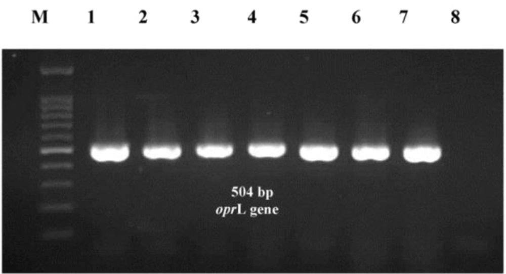
Our study observed 41 antibiotic resistance patterns and eight E-types among
P. aeruginosa isolates. This study observed the highest P. aeruginosa prevalence among ICU patients; most were isolated from throat samples. One reason for these findings can be related to the high sickness of patients hospitalized in ICUs with weak immune systems. Moreover, abuse or overuse of antibiotics and medical instruments in ICUs intensifies the circumstances. Other studies in Iran also proved that the emergence of
P. aeruginosa is significantly higher in ICUs than in other hospital units (
10,
11). The most probable specimens contaminated with
P. aeruginosa in our study were throat, followed by urine samples. In the study by Vaez et al., however, the highest frequency of
P. aeruginosa was related to ICU patients, and urine samples were primarily infected with
P. aeruginosa (
11). In another study by Izadi Pour Jahromi et al., burn patients and pediatrics were reported as high-risk groups for
Pseudomonas infections, and isolates showed notable and drastic resistance to various antibiotics (
12). Some other countries reported that the most frequent
Pseudomonas infections occurred in respiratory tracts, most of which were among ICU patients (
13-
15).
Our isolates showed high resistance to aminoglycosides, carbapenems, cephalosporins, and fluoroquinolones. The most effective antibiotics belonged to polymyxins, especially polymyxin B. The MDR and XDR were detected in 86.8% and 81.5% of our isolates, respectively, which proves the high antibiotic resistance rate among
P. aeruginosa strains. Although different rates have been reported in Iran and other countries for MDR and XDR
P. aeruginosa, all agree with the upward trend of resistant strains emergence, especially XDR strains (
16-
18). In our study, four strains were classified as PDR, showing the increasing emergence of PDR strains. The emergence of PDR strains was not reported in previous studies, or at least it was very low (
13,
15,
16). In contrast, in our study, the detection of four PDR strains out of 76 isolates proves that we are near the end of the antibiotic era.
Studies of genotyping and molecular epidemiology of
P. aeruginosa strains showed that the genetic diversity is catastrophically high among clinically resistant
P. aeruginosa strains than in susceptible or environmental strains (
19). The molecular epidemiologic studies warned about the increasing emergence and spread of
P. aeruginosa high-risk clones worldwide, a major concern in preventing and controlling nosocomial infections (
20). Thus, the source tracking and molecular epidemiologic information of resistant
P. aeruginosa play a key role in infection treatment and control.
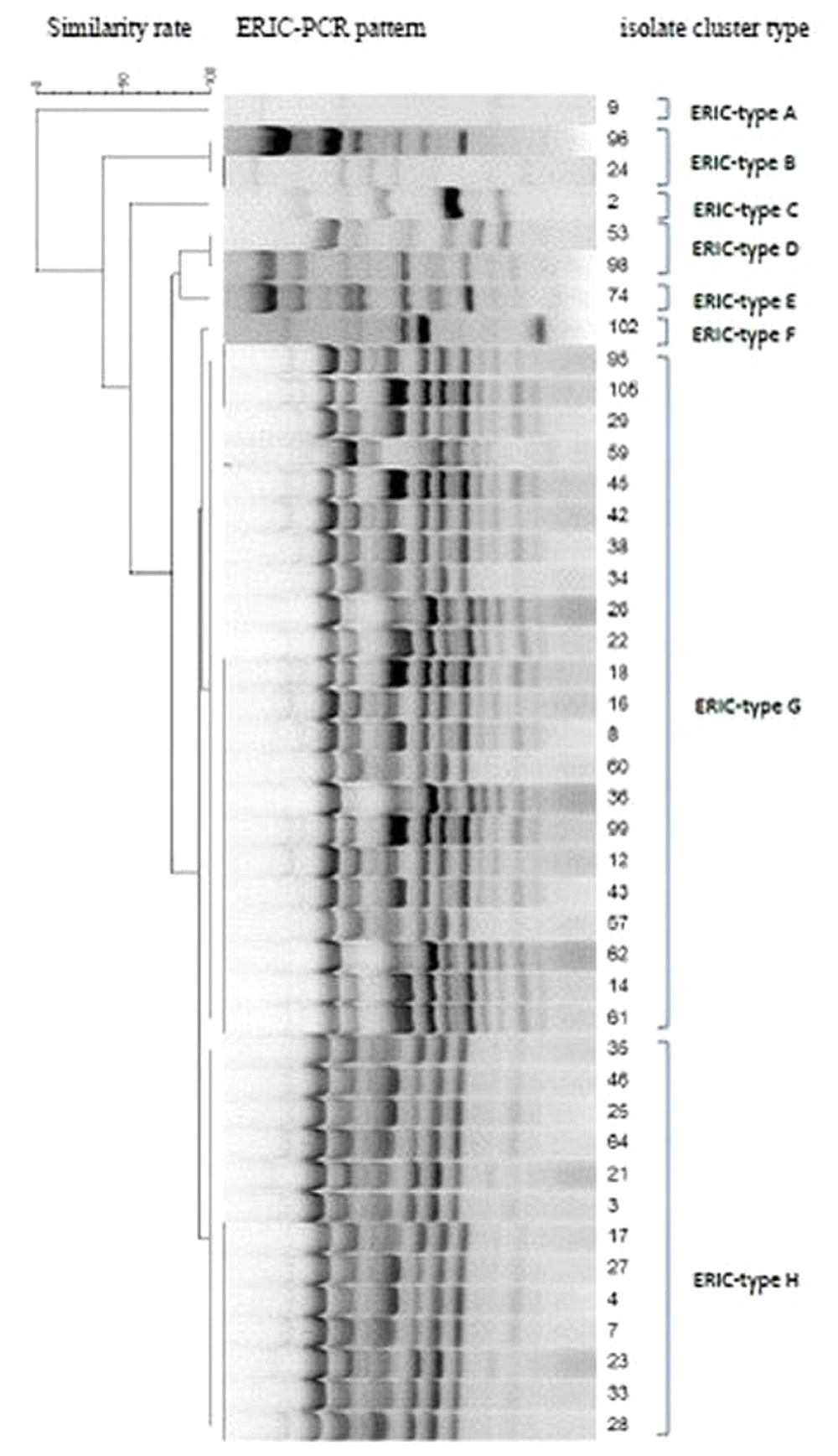
The present study observed eight E-types among resistant
P. aeruginosa named from A to H alphabetic letters. The dominant E-types among our isolates belonged to G and H, composed of strains isolated from ICU patients. Although a high diversity in resistance patterns was observed among strains belonging to these two E-types, all were resistant to ticarcillin-clavulanic acid and fosfomycin and showed almost 90% similarities in their ERIC patterns. The isolates related to these two E-types can be considered dominant resistant strains spread in the internal ICU. The E-type A was unique among isolates with no similarity with other E-types from a patient hospitalized in the surgical ICU. Moreover, E-types C, E, and F were each composed of only one strain isolated from throat samples and showed a 50% similarity with each other. Notably, although the former E-types showed only 50% similarity, they showed resistance to some antibiotics in common. According to the analysis, no correlations were observed between E-types and sample types or antibiotic resistance patterns (P ≥ 0.05), while the strains with the same E-types were almost related to the same ward in the hospital. In the study by other researchers from Iran, the ERIC-PCR results of
P. aeruginosa isolates could not be useful for predicting antibiotic resistance patterns because the strains with the same E-types could possess different antibiotic resistance profiles (
21).
On the one hand, the current study showed several orphan clusters indicating the high rate of genetic diversity among isolates; on the other hand, we found a dominant cluster with two different E-types with considerable isolate numbers, all colonized and isolated from patients in the internal ICU. The diversity among E-types for
P. aeruginosa is not rare, and most of the studies worldwide reported some sort of diversity (
22-
27). The point is that among these various genotypes, some can dominate in a single ward and spread to other hospital sites, making it much more difficult to eradicate from hospitals. The main threat here is regarding the PDR strains since all were isolated from the same ward and belonged to the same genetic cluster. They can be considered nosocomial pathogens and should be deliberated as a critical threat in an emerging outbreak in a hospital.
5.1. Conclusions
We observed a heterogeneous population of dominant P. aeruginosa strains in different hospital wards. However, they possessed different resistance patterns, making infection control and patient treatments more difficult and time-consuming for the healthcare system. In order to disclose and eradicate the resistant P. aeruginosa strains, the genetic and antibiotic profiling of any dominant local colon must be considered a critical strategy and guideline for the hospital infection control committee.