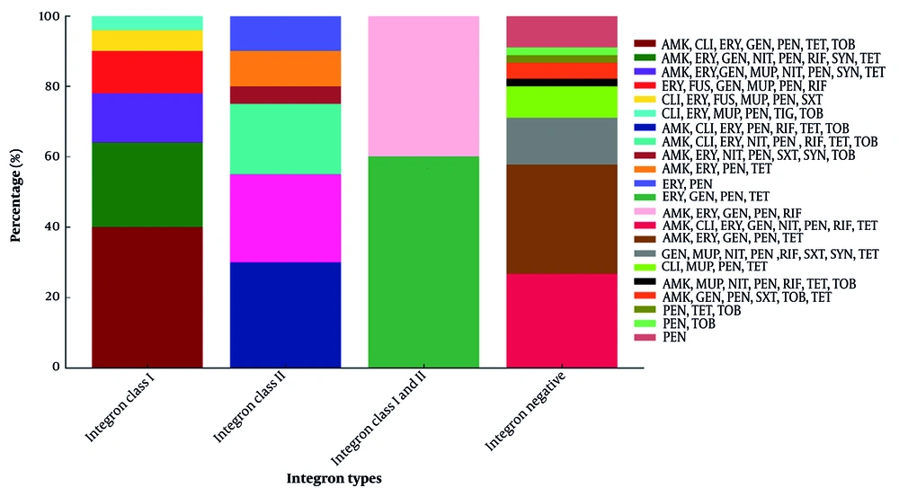
The analysis revealed that antibiotics such as tobramycin, fusidic acid, tigecycline, and quinupristin-dalfopristin exhibited the highest levels of activity against the tested
S. aureus isolates. These results align with previous studies, including those by Rafati Zomorodi et al., who reported strong efficacy for linezolid and vancomycin (
11), and Saedi et al., who observed high susceptibility rates (over 90%) for linezolid, vancomycin, and trimethoprim-sulfamethoxazole (
12). Mupirocin continues to serve as a key topical antimicrobial for curbing
S. aureus transmission and mitigating infection risks across both healthcare and community environments, as supported by earlier studies. In the current investigation, 24.2% of the isolates exhibited resistance to mupirocin, with the majority (79.3%) categorized as high-level mupirocin-resistant (HLMUPR) and the remaining 20.7% classified as low-level mupirocin-resistant (LLMUPR). The detected prevalence of HLMUPR-methicillin-resistant
S. aureus strains in this study notably surpasses previously reported rates in countries such as China (7%), France (0.8%), and Canada (4.3%) (
13-
15). This considerable discrepancy underscores the geographic variability in mupirocin resistance patterns among methicillin-resistant
S. aureus isolates and emphasizes the critical role of regional surveillance in informing effective infection prevention and antimicrobial stewardship efforts.
The distribution of iMLS
B resistance phenotype among
S. aureus isolates demonstrates considerable variability across different geographical regions and healthcare systems. In the present study, the prevalence of iMLS
B was identified at 37.5%, which is higher than rates reported in Turkey (7.8%) (
16), Egypt (7.7%) (
17), Iran (8.6%) (
18), and Nepal (11.48%) (
19), but similar to the prevalence observed in India (37.5%) (
20). Prior investigations in Iran have highlighted notable regional differences in iMLS
B rates, ranging from 6% to 32.3%, reflecting the impact of localized antibiotic prescribing practices and diagnostic approaches. In addition, the cMLS
B phenotype was observed in 46.7% of isolates. This finding aligns with previous studies, such as those by Delialioglu et al. (24.3%) (
16) and Eksi et al. (20.4%) (
21). Nevertheless, other investigations have documented widely differing rates, ranging from 13.1% to as high as 82.9%, depending on the location and study methodology. Such variation likely stems from differences in community and hospital-based macrolide consumption, study population characteristics, and the dissemination of specific clonal lineages. According to our data, the cMLS
B phenotypes were relatively frequent among methicillin-resistant
S. aureus strains (46.7%), a finding consistent with earlier studies from Nepal, Iran, Egypt, and Turkey (
16-
19).
Regarding fusidic acid resistance, recent reports from various Asian countries indicate low resistance levels, typically below 10%. Our study identified a fusidic acid resistance rate of 7.5%. These results are consistent with findings from a large multicenter Iranian study, which reported a 3% prevalence of fusidic acid-resistant methicillin-resistant
S. aureus among 726
S. aureus isolates. An upward trend in fusidic acid resistance among methicillin-resistant
S. aureus isolates has been documented. For instance, data from Kuwait revealed a dramatic rise in resistance rates, escalating from 22% in 1994 to 92% by 2004 (
22). While earlier reports from Iran suggested lower resistance levels ranging between 2.5% and 8% (
23). Globally, resistance rates differ substantially, ranging from 62.4% in Greece to as low as 0.3% in the United States (
24).
It is well established that integrons play a pivotal role in the dissemination of multidrug resistance among pathogenic bacteria (
6). In the current study, class 1 integrons were the most frequently detected type, identified in 34.1% of the isolates. Class 2 integrons were present in 14.3% of samples, and a smaller proportion (5.6%) harbored both class 1 and class 2 integrons simultaneously. These findings, consistent with previous literature, emphasize the predominance of class 1 integrons over class 2 in
S. aureus populations. Our data closely align with the report by Mohammadi et al. in Iran, where class 1 integrons were found in 24.8% of
S. aureus isolates (
25). Similar trends have been reported from clinical samples in Iran. In a multi-center study conducted by Zomorodi et al. on 183
S. aureus isolates, integron class 1 was detected in 7.6% of the tested isolates, while none were positive for class 2 (
11). In a study performed in Iran on 139
S. aureus isolates, Mostafa et al. reported a prevalence of 72.6% and 35.2% of integron class 1 and 2, respectively (
9). A study conducted in India in 2015 by Marathe et al. reported that 71% of the identified methicillin-resistant
S. aureus strains harbored class 1 integrons, while none of the isolates tested positive for class 2 or class 3 integrons, indicating the dominant presence of class 1 integrons in the studied population (
26). In contrast, research conducted by Guney in Turkey reported an absence of class 1 integrons among the examined isolates, emphasizing the substantial regional and bacterial diversity in integron distribution (
27). Growing evidence points to class 1 integrons as major contributors to the dissemination of antibiotic resistance, especially in methicillin-resistant
S. aureus strains. Variations in the occurrence of these genetic elements may stem from factors such as geographic location, clonal differences among isolates, and disparities in antibiotic prescription practices and overuse across regions.
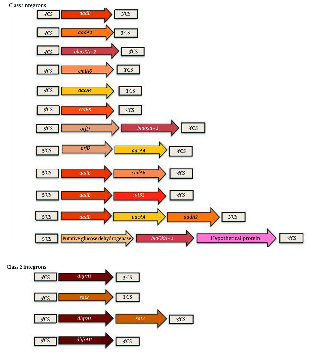
In the current investigation, the most frequently detected gene cassettes within class 1 integrons were
aadB–
aacA4–
aadA2, bla_OXA-2, and
aadB–
cmlA6, followed by the
aadB–catB3 cassette. A study conducted by Mostafa et al. in 2015 similarly identified
aadB,
aadA2, and
dhfrA1–
sat2–aadA1 as the predominant cassette arrays. Their research also reported several novel gene cassettes in
S. aureus isolates harboring class 1 integrons, including
aadB, oxa2,
aacA4, orfD–
aacA4–
catB8,
aadB–catB3, orfD–
aacA4, and
aadB–aadA1–
cmlA6. In contrast, among class 2 integrons, the most common arrays were
dhfrA1–
sat2–aadA1,
dhfrA11, and
dhfrA1–
sat2 (
9). Between 2001 and 2006, Xu et al. analyzed a collection of nosocomial methicillin-resistant
S. aureus isolates and found that 76 out of 179 strains (42.5%) carried class 1 integrons (
28). The most common resistance determinants were organized into four distinct gene cassette arrays:
aadA2,
aacA4–cmlA1,
dfrA17–aadA5, and
dfrA12–orfF–aadA2. Class 1 integrons are recognized for their ability to capture and disseminate diverse antimicrobial resistance genes, with aminoglycoside resistance cassettes being particularly frequent (
9). Notably, these integrons have been associated with
Tn3 family transposons, such as
Tn21 and
Tn1696, suggesting that the extensive distribution of class 1 integrons is likely driven by the mobility of integron-containing transposons (
9,
29). Moreover, the distribution of
Tn7 among clinical isolates has been shown to correlate with elevated rates of trimethoprim resistance, a phenotype conferred by the dihydrofolate reductase enzyme encoded by the
dhfr gene located within
Tn7 (
9).
In our analysis, plasmid-borne integrases were detected in 13.3% (10/75) of the isolates, which is lower than those previous reports by Mostafa et al. (36%) and Goudarzi et al. (80%) in
S. aureus (
9,
29). It should be noted, however, that this estimate may be subject to bias, as plasmid and chromosomal DNA can exhibit similar electrophoretic mobility patterns. In a previous investigation, Goudarzi et al. characterized the genetic composition of integrons in
S. aureus isolates and reported six distinct gene cassettes
aadA,
aadB,
bla_OXA,
aacA4,
cmlA6, and
catB within class 1 integrons. In addition, three gene cassettes
dfrA1,
aadA1, and
sat2 were detected in class 2 integrons (
29). These findings highlight the genetic diversity of resistance determinants harbored by integrons and underscore their potential role in facilitating the dissemination of multidrug resistance among clinical strains.
Despite the valuable insights provided, this study has several limitations. First, the study was conducted in only two tertiary care hospitals affiliated with Shahid Beheshti University of Medical Sciences, which may limit the generalizability of the findings to other hospitals or regions. Second, only integron-positive S. aureus isolates were examined, so the overall prevalence of antimicrobial resistance in the total S. aureus population in hospital wastewater may not be fully represented. Third, molecular analysis focused on gene cassette arrays within integrons, but other resistance mechanisms, such as plasmids or transposons outside of integrons, were not evaluated. Finally, environmental factors and temporal variations beyond the 15-month sampling period may influence integron distribution, which were not comprehensively assessed.
5.1. Conclusions
This study shows that a considerable proportion of S. aureus isolates in the surveyed hospital wastewater harbor integrons, contributing to the potential spread of multidrug resistance. These findings reinforce the urgency of revising current infection control strategies and highlight that, in some cases, resistance may arise through mechanisms independent of integron carriage.