Arch Clin Infect Dis

Image Credit:Arch Clin Infect Dis

Investigating the Microbiota of Ovarian Follicular Fluid and Vaginal Secretions in Infertile Women with Polycystic Ovary Syndrome: A Prospective Cohort Study

Author(s):
Nikou BahramiNikou BahramiNikou Bahrami ORCID1, Zahra Shams MofaraheZahra Shams MofaraheZahra Shams Mofarahe ORCID1,*, Hamid NazarianHamid NazarianHamid Nazarian ORCID1, Marefat Ghaffari NovinMarefat Ghaffari NovinMarefat Ghaffari Novin ORCID1, Zahra HeidarZahra HeidarZahra Heidar ORCID2, Seyed Davar SiadatSeyed Davar SiadatSeyed Davar Siadat ORCID3, Latif GachkarLatif GachkarLatif Gachkar ORCID4, Pourya RaeePourya RaeePourya Raee ORCID1, Jalil PakraveshJalil PakraveshJalil Pakravesh ORCID5, Masood SoltanipurMasood SoltanipurMasood Soltanipur ORCID3
1Department of Biology and Anatomical Sciences, School of Medicine, Shahid Beheshti University of Medical Sciences, Tehran, Iran
2Clinical Research Development Center, Mahdiyeh Educational Hospital, Shahid Beheshti University of Medical Sciences, Tehran, Iran
3Department of Mycobacteriology and Pulmonary Research, Pasteur Institute of Iran, Tehran, Iran
4Infectious Diseases and Tropical Medicine Research Center, Shahid Beheshti University of Medical Sciences, Tehran, Iran
5Department of Obstetrics and Gynecology, Aban General Hospital, Tehran, Iran

Archives of Clinical Infectious Diseases:Vol. 20, issue 6; e167638
Published online:Dec 31, 2025
Article type:Research Article
Received:Oct 06, 2025
Accepted:Dec 27, 2025
How to Cite:Bahrami N, Shams Mofarahe Z, Nazarian H, Ghaffari Novin M, Heidar Z, et al. Investigating the Microbiota of Ovarian Follicular Fluid and Vaginal Secretions in Infertile Women with Polycystic Ovary Syndrome: A Prospective Cohort Study. Arch Clin Infect Dis. 2025;20(6):e167638. doi: https://doi.org/10.5812/archcid-167638

Abstract

Background:

Polycystic ovary syndrome (PCOS) is a complex endocrine disorder that affects a significant proportion of women during their reproductive years. It is characterized by hormonal and metabolic dysregulation. Growing evidence suggests that the microbiota present in ovarian follicular fluid and vaginal secretions may significantly influence reproductive outcomes.

Objectives:

The primary objective of this study is to investigate and compare the microbiota of ovarian follicular fluid and vaginal secretions in infertile women with PCOS, specifically to analyze compositional differences between patients who achieve clinical pregnancy and those who do not.

Methods:

In this prospective cohort study, follicular fluid and vaginal swabs were collected from 30 women with PCOS enrolled for intracytoplasmic sperm injection (ICSI) cycle at Mahdiyeh Hospital, Tehran, between September 2023 and August 2024. None of the patients had taken metformin or antibiotics for 3 months prior to oocyte retrieval. Quantitative real-time polymerase chain reaction (RT-PCR) was employed to assess the Lactobacillus group, Bifidobacterium spp., Streptococcus spp., Staphylococcus spp., Gardnerella vaginalis, Mycoplasma hominis, Escherichia coli, Bacteroides group, and Prevotella spp. Also, the number and maturity of eggs, fertilization and cleavage rates, number and quality of transferred embryos, male partner's semen parameters, infertility duration, cause and type of infertility, and transfer cycle details were recorded. GraphPad Prism 10 software was used for statistical analysis of the data.

Results:

While most bacterial profiles showed no significant differences between pregnant and non-pregnant groups, M. hominis levels in left follicular fluid were significantly higher in the non-pregnant group (P-value: 0.0225). There was no statistically significant difference in the number and maturity of eggs, fertilization and cleavage rates, the number and quality of transferred embryos, male partner's semen parameters, infertility duration, cause and type of infertility, and type of embryo transfer cycle between study groups.

Conclusions:

This study compared the reproductive microbiota in women with PCOS undergoing ICSI cycles and found significantly higher levels of M. hominis in left-ovary follicular fluid in non-pregnant women. Other bacterial and clinical parameters showed no significant differences. Future larger-scale studies using metagenomic sequencing are warranted to confirm this association and further characterize the follicular microenvironment's role in in vitro fertilization (IVF)/ICSI outcomes.

1. Background

Infertility is characterized by the failure to achieve a clinical pregnancy following at least 12 months of consistent, unprotected sexual activity (1). The World Health Organization's most recent report on infertility, published in April 2023, estimates that about 17.5% of the global population is affected by infertility. This suggests that roughly one out of every six adults worldwide faces challenges related to infertility (2). Females account for half of all infertility cases; however, they are the primary factor in approximately 70 - 80% of these instances (3). The primary factors contributing to female infertility include anovulation, disorders of the fallopian tubes, pelvic adhesions, endometriosis, and cases where the cause remains unidentified (4). Polycystic ovary syndrome (PCOS) represents the most prevalent endocrine disorder among women and is responsible for approximately 80% of cases of anovulatory infertility (5). The estimated prevalence of PCOS is between 7 and 15% (6-8). Worldwide, the number of infertility cases linked to PCOS in women of reproductive age (15 - 49 years) increased twofold between 1990 and 2019, rising from 6.00 million cases in 1990 to 12.13 million in 2019 (9). In 2003, the diagnostic criteria for PCOS were updated by both the European Society for Human Reproduction and Embryology (ESHRE) and the American Society for Reproductive Medicine (ASRM). Based on the Rotterdam consensus, a diagnosis of PCOS can be established when at least two out of the following three criteria are met: Oligo- or anovulation, clinical and/or biochemical evidence of hyperandrogenism, and polycystic ovarian morphology (PCOM). Polycystic ovarian morphology is characterized by the presence of at least 12 follicles, each measuring between 2 and 9 mm in diameter, distributed throughout the ovary or when the ovary's volume is equal to or greater than 10 cubic centimeters (10, 11). Despite extensive research into the etiology of PCOS, the exact causes remain unclear. PCOS is thought to develop due to multiple factors, meaning it has many causes. Key areas of investigation include genetic, environmental, metabolic, and immunologic factors (12).
The human body hosts a vast array of microorganisms, collectively known as the microbiota. These microorganisms, including bacteria, archaea, fungi, viruses, and protozoa, significantly outnumber human somatic cells — estimates suggest that there are approximately ten times more microbial cells than human cells in the body (13). Human microbes can generally be classified into three primary categories according to their interactions with the host: Beneficial, pathogenic, and opportunistic. Each of these groups contributes significantly to human health maintenance (14). Alterations in the diversity of microorganisms residing in the human body are strongly associated with a range of diseases that impact multiple systems, including the skin, gastrointestinal tract, urinary tract, reproductive system, and other areas susceptible to illness (15). Chen et al. found that different parts of the female reproductive system, such as the cervical canal, uterus, fallopian tubes, and peritoneal fluid, each have their own unique communities of microbes. These microbial populations are quite different from those found in the vagina, indicating a continuous, varied microbiota along the reproductive tract. This means the female reproductive system is not sterile (16). The detection of opportunistic pathogens in the lower female reproductive tract has been linked to negative pregnancy outcomes following both spontaneous and in vitro fertilization (IVF)-assisted conceptions (17). The microbiota has a significant influence on the reproductive endocrine system in women, interacting with various hormones such as estrogens, androgens, and insulin throughout different stages of life (18). The human microbiome plays a significant role across multiple phases of female reproduction, impacting processes such as follicle and oocyte maturation in the ovaries, fertilization, embryo transport, implantation, the entire pregnancy, and even labor (19). An imbalance in gut bacteria can cause problems with the ovaries, including egg development issues, irregular reproductive cycles, and abnormal ovulation (20). In 2012, the gut microbiota dysbiosis hypothesis proposed that an imbalance in intestinal microbial communities may elevate intestinal permeability, allowing lipopolysaccharide (LPS) to enter the systemic circulation. This process can trigger immune activation and inflammatory responses, ultimately contributing to the development of insulin resistance (21). Moreover, recent research has begun to explore the significance of the vaginal microbiome (22). The vaginal microbiome is indeed characterized by a distinct and unique microbial community. In healthy people, Lactobacillus bacteria are the main type found, and they are essential for keeping the vagina healthy. Other bacterial species constitute less than 10% of the total microbial community and are found in minimal quantities (23). The microbiome in follicular fluid plays a significant role in the context of in vitro fertilization outcomes. Furthermore, the existence of certain microorganisms, particularly species of Lactobacillus, has been linked to improved embryo quality as well as increased rates of successful embryo transfer and pregnancy (24). A receptive endometrium characterized by a microbiota not dominated by Lactobacillus species is linked to notably reduced rates of implantation, pregnancy, ongoing pregnancy, and live births. However, the specific pathogens involved and the underlying mechanisms through which they disrupt embryonic implantation are not yet fully understood (25).

2. Objectives

We focused on women diagnosed with PCOS due to its multifaceted nature as the most common endocrine disorder among women during their reproductive years, and its recognized contribution to female infertility. While the impact of PCOS on ovarian physiology, follicular development, and oocyte quality is well-documented, the direct role of the local reproductive tract environment, specifically the follicular fluid and vaginal microbiota in contributing to the lower success rates observed during Assisted Reproductive Technology cycles, such as intracytoplasmic sperm injection (ICSI), remains inadequately explored in this specific population.
By comparing the microbiota profiles in women with PCOS, who achieve pregnancy versus those who do not, this study aims to identify distinct microbial signatures within the follicular fluid that may compromise oocyte quality or implantation success in the context of PCOS and establish whether a deviation from a favorable microbial community (e.g., Lactobacillus-dominant flora) is linked to poorer ICSI outcomes specifically in women with the underlying PCOS diagnosis. Therefore, the PCOS cohort serves as a high-interest population in which understanding subtle microbial influences could lead to targeted interventions, such as probiotics or antimicrobial treatments, to help restore a healthy microbiota balance, potentially enhancing fertility outcomes in affected women.
The decision to separately aspirate and analyze the follicular fluid from both the left and right ovaries, even within the same patient undergoing a single assisted reproductive technology (ART) cycle, is rooted in the need to comprehensively characterize the reproductive microenvironment and control for potential confounding factors. We employed bilateral sampling to assess localized asymmetry. At the same time, systemic factors affect both ovaries, localized conditions, such as variations in blood supply, inflammatory status, or the presence of subtle, unilateral subclinical infection or inflammation, could lead to dissimilar microbial or biochemical compositions in the follicular fluid of the left versus the right ovary. Analyzing them separately allows us to determine if the observed microbial profiles are systemic or localized.

3. Methods

3.1. Study Population and Specimen Collection
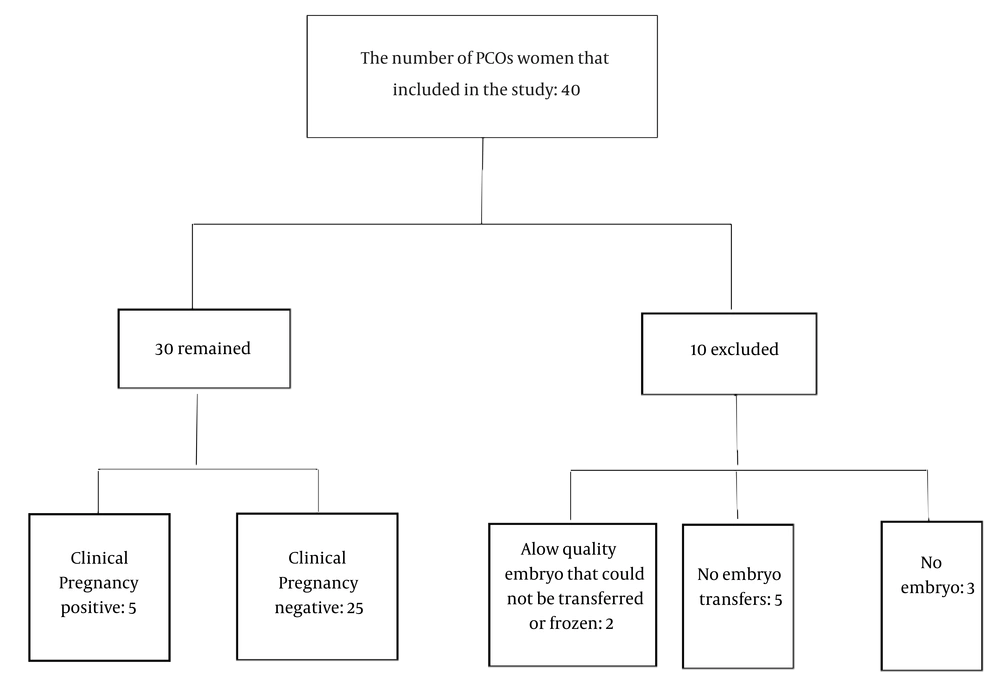
In this prospective cohort study, 40 consenting couples commencing fully stimulated IVF cycles at Mahdiyeh Hospital (Tehran) were enrolled from September 2023 to August 2024. Ten patients were excluded from the study due to factors such as lack of embryo formation, poor-quality embryos for transfer, and no transfer. In total, 30 participants remained eligible and were included in the statistical analysis (Figure 1).
The diagram of study population. It shows the total number of screened individuals, the number meeting the inclusion criteria, exclusions with reasons, and the final number of women with polycystic ovary syndrome (PCOS) enrolled in the analysis.
Figure 1.

The diagram of study population. It shows the total number of screened individuals, the number meeting the inclusion criteria, exclusions with reasons, and the final number of women with polycystic ovary syndrome (PCOS) enrolled in the analysis.

Study inclusion criteria were as follows: Infertile women with PCOS, aged from 18 to 40 years, were enrolled in the study. All participants had negative serological tests for human immunodeficiency virus (HIV), hepatitis B virus (HBV), hepatitis C virus (HCV), and syphilis. All participants were of Iranian nationality. The diagnosis of PCOS was established according to the revised 2003 Rotterdam criteria (10).
The following exclusion criteria were applied: The use of antibiotics, metformin, or probiotics since 3 months before oocyte retrieval, cycle with testicular sperm extraction (TESE), uterine malformations, genital tract infection, renal disease, cardiovascular disease, lung disease, nervous disease, diabetes mellitus, liver disease, patients with Cushing's disease or Addison's disease, cases of excessive obesity, smoking, and alcohol use.
Potential confounding variables, including participant age, Body Mass Index (BMI), and the male partner's semen quality, were systematically controlled for by conducting all procedures in a reputable university embryology laboratory, applying rigorous matching of demographic variables, and enforcing strict inclusion and exclusion criteria throughout the study.
This study was designed as a prospective cohort study, including two groups of women diagnosed with PCOS who underwent in vitro fertilization with ICSI. One group consisted of women whose treatment cycles resulted in clinical pregnancy, while the other included those whose cycles did not. Each participant provided two types of samples: Follicular fluid was obtained from both the left and right ovaries (n = 60), and vaginal swab specimens were also collected (n = 30). The follicular fluid samples were carefully moved under sterile conditions into a clean culture dish to assess the presence of oocytes. After the oocytes were removed, the remaining follicular fluid was transferred into a sterile 15 mL Falcon tube and preserved at -80°C. Vaginal swabs were obtained from the posterior fornix before transvaginal oocyte retrieval, rapidly frozen using liquid nitrogen, and subsequently stored at -80°C until DNA extraction. Also, the number of eggs, maturity of eggs, fertilization rate, cleavage rate, embryo quality (based on the criteria of the document of the Istanbul Assembly in 2011 [Embryology Group of ASRM and ESHRE]), transferred embryo number, husband's semen analysis, infertility duration, cause and type of infertility, and the type of transfer cycle were checked for each patient.
This study was approved by the Medical Ethics Committee of the School of Medicine at Shahid Beheshti University of Medical Science (approval identifier: IR.SBMU.MSP.REC.1402.622).

3.2. DNA Preparation, Quantitative Real time-Polymerase Chain Reaction assays

Total DNA was isolated using a FavorPrepTM Tissue Genomic DNA Extraction Mini Kit (FAVORGEN, TAIWAN) from follicular fluid and vaginal swabs. The follicular fluid sample underwent centrifugation at 12,000 revolutions per minute for a duration of 10 minutes, after which the resulting pellet was collected for DNA extraction. The swab was placed in a 2 mL sterile centrifuge tube, to which 2 mL sterile phosphate-buffered saline (PBS) buffer was added to submerge the swab. After 120 minutes, shaking was performed at room temperature and the PBS solutions were centrifuged at 12,000 rpm for 10 minutes. The sediment was used for DNA extraction. DNA was isolated utilizing the FavorPrepTM Tissue Genomic DNA Extraction Mini Kit, following the manufacturer's protocols. DNA yield was assessed using NanoDrop (BioTech Epoch Microplate Spectrophotometer, United States).

3.3. Real time-Polymerase Chain Reaction assays

All real time-polymerase chain reaction assays (RT-PCR) reactions were performed on a Rotor-Gene Q 5PLEX HRM (QIAGEN, Germany). Reaction mixtures contained 2 µL DNA template, 10 µL 2x SYBR Green real-time PCR master mix (Parstous, Iran), 7 pmol forward and reverse primers (metabion, international AG, Germany) (Table 1), resulting in a final reaction volume of 20 µL.
Table 1.List of Primers Used for Polymerase Chain Reaction Amplification of Selected Microorganisms
Bacteria and PrimersAmplicon Length (bp)
Lactobacillus group341
F: AGCAGTAGGGAATCTTCCA
R: CACCGCTACACATGGAG
Bifidobacterium spp.243
F: TCGCGTCYGGTGTGAAAG
R: CCACATCCAGCRTCCAC
Streptococcus spp.197
F: GTACAGTTGCTTCAGGACGTATC
R: ACGTTCGATTTCATCACGTTG
Staphylococcus spp.118
F: CAGGAGAAGTTAAAGAACAAGAAG
R: GTGAACGAACTAATTGAGATACG
Gardnerellavaginalis320
F: TTACTGGTGTATCACTGTAAGG
R: CCGTCACAGGCTGAACAGT
Mycoplasmahominis129
F: CATGCATGTCGAGCGAGGTT
R: CCATGCGGTTCCATGCGT
Escherichiacoli190
F: CATTGACGTTACCCGCAGAAGAAGC
R: CTCTACGAGACTCAAGCTTGC
Bacteroides group140
F: GGTGTCGGCTTAAGTGCCAT
R: CGGAYGTAAGGGCCGTGC
Prevotella spp.283
F: CACCAAGGCGACGATCA
R: GGATAACGCCYGGACCT
The cycling program for Streptococcus spp., Staphylococcus spp., Gardnerella vaginalis, and Mycoplasma hominis was as follows: Initial denaturation at 95°C for 10 minutes; amplification for 45 cycles of 10 seconds at 95°C, 10 seconds at 57°C, and 50 seconds at 72°C; melting curve analysis for 5 seconds at 95°C, 15 seconds at 65°C and a temperature continuous acquisition up to 95°C; and cooling for 30 seconds at 40°C.
The cycling program for Escherichia coli, Bacteroides group, and Prevotella spp. was as follows: Initial denaturation at 95°C for 5 minutes; amplification for 40 cycles of 15 seconds at 95°C, 30 seconds at 59°C, and 40 seconds at 72°C; melting curve analysis for 5 seconds at 95°C, 15 seconds at 70°C and a temperature continuous acquisition up to 95°C; and cooling for 30 seconds at 40°C.
The cycling program for Bifidobacterium spp. was as follows: Initial denaturation at 95°C for 5 minutes; amplification for 45 cycles of 10 seconds at 95°C, 30 seconds at 58°C, and 30 seconds at 72°C; melting curve analysis for 5 seconds at 95°C, 15 seconds at 75°C and a temperature continuous acquisition up to 95°C; and cooling for 30 seconds at 40°C.
The cycling program for the Lactobacillus group was as follows: Initial denaturation at 95°C for 5 minutes; amplification for 40 cycles of 10 seconds at 95°C, 30 seconds at 58°C, and 30 seconds at 72°C; melting curve analysis for 5 seconds at 95°C, 15 seconds at 65°C and a temperature continuous acquisition up to 95°C; and cooling for 30 seconds at 40°C.
Each experiment used both negative and positive controls to check for contamination and ensure the results were specific. Positive control included E. coli 16S rRNA gene at concentrations of 100, 10, 1, 0.1, and 0.01 ng/µl. Negative controls included all reaction mixtures except DNA templates.
A melting curve analysis was performed after amplification to distinguish the targeted PCR products from nonspecific PCR products. The concentration of microorganisms in each sample was calculated by comparing the crossing point PCR-cycle (Cp) values obtained from the sample with the Cp values of the positive controls.

3.4. Statistical Analysis

GraphPad Prism 10 software was used for statistical analysis of the data. Tables were used to display the frequency distributions of the variables. The Shapiro-Wilk test was employed to assess whether the data conformed to a normal distribution. For datasets exhibiting normality, Welch's t-tests were applied, whereas for those not meeting normality assumptions, the non-parametric Mann-Whitney tests were utilized. Fisher's Exact test was used to assess the cause and type of infertility, the quality of the transferred embryo, and the embryo transfer cycle between the case and control groups. By carefully matching patients, setting strict inclusion and exclusion criteria, and applying specific restrictions, we ensured that confounding variables did not affect the study results. A P-value < 0.05 (95% confidence interval) was considered as statistically significant.

4. Results

Among the 40 women with PCOS who underwent ICSI procedures and participated in the study, 75% (n = 30/40) completed the study. The participants had a mean age of 33.6 ± 5.4 years, and a mean BMI of 26.9 ± 4.7 kg/m², while 40% (n = 12/30) were overweight and 23.3% (7/30) were obese. The median duration of infertility was 8 years (IQR: 5 - 11 years) in the case group and 7 years (IQR: 2.5 - 10.5 years) in the control group. 26.66% (n = 8/30) had secondary infertility. 16.66% (5/30) had infertility with female factor, and 83.33% had male and female factor. There was no statistically significant difference in variables such as age, body mass index, duration of infertility, type of infertility and its cause, sperm count and morphology and motility of the partner's semen, total number of eggs, number of mature and immature eggs and their rate, fertilization rate and cleavage rate, embryo quality, day of transfer, number of transferred embryos and type of transfer cycle between the case and control groups (Table 2).
Table 2.Clinical Characteristics Information of the Two Groups of Study a
VariablesPCOs Women with Negative Pregnancy (N = 25)PCOs Women with Positive Pregnancy (N = 5)TestP-Value
Age (y) Mann-Whitney test0.0620
Median 3632
IQR (Q3 - Q1)8.511
BMI Mann-Whitney test0.7375
Median28.1028.72
IQR (Q3 - Q1)6.6311.15
Infertility duration (y) Mann-Whitney test0.5609
Median 87
IQR (Q3-Q1)68
Type of infertilityFisher's exact test0.2868
Primary 17 (68)5 (100)
Secondary8 (32)0
Cause of infertilityFisher's exact test0.1833
Female factor3 (12)2 (40)
Male and Female factor22 (88)3 (60)
Sperm concentration (million)62.64 ± 41.4375 ± 42.13Welch's T test0.5714
Normal sperm morphology (%)Mann-Whitney test0.0710
Median 23
IQR (Q3 - Q1)2.52
Total motility (%)Mann-Whitney test0.2678
Median 6575
IQR (Q3 - Q1)4522.5
Total oocyte countMann-Whitney test0.1917
Median 1218
IQR (Q3 - Q1)11.511
MII oocyte count Mann-Whitney test0.1825
Median 916
IQR (Q3 - Q1)1211.5
MII oocyte rate (%)Mann-Whitney test0.0587
sMedian 73.3386.36
IQR (Q3 - Q1)23.698.61
Immature oocyte (GV+MI) count Mann-Whitney test0.0936
Median 33
IQR (Q3 - Q1)11
Immature oocyte (GV+MI) rate (%)Mann-Whitney test0.0587
Median 26.6713.64
IQR (Q3 - Q1)23.698.61
2PN count (fertilized oocyte count)9.72 ± 5.74814.80 ± 6.573Welch's T test0.1651
Fertilization rate80.29 ± 14.7186.80 ± 10.94Welch's T test0.2906
Embryo qualityFisher's exact test0.6221
Good (A) 11 (44)1 (20)
Good+Fair (A+B)14 (56)4 (80)
Cleavage rate (%)Mann-Whitney test0.9717
Median 100100
IQR (Q3 - Q1)12.516.78
Day of embryo transferMann-Whitney test0.8593
Median 43
IQR (Q3 - Q1)11.5
Transferred embryo numberMann-Whitney test0.3320
Median 33
IQR (Q3 - Q1)10.5
Embryo transfer cycle Fisher's exact test0.2868
Fresh 8 (32)0 (0)
FET17 (68)5 (100)

Abbreviations: PCOS, polycystic ovary syndrome; BMI, Body Mass Index.

a Values are expressed as mean ± SD or No. (%) unless otherwise indicated.

The vaginal and follicular fluid samples of 30 female participants were included in the study. Here, quantitative RT-PCR was used to compare the microbial composition of patients. Based on pregnancy outcomes following embryo transfer, infertile women with PCOS were categorized into two subgroups: Those who achieved pregnancy and those who did not. We examined the microbial composition of both the vaginal and follicular fluid samples from these groups. Out of 30 infertile PCOS patients, 8 exhibited a positive biochemical pregnancy, while 22 did not. In terms of clinical pregnancy, 5 patients were positive, and 25 were negative. Detailed clinical characteristics for these groups are summarized in Table 2. Table 3 presents comparative abundances at both the species and genus levels across various sampling sites within each group.
Table 3.Bacterial Data from Vaginal Swab and Follicular Fluid (Right and Left) a
Bacteria and CategoriesCP Negative (n = 25)CP Positive (n = 5)P-Value
Median (95% CI of Median)IQR(Q3 - Q1)Median (95% CI of Median)IQR (Q3 - Q1)
Gardnerella vaginalis (16S rRNA)
VS9.766 (1.673 - 1189)5217.171845.18 (3.548 - 454.6)256.9590.7462
RFF0.09099 (0.01433 - 0.3879)0.6086330.01703 (0.007577 - 0.1849)0.135050.6268
LFF0.09057 (0.01791 - 0.5334)0.5477720.01203 (0.0005920 - 0.1479)0.1384650.3005
Mycoplasma hominis (16S rRNA)
VS0.2740 (0.07620 - 39.81)98.234620.05222 (0.01764 - 12.70)9.314880.2513
RFF0.1891 (0.05595 - 0.7475)0.899410.05390 (0.01876 - 6.040)3.092060.7056
LFF0.1672 (0.08361 - 1.035)1.669010.04676 (0.004425 - 0.2654)0.1510810.0225 b
Staphylococcus species (rpoB)
VS4.160 (3.120 - 11.46)14.6032.924 (2.041 - 3372)1731.710.8716
RFF0.1949 (0.08539 - 1.948)2.451010.1191 (0.05549 - 145.2)72.593650.5518
LFF0.1357 (0.06528 - 1.578)1.703540.04455 (0.02109 - 131.1)65.612280.5518
Streptococcus species (tuf)
VS25.98 (2.950 - 37.57)109.98612.31 (0.4403 - 230.8)142.304> 0.9999
RFF0.2058 (0.09963 - 0.3861)0.348020.04379 (0.02552 - 0.6884)0.454180.4812
LFF c0.2808 (0.2017 - 0.3598)0.19150.1468 (0.01487 - 0.2788)0.10630.1438
Escherichia coli (16S rRNA)
VS756.6 (440.7 - 1234)1334.4648.7 (140.8 - 38695)23091.80.9570
RFF2244 (550.8 - 3457)3493.22743 (2599 - 4636)12370.3553
LFF1838 (533.7 - 3032)3271.52917 (1740 - 5088)22700.2947
Lactobacillus group (16S rRNA)
VS28692 (5399 - 53888)6275918960 (6510 - 312528)1778200.4478
RFF11.84 (5.287 - 19.44)18.4834.343 (1.015 - 21.78)20.2510.4762
LFF9.571 (2.315 - 14.75)19.4357.204 (0.1654 - 35.87)19.3160.7462
Bifidobacterium (16S rDNA)
VS13.47 (3.697 - 1348)4959.07636.14 (1.885 - 494.1)305.279> 0.9999
RFF0.7436 (0.1522 - 10.12)1626.86961.807 (0.8157 - 21.15)10.6590.5888
LFF1.275 (0.4168 - 6.551)1038.73151.637 (0.4229 - 9.279)5.32250.9142
Bacteroides group(16S rDNA)
VS53.28 (9.206 - 306.7)334.2758.80 (7.401 - 1074)575.1920.9570
RFF17.50 (4.009 - 28.73)31.30310.93 (1.302 - 90.57)67.995> 0.9999
LFF17.59 (1.644 - 32.26)37.7558.446 (0.9389 - 48.34)30.9030.9570
Prevotella species (16S rRNA)
VS6.826 (2.136 - 96.72)179.3126.433 (0.9851 - 234.5)145.8450.9142
RFF3.868 (2.995 - 19.96)35.7713.889 (1.402 - 9.950)4.9040.6268
LFF4.772 (3.584 - 9.520)30.5826.186 (3.755 - 22.05)9.3690.7875

Abbreviations: CP, clinical pregnancy; VS, vaginal swab; RFF, right follicular fluid; LFF, left follicular fluid.

a The number of bacteria is reported based on colony count formation in microliters (CFU/λ).

b Statistically significant difference between the two groups.

c Due to the normal distribution data of the Streptococcus Species in the left follicular fluid, the mean and standard deviation are reported.

The only statistically significant difference in the bacterial examination of the two groups was in M. hominis in the left follicular fluid, which was higher in the negative clinical pregnancy group than in the control group (P-value: 0.0225). For the remaining bacterial species investigated, including Lactobacillus group, Bifidobacterium spp., Streptococcus spp., Staphylococcus spp., G. vaginalis, E. coli, Bacteroides group, and Prevotella spp., no statistically significant differences were found between the study groups (PCOS patients undergoing ICSI with negative clinical pregnancy versus controls). The lack of significant association suggests that, within the scope of this study, these specific microorganisms do not appear to be differentially represented in the follicular fluid or vaginal microbiota of the cohort examined, and thus their potential role in the reproductive outcomes measured remains inconclusive (Table 3).

5. Discussion

This study conducted a comprehensive analysis of the microbial communities within both the vaginal tract and the ovarian follicular fluid of infertile patients presenting with PCOS. Our findings confirm that human follicular fluid is not invariably sterile, as it harbors detectable microorganisms. Furthermore, we compared the distinct microbiota profiles between the vaginal environment and the follicular fluid in relation to various ICSI outcomes. This study identified a unique follicular fluid microflora signature, in which an increase in M. hominis appears to correlate with female infertility in the PCOS cohort. Samples of bacteria were isolated separately from the right and left follicular fluid collected via transvaginal oocyte retrieval during ICSI cycles. Specifically, the presence of M. hominis in the left follicular fluid was associated with a statistically significant difference in clinical pregnancy outcome.
Other studies have also reported the presence of microorganisms in human follicular fluid (24, 26, 27). Several of the microorganisms detected in this study, such as Streptococcus, Staphylococcus, and Lactobacillus species, were also reported by Pelzer et al. and Usman et al. (24, 27). The microbiome of the female reproductive tract is crucial for maintaining health and preventing dysbiosis, and it may also influence successful fertilization and pregnancy outcomes (28). Bacteria other than Lactobacillus species such as E. coli, Streptococcus spp., other Enterobacteriaceae, Staphylococcus spp., Haemophilus, and mixed bacterial communities found in the cervicovaginal area have been linked to fewer successful IVF pregnancies and higher rates of early miscarriage (29-31).
This study focuses on investigating and comparing the microbiota in these environments of infertile women with PCOS, specifically analyzing differences between those who achieve pregnancy and those who do not.
One of the major strengths of this study is its novel investigation of the microbiota of ovarian follicular fluid alongside vaginal swabs in Iranian women with PCOS undergoing ICSI. By simultaneously assessing microbial communities at both the upper and lower sites of the female reproductive tract, this study provides a more comprehensive understanding of the reproductive microbiome in this specific population. Furthermore, the study was conducted at a well-established and highly experienced IVF center, ensuring standardized clinical protocols, high-quality laboratory procedures, and optimal sample handling throughout all stages of treatment. This substantially reduced the likelihood of technical or facility-related biases that could affect the outcomes. In addition, the use of clearly defined inclusion and exclusion criteria allowed for effective control of potential confounding variables, such as underlying infections, recent antibiotic use, and systemic conditions, thereby enhancing the internal validity and reliability of the findings. Collectively, these strengths increase the robustness of the study results and support their relevance for future research and clinical applications in the context of assisted reproductive technologies in women with PCOS.
Several limitations of this study should be acknowledged. First, the relatively small sample size reduces statistical power and may limit the detection of subtle, yet potentially clinically relevant, differences between groups. This constraint also limits the ability to perform robust subgroup analyses or adjust for potential confounding variables. Second, the exclusive use of quantitative polymerase chain reaction (qPCR) as the analytical method represents an important methodological limitation. While qPCR is a sensitive and targeted approach for the detection of predefined microorganisms, it inherently restricts analysis to selected bacterial species and does not provide a comprehensive profile of the microbial community. Consequently, the complete exclusion of non-target and low-abundance taxa may have led to an underestimation of overall microbial diversity and overlooked potentially relevant microbial interactions. In addition, the lack of access to advanced next-generation sequencing (NGS) based techniques, including shotgun metagenomics, metabolomics, and metatranscriptomics, further limits the scope of microbial characterization. These approaches could have provided insights into functional activity, metabolic pathways, and host-microbe interactions, which are not captured by DNA-based targeted assays alone. Potential sources of bias should also be considered. Selection bias may have been introduced due to the restricted study population, which comprised only Iranian women with PCOS undergoing assisted reproductive treatment. Moreover, unmeasured variables such as diet, sexual behavior, hormonal fluctuations, and lifestyle factors could have influenced the composition of the reproductive tract microbiota and were not fully accounted for in the analysis.
Despite these issues, the study still allows comparisons between vaginal and follicular fluid communities assessed by different methods. The results contribute valuable insights into the impact of follicular fluid and vaginal microbiota on IVF/ICSI outcomes within our environment, a gap in current knowledge. These findings are particularly relevant for gynecologists and fertility specialists, especially during pre-treatment counseling. However, it underscores the urgent need for large, multicenter studies to further elucidate the role of microorganisms in fertility treatments, building on the foundation this study provides.
Future research may consider using other techniques (such as metabolomics, metaproteomics, and whole-genome sequencing) for more comprehensive microbial identification. Additionally, differentiating follicular fluids from the left and right ovaries and stratifying analyses by specific microorganisms could yield more profound insights into the associations. Special attention should be given to women undergoing IVF, particularly those with previous failed cycles or unexplained infertility. Microbial screening of vaginal swabs prior to IVF/ICSI, along with assessment of follicular fluid collected during transvaginal oocyte retrieval, may help identify abnormal flora and potentially improve treatment outcomes.

5.1. Conclusions

This study compared the reproductive microbiota of 30 women with PCOS undergoing ICSI between those who did or did not achieve pregnancy. The primary finding was a significantly higher level of M. hominis in left-ovary follicular fluid in the non-pregnant group, while other bacterial targets and standard clinical parameters were similar. Future multicenter studies with larger cohorts and metagenomic sequencing are needed to confirm this association and to explore the broader taxonomic and functional landscape of the follicular microenvironment in relation to IVF outcomes.

Acknowledgments

Footnotes

References


Crossmark
Crossmark
Checking
Share on
Cited by
Metrics

Purchasing Reprints

  • Copyright Clearance Center (CCC) handles bulk orders for article reprints for Brieflands. To place an order for reprints, please click here (   https://www.copyright.com/landing/reprintsinquiryform/ ). Clicking this link will bring you to a CCC request form where you can provide the details of your order. Once complete, please click the ‘Submit Request’ button and CCC’s Reprints Services team will generate a quote for your review.
Search Relations

Author(s):

Related Articles