


1. Background
In Iran, approximately 46,000 people are living with human immunodeficiency virus (HIV), based on estimates from the Joint United Nations Programme on HIV/AIDS and the World Health Organization, as well as unofficial statistics (1-3). Among adults aged 15 years and older, nearly 30,000 men and 15,000 women are living with HIV, indicating a predominance of male cases in the HIV epidemic. These patients are at a significantly higher risk of infection with various microorganisms, including filamentous bacteria.
Filamentous bacteria belong to the phylum Actinomycetota, formerly Actinobacteria, which includes several families of medical and ecological importance. The most ecologically and medically important genera within these families are Actinomyces, Bifidobacterium, Nocardia, Nocardiopsis, Propionibacterium, and Streptomyces. These gram-positive, anaerobic or microaerophilic, non-acid-fast, nonmotile, slow-growing bacteria are sometimes reported to cause opportunistic endogenous infections in humans and animals (4, 5). For example, Actinomyces spp. can cause actinomycosis, granulomatosis, multiple abscesses, and tissue fibrosis. Species such as A. meyeri, A. naeslundii, and A. georgiae are associated with periodontal diseases (5). Although Streptomyces produces a wide range of secondary metabolites, including nearly 70% of known antibiotics, it can also cause opportunistic infections (6). Nocardia is another genus within the Actinomycetes and can cause nocardiosis, pneumonia, encephalitis, cellulitis, and abscesses in various organs (7). Medically, people with dysfunctional immune systems, such as patients with cancer, transplant recipients, immunosuppressant drug users, and HIV-infected patients, are at higher risk of acquiring infections with Nocardia spp. than others (8).
The presence of Actinomycetes in the oral cavity of HIV-infected patients can directly or indirectly cause oral and dental infections (9). For example, these bacteria can facilitate the adhesion of anaerobic gram-negative bacteria, such as Porphyromonas gingivalis, to dental plaque (10). In HIV-infected patients, oral infections caused by Actinomycetes are routinely treated with beta-lactams, cyclines, macrolides, quinolones, and sulfonamides (11-15). The prevalence of Actinomycetes in healthy people and HIV-infected patients typically ranges from 1.5% to 3% and from 3% to 6%, respectively.
2. Objectives
Given the importance of oral infections in HIV-infected patients, the primary aims of the present study were to isolate Actinomycetes from the oral cavity of HIV-infected participants, characterize the isolates phenotypically and genotypically, and assess their antimicrobial susceptibility (AMS).
3. Methods
3.1. Ethical Considerations
The present study was conducted in full accordance with the Declaration of Helsinki. All participants were informed about the study objectives and benefits and subsequently provided written informed consent. All participants’ personal information was archived as strictly confidential. The study was approved by the Ethics Committee of Islamic Azad Medical Sciences University (IR.IAU.PS.REC.1402.385).
3.2. Sampling
This cross-sectional study was conducted from March 2022 to August 2024 and included 200 oral cavity samples from HIV-infected patients referred to the Southern Health Center of Tehran University of Medical Sciences, Tehran, Iran. Eligible participants were required to have no history of antimicrobial, antifungal, or mouthwash use during the two weeks before sampling and to have a CD4 count of 200 cells or more per microliter of blood. Participants were also required not to have severe infections or systemic diseases such as diabetes or cancer. Buccal swabs, including samples from periodontal and periapical lesions, were collected from participants into sterile tubes. The tubes were appropriately coded and sealed. Samples were transported under cold conditions to the Filamentous Bacteria Laboratory, Department of Pathobiology, School of Public Health, Tehran University of Medical Sciences, Tehran, Iran.
3.3. Materials
In the present study, all culture media and biochemicals were purchased from Merck, Germany, and were of analytical grade.
3.4. Culture
To facilitate bacterial growth, incubation was performed at 37°C for 7 days on blood agar (BA) (1, 16). In addition, BA and type-A gas packs were used for the growth of anaerobic bacteria. Differential culture media, including brain-heart infusion agar, were also used for bacterial cultivation. After primary isolation on BA, samples were subcultured on paraffin agar media and incubated for 14 days (17). Morphological characteristics of Streptomyces spp. included dry, powdery, and sometimes gray colonies, whereas Nocardia spp. formed wet colonies with prominent borders. For further phenotypic identification, samples were stained using a Gram stain (1).
3.5. Biochemical Assessment
To identify Nocardia spp., suspected colonies were cultured on agar media, and biochemical assays, including tyrosine degradation and casein, xanthine, and gelatin hydrolysis assays, were performed (17). Actinomyces spp. were identified using biochemical assays such as catalase, nitrate reduction, indole production, and gelatin, urea, and cellobiose hydrolysis assays (2). For Streptomyces spp., identification was based on casein, xanthine, and urea hydrolysis assays, as well as various carbohydrate fermentation assays (18).
3.6. Scanning Electron Microscopy
To confirm the isolates and appropriately differentiate Actinomycetes from fungi, three isolates of each genus were examined using a scanning electron microscope (FEI Quanta 200 ESEM, USA) at 2500× magnification.
3.7. Antimicrobial Susceptibility Test
To investigate the AMS patterns of the isolates, antimicrobial susceptibility testing (AST) was performed using the Kirby-Bauer method, also known as the agar or disk diffusion method. The turbidity of the bacterial suspension was adjusted to the 0.5 McFarland standard using broth media or sterile normal saline (19). A sterile cotton swab dipped in the bacterial suspension was used to inoculate Mueller-Hinton agar plates by lawn culture, followed by incubation at 35°C for 18 - 24 hours. In general, AST of the isolates was performed against amikacin (30 μg), ciprofloxacin (50 μg), amoxicillin (25 μg), clavulanic acid (20 μg), linezolid (30 μg), penicillin G (10 μg), tetracycline (30 μg), clindamycin (30 μg), erythromycin (15 μg), nitrofurantoin (25 μg), rifampin (5 μg), nalidixic acid (30 μg), and gentamicin (10 μg) (Padtan Teb, Iran) (20, 21).
3.8. Genotypic Identification
To accurately confirm Actinomycetes, molecular methods, including polymerase chain reaction (PCR) and sequencing, were used. First, genomic DNA (gDNA) was extracted and used for PCR, and the PCR products were then sequenced. Actinomycetes-specific primers were used for molecular identification of the bacteria (16). In this study, the phenol-chloroform method and commercial extraction kits were used to extract gDNA from Actinomycetes isolates (SinaClon, Iran). The quality and concentration of the extracted gDNA samples were assessed using a NanoDrop spectrophotometer (Thermo Fisher Scientific, USA) by measuring absorbance at 260/280 nm. Universal primers for 16S rRNA loci were selected based on literature reviews, and PCR was performed using a thermal cycler (Bio-Rad, USA). The PCR cycling program included 35 cycles of denaturation at 94°C for 1 minute, annealing at 60°C for 1 minute, and elongation at 72°C for 1 minute. To analyze the PCR products, electrophoresis was performed on 1.5% agarose gels with ethidium bromide staining, and the results were examined under ultraviolet light using a gel documentation apparatus (UV transilluminator). The PCR products were then sequenced using the Sanger sequencing method (SinaClon, Iran). After editing the raw data using BioEdit software, sequences were compared with those in the GenBank database (https://www.ncbi.nlm.nih.gov/genbank) for possible similarities.
The amplified gene fragments corresponded to the 16S rRNA gene. Raw nucleotide sequences were edited using bioinformatics software and subsequently used to construct phylogenetic trees. Homologous 16S rRNA gene sequences from closely related strains, including six Nocardia and five Streptomyces strains from other geographical regions, were retrieved using the BLAST online tool (https://blast.ncbi.nlm.nih.gov/Blast.cgi). The genomic sequences of Actinomycetes were used for phylogenetic analysis using trial versions of Molecular Evolutionary Genetics Analysis (MEGA7) and CLC Genomics Workbench (Qiagen, Germany).
3.9. Statistical Analysis
Data were statistically analyzed using the Statistical Package for Social Sciences software for Windows (IBM, USA) and the chi-square test. P values of 0.05 or less were considered significant.
4. Results
In this study, phenotypic and genotypic methods were used to isolate and characterize Actinomycetes from buccal samples collected from 200 HIV-infected participants referred to the Southern Health Center of Tehran University of Medical Sciences, Tehran, Iran. Among the HIV-infected participants, most were male, and all were middle-aged. Actinomycetes were isolated more frequently from male participants. However, no statistically significant association was observed between bacterial isolation and the participants’ sex (P = 0.9772) or age (P = 0.6434). The study cohort comprised 47.1% males, with a mean age of 45.2 years (SD = 15.8). The cohort predominantly consisted of individuals with low (40.6%) and middle-class (47.8%) incomes. Although bacterial isolation was most common in the middle-income group (52.1% of cases), the association between income level and bacterial isolation was not statistically significant (P = 0.527). All HIV-positive participants (n = 18) were receiving antiviral treatment. Most of these participants (83.3%) reported no significant side effects; however, this proportion was not statistically significant (P = 0.344).
An assessment of oral hygiene revealed that most participants had poor hygiene (78.4%), followed by fair (15.2%) and good (6.4%) hygiene. Although Actinomycetes isolation occurred most commonly in the poor-hygiene group, this association was not statistically significant (P = 0.071). Similarly, there was no significant association with regular dental visits (regular visits: 34.1%; P = 0.484). Of 200 samples collected from HIV-infected participants, 19 Actinomycetes strains were isolated (isolation rate: 9.5%). The taxonomic distribution was as follows: Streptomyces (six strains, 31.6%), Nocardia (six strains, 31.6%; all N. farcinica), Nocardiopsis (six strains, 31.6%; including two N. alba), and Saccharopolyspora (one strain, 5.3%). All primary results were subsequently verified using phenotypic methods (scanning electron microscopy) and molecular methods (PCR and sequencing) (Figures 1 and 2). The nucleotide sequences were submitted to GenBank after primary editing (Table 1).
Table 1.GenBank Accession Numbers of the Isolated Actinomycetes in the Current Study
| Isolate | Accession No. |
|---|---|
| Nocardiopsis alba strain 8a | PQ634397.1 |
| Nocardiopsis flavescens | PQ633385.1 |
| Nocardiopsis sp. | PQ634038.1 |
| Streptomyces sp. | PQ634381.1 |
| Nocardiopsis sp. | PQ600881.1 |
| Nocardiopsis sp. | PQ600897.1 |
| Nocardiopsis sp. | PQ600898.1 |
| Saccharopolyspora sp. | PQ600917.1 |
| Nocardiopsis sp. | PQ601059.1 |
| Nocardiopsis alba | PQ601058.1 |
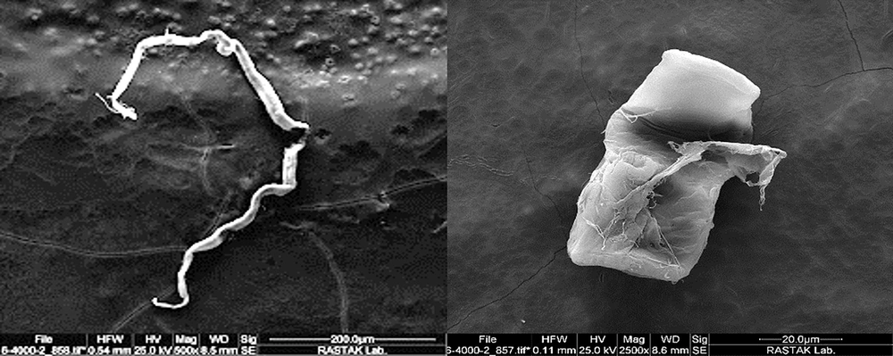
Figure 1.
Phenotypes of Actinomycetes from HIV-infected participants using scanning electron microscopy at 2500×. Left, Streptomyces sp.; right, Nocardia sp.
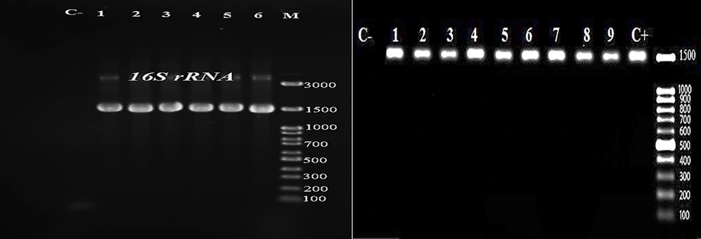
Figure 2.
Electrophoresis of bacterial gDNA on 1.5% agarose gels using ethidium bromide staining. Right) C-, negative control; C+, positive control; lanes 1 - 9, 1500-bp 16S rRNA gene PCR products; 100-bp DNA ladder; and left) C-, negative control; lanes 1 - 6, 1500-bp 16S rRNA gene PCR products; M, 1-kb DNA ladder.
AST results for Actinomycetes isolated from HIV-infected participants showed that Streptomyces spp. and N. farcinica were multidrug-resistant (MDR) to penicillin G, amoxicillin and clavulanic acid, tetracycline, erythromycin, nalidixic acid, rifampin, and clindamycin, but were susceptible to other antimicrobials. Using the BLAST online tool, other filamentous bacterial species were identified with 99% similarity to the isolated bacteria (Figure 3). The isolates in this study were genetically similar to those from China, India, Indonesia, and the USA.
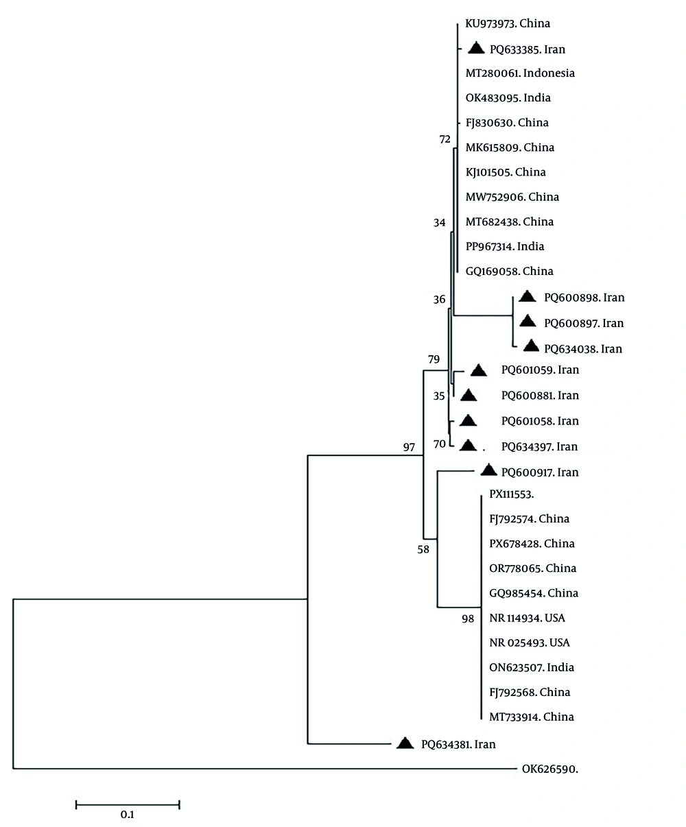
Figure 3.
Phylogenetic tree of the Streptomyces and Nocardia species isolated in the current study using the neighbor-joining method and bootstrap replicates of 1000.
Table 2.Summary of Actinomycetes Isolation and Antibiotic Resistance Profiles in HIV-Infected Patients from Other Studies
| Study (y) | Study Subject | Organism | Antimicrobial | Major Finding |
|---|---|---|---|---|
| Wolff et al. (2022) (35) | Oral clinical isolates | Actinomyces spp. (n = 100) | Penicillin, ampicillin-sulbactam, meropenem, clindamycin, moxifloxacin, linezolid, tigecycline | Low resistance to β-lactams; high resistance to moxifloxacin and daptomycin; good susceptibility to linezolid |
| Smith et al. (2005) (36) | Clinical isolates | Actinomyces spp. (n = 87) | Penicillin, amoxicillin, ceftriaxone, tetracycline, clindamycin, ciprofloxacin | High susceptibility to β-lactams; resistance to ciprofloxacin and macrolides |
| Steininger and Willinger (2016) (37) | Retrospective clinical isolates | Pathogenic Actinomyces spp. (n = 392) | β-lactams, carbapenems, tetracyclines, metronidazole | Susceptible to β-lactams and carbapenems; intrinsic resistance to metronidazole |
| Brooks et al. (2022) (38) | Systematic review of oral microbiome studies | Oral microbiota (ARGs) | Multiple antibiotic classes | Widespread antibiotic resistance genes detected in the oral cavity |
| Dollas et al. (2025) (39) | Saliva samples from adults | Oral bacterial flora | Multiple antibiotic classes | High prevalence of antimicrobial resistance in salivary isolates |
5. Discussion
In the present study, Actinomycetes were isolated from 19 of 200 HIV-infected participants (9.5%). Nocardia and Streptomyces were the most common genera, with prevalence rates of 63.2% and 36.8%, respectively. Similarly, Steinbrink et al. showed that the frequency of nocardiosis in HIV-infected patients was 1.85% (22). Fathi et al. reported that Candida spp. were isolated from 47.2% of HIV-infected patients (23). A study by Abbasian et al. highlighted that N. farcinica infections represented an important opportunistic infection in HIV-infected patients (24). Vujkovic-Cvijin et al. reported that Proteobacteria, particularly members of Enterobacteriaceae, were among the most common bacterial groups in infected patients (25).
In a study by Larijanian et al., two cases of A. naeslundii, one case of A. israelii, one case of A. viscosus, three cases of N. asteroides, and one case of N. brasiliensis were reported (4). In a 6-month study by Eshraghi et al. in 2003 on 100 patients with periodontal infections, three positive samples of A. viscosus and two samples of A. naeslundii, with symptoms of gingivitis and periodontitis, were reported (26). In a study by Borssen et al., A. naeslundii and A. israelii were isolated from 72 abscess samples collected from the roots of oral canals (27). Another study by Khatibi et al. in 2019 on the pathogenicity of Actinomycetes, which caused stomatitis in 24% - 60% of denture wearers, showed that the causative agents included C. albicans and filamentous bacteria such as Actinomyces spp. The results showed that Actinomycetes were isolated from five of the 15 control samples and 11 of the 15 patient samples (28). In a study by Abtahi et al. in 2003 in Arak City, Iran, on the prevalence of nocardiosis in patients with pulmonary infections, nocardiosis was diagnosed in 4.32% of patients (29). In a study by Hashemzadeh et al., PCR and sequencing results showed that 27 samples were positive for nocardiosis, including isolates of N. nova, N. farcinica, N. cyriacigeorgica, and N. asteroides (30). A study by Fatahi Bafghi indicated that the epidemiology of the disease was rapidly expanding globally (31). Findings showed that most oral microflora in these patients consisted of filamentous bacteria, causing destruction of gum tissue integrity and pathological changes. In a study by Kenga et al. on 808 HIV-infected children, infections with Staphylococcus aureus, Salmonella spp., Escherichia coli, and Klebsiella spp. were reported (32).
In the present study, AST was performed on 19 Actinomycetes isolates, including Nocardia and Streptomyces spp., against 13 antimicrobial agents using the Kirby-Bauer method. The isolates generally showed resistance to penicillin G, amoxicillin and clavulanic acid, erythromycin, tetracycline, rifampin, nalidixic acid, and clindamycin, but were susceptible to gentamicin, ciprofloxacin, amikacin, linezolid, trimethoprim-sulfamethoxazole, and nitrofurantoin. A comparison with the findings of a study by Abbasian et al. revealed consistent susceptibility to trimethoprim-sulfamethoxazole and amikacin in the two studies (24). In a study by Osamu et al. on clinical samples, A. israelii was resistant to ofloxacin and sodium fluoride compared with other Actinomyces strains. Naturally, Actinomyces spp. are susceptible to penicillin, cephalosporins, clindamycin, carbapenem, and tetracycline; however, they are exceptionally resistant to penicillin (33). Salehipour et al. reported that, among 31 isolates of N. asteroides, all were fully susceptible to trimethoprim-sulfamethoxazole and linezolid and moderately susceptible to amoxicillin and clavulanic acid, cefepime, ceftriaxone, ciprofloxacin, imipenem, moxifloxacin, and tobramycin (34). A summary of other similar studies is provided in Table 2 (35-39).
In a study by Ezeonwumelu et al. in England, bacteria were isolated from the oral cavities of HIV-infected patients. Their results indicated that trimethoprim-sulfamethoxazole had the weakest inhibitory effect against S. aureus and the strongest inhibitory effect against E. coli ATCC 25922 and Pseudomonas aeruginosa ATCC 27853 (40).
In the present study, PCR amplification of the 16S rRNA gene enabled accurate species identification of the Actinomycetes. Nucleotide sequences showed 99% similarity with reference sequences in GenBank for Streptomyces and Nocardia isolates, indicating close phylogenetic relationships. Phylogenetic analysis of the isolates, including N. farcinica and Streptomyces sp., revealed close relationships with strains from neighboring and distant countries. The rooted phylogenetic tree illustrated evolutionary relationships among various Streptomyces and N. farcinica strains, allowing inference of their divergence from a common ancestor. The N. flavescens (PQ633385.1) identified in the present study clustered closely with strains isolated from China, India, and Indonesia. Saccharopolyspora sp. (PQ600917.1) was genetically close to bacteria isolated from China, the United States, and India. Seven bacterial strains, including Nocardiopsis sp. (PQ600898.1), Nocardiopsis sp. (PQ600897.1), Nocardiopsis sp. (PQ634038.1), Nocardiopsis sp. (PQ601059.1), Nocardiopsis sp. (PQ600881.1), Nocardiopsis sp. (PQ634397.1), and N. alba (PQ601058.1), isolated in this study were less similar to filamentous bacteria isolated from China, India, and Indonesia. Furthermore, a bacterial strain of Streptomyces sp. (PQ634381.1) isolated in this study showed no similarity to filamentous bacteria isolated from other countries.
Overall, the reported similarities may be attributable to developed transportation networks and frequent travel between geographical regions for vocational, educational, occupational, official, and commercial purposes. However, infections transmitted through food, devices, and especially prosthetics cannot be dismissed. This molecular identification approach was consistent with modern standards, as highlighted by Kim et al., who described molecular methods as the gold standard for bacterial identification (41).
5.1. Study Limitations
This study had a few limitations that should be addressed. First, some potentially important confounding variables were not investigated. Factors such as individual oral hygiene practices and smoking status may affect the oral microbial composition and antimicrobial resistance patterns observed in HIV-positive patients. Second, the prices of laboratory materials increased dramatically during the study. Third, variability in personal oral health behaviors and access to dental care among HIV-positive participants might have contributed to inter-individual differences that were not fully assessed in the analysis. Fourth, citations were limited because of the limited number of studies on oral AMS schemes among HIV-positive patients in Iran and other regional countries.
5.2. Conclusions
This study revealed that Actinomycetes, primarily Nocardia and Streptomyces spp., were highly prevalent in Iranian HIV-infected patients. The current findings also revealed that most of these isolates were MDR isolates. Accurate molecular characterization of Actinomycetes and assessment of their AMS profiles can significantly improve the targeted treatment of Actinomycete-associated oral infections.
