Strain-Dependent Apoptotic Gene Expression in Influenza-Infected Lung Epithelial Cells

Author(s):
Arash LetafatiArash LetafatiArash Letafati ORCID1, Simin AbbasiSimin Abbasi1, Talat Mokhtari AzadTalat Mokhtari Azad1, Sevrin ZadheidarSevrin Zadheidar1, Jila YavarianJila Yavarian1, 2,*
1Department of Virology, School of Public Health, Tehran University of Medical Sciences, Tehran, Iran
2Research Center for Antibiotic Stewardship and Antimicrobial Resistance, Tehran University of Medical Sciences, Tehran, Iran

Archives of Clinical Infectious Diseases:Vol. 21, issue 1; e171057
Published online:Feb 28, 2026
Article type:Research Article
Received:Jan 14, 2026
Accepted:Feb 15, 2026
How to Cite:Letafati A, Abbasi S, Mokhtari Azad T, Zadheidar S, Yavarian J. Strain-Dependent Apoptotic Gene Expression in Influenza-Infected Lung Epithelial Cells. Arch Clin Infect Dis. 2026;21(1):e171057. doi: https://doi.org/10.5812/archcid-171057

Abstract

Background:

Influenza viruses induce multiple programmed cell death pathways in respiratory epithelial cells. These pathways are associated with host antiviral defense but may also contribute to tissue damage through inflammation and increased disease severity, thereby functioning as a double-edged sword. Apoptosis is a major cell death pathway that regulates the fate of infected cells following viral infection.

Objectives:

This study evaluated the mRNA expression of BAX, BAK1, and Bcl-2, three key apoptosis-regulating genes, in A549 lung epithelial cells infected with influenza A/H1N1, A/H3N2, and influenza B viruses.

Methods:

A549 cells were cultured and inoculated with 100 TCID50 of influenza A/H1N1, A/H3N2, and influenza B viruses for 48 hours. Results were compared with those for uninfected control cells. At 48 hours post-infection, RNA extraction (RNJIA, ROJE) and cDNA synthesis were performed, and the mRNA expression levels of BAX, BAK1, and Bcl-2 were assessed using SYBR Green reverse transcription quantitative PCR. All analyses were based on log fold change (logFC). Differential gene expression versus control was evaluated using the Wilcoxon signed-rank test and a one-sample t test, whereas time-dependent changes within each virus group were assessed using the Kruskal-Wallis test and one-way analysis of variance, followed by pairwise post-hoc tests with Benjamini-Hochberg correction.

Results:

At 48 hours post-infection, BAX expression was significantly upregulated in A/H1N1-infected cells (log fold change = 2.85, P = 0.031). Although A/H3N2 also upregulated BAX expression (log fold change = 2.35, P = 0.061), this change was not significant. Minor downregulation of BAX was observed in influenza B-infected cells (log fold change = -0.26, P = 0.463). BAK1 expression was significantly upregulated in A/H3N2-infected cells (log fold change = 6.20, P = 0.019). A/H1N1-infected cells also showed BAK1 upregulation (log fold change = 2.10, P = 0.431), although this change was not significant, whereas influenza B-infected cells showed significant BAK1 upregulation (log fold change = 6.21, P = 0.025). Bcl-2 was significantly downregulated in all three infected groups (A/H1N1: log fold change = -6.05, P = 0.040; A/H3N2: log fold change = -4.02, P = 0.002; influenza B: log fold change = -0.70, P = 0.032).

Conclusions:

This experiment demonstrated strain-dependent regulation of apoptosis-related genes in A549 cells following infection with different influenza viruses. Significant upregulation of BAX and BAK1 after A/H1N1 infection, upregulation of BAK1 after A/H3N2 and influenza B infection, and downregulation of Bcl-2 in all infected groups suggest activation of pro-apoptotic signaling. These findings are preliminary transcriptional observations from an in vitro system and may provide insight into mechanisms involved in the regulation of pro-apoptotic genes in the apoptosis pathway.

1. Background

Influenza viruses are enveloped, negative-sense, single-stranded RNA viruses belonging to the Orthomyxoviridae family. Based on genetic and antigenic differences, influenza viruses are divided into four types: A, B, C, and D (1, 2). Influenza A/H1N1, A/H3N2, and influenza B are among the most important viral respiratory pathogens responsible for regional epidemics in humans and can spread rapidly within populations. Viral subtypes are classified according to the surface glycoproteins hemagglutinin and neuraminidase, which play key roles in viral attachment, entry, and release from host cells (3, 4). The influenza genome comprises eight RNA segments, each associated with nucleoprotein and the RNA-dependent RNA polymerase complex. This structure enables efficient viral replication, genetic reassortment, and antigenic variation. The segmented genome contributes to the frequent emergence of novel strains that can evade pre-existing antibodies in the host (4-7).
Influenza viruses are transmitted via droplets and aerosols generated during coughing and sneezing and can also spread indirectly through contact with contaminated surfaces or fomites (8). After inhalation, the virus attaches to epithelial cells in the upper and lower respiratory tract and initiates replication and spread within the body (8, 9). Influenza virus infection induces immune activation, cytokine release, and inflammation. Immune activation helps control viral replication; however, host–pathogen interactions often result in tissue damage, with the lungs being the primary affected organ.
Multiple cell death pathways, including apoptosis, pyroptosis, and other forms of programmed cell death, may be activated during infection (10). In severe infection, excessive immune activation and cytokine release can result in a cytokine storm, lung tissue damage, and severe respiratory complications (11).
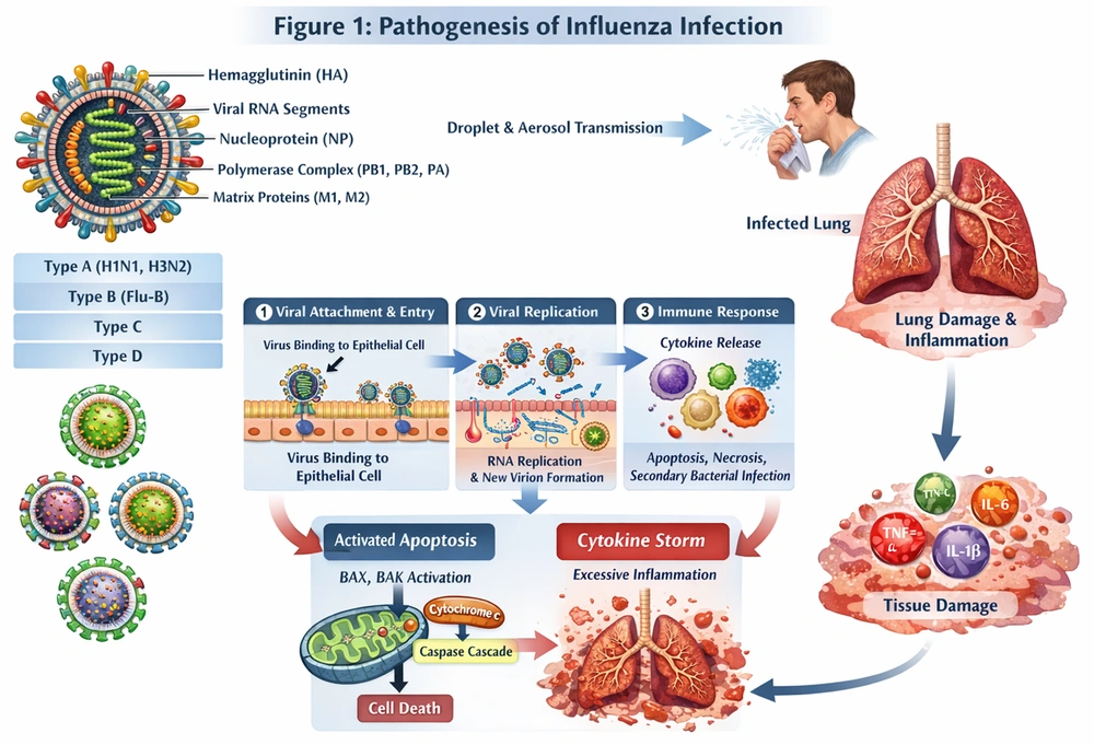
Influenza viruses can modulate key apoptotic genes, including BAX, BAK1, and Bcl-2, to either promote or delay cell death in a stage-dependent manner that benefits the virus (12, 13). Activation of BAX and BAK1 can lead to mitochondrial permeabilization, cytochrome c release, and subsequent activation of the caspase cascade, ultimately inducing apoptosis within the cell (14, 15). Apoptosis can restrict viral replication; however, excessive or dysregulated apoptosis may contribute to tissue injury and increased infection severity. Following apoptosis and immune system activation, cytokines and chemokines are released, which are associated with inflammation after infection (Figure 1) (14, 15).
Overview of influenza virus structure, transmission, and host-pathogen interactions
Figure 1.

Overview of influenza virus structure, transmission, and host-pathogen interactions

2. Objectives

In this study, we investigated the mRNA expression levels of BAX, BAK1, and Bcl-2 in A549 cells infected with the A/H1N1, A/H3N2, and influenza B viruses.

3. Methods

3.1. Cell Culture

A549 cells were used as the in vitro model in this study. Three groups of influenza viruses, including A/H1N1, A/H3N2, and influenza B, were selected to assess the mRNA expression of BAK1, Bcl-2, and BAX relative to the uninfected control group. The A549 cell line was cultured in Dulbecco Modified Eagle Medium (DMEM; Gibco) containing 10% inactivated fetal bovine serum (Gibco), 100 U/mL penicillin, and 100 μg/mL streptomycin until the cells reached 80% confluency.
Cell counting was performed using A549 cells at approximately 80% confluency. A 10-µL aliquot of the cell suspension was placed in a Neubauer hemocytometer, and total cells were counted without staining. The total count from four squares was 2 million cells. Given that each well of an 8 × 12 plate was estimated to require 15,000 cells, approximately 1.5 million cells needed to be prepared. As 2 million cells were suspended in 7 mL of medium, 5 mL of the suspension was required. All comparative infection experiments were performed using a consistent inoculum definition and the same effective-dose basis across all three viral strains (A/H1N1, A/H3N2, and influenza B). Before the TCID50 assay, the number of cells in each well was standardized, and all viral strains had comparable initial cycle threshold values. This ensured uniform starting conditions for titration. Infection conditions, including cell number, viral input, and experimental setup, were fully standardized.
A final volume of 200 µL was set for each well; therefore, 20 mL was required for the entire TCID50 experiment. Parafilm and aluminum foil were used to cover the plate and prevent evaporation, and the plate was incubated for 24 hours at 37°C. The next day, the plate was examined, and the cells were confirmed to have properly settled at the bottom of the wells and appeared healthy. Using a multichannel pipette, the medium in each well was removed, and the wells were washed once with serum-free DMEM. The wash volume was set at 100 µL per well.
Viral dilutions were then prepared. Microtubes containing A/H1N1, A/H3N2, and influenza B viruses, which were processed on different days and plates to avoid cross-contamination, were collected. Then, 450 µL of serum-free DMEM and 50 µL of each virus were added to a microtube. A dilution series was then prepared to a total dilution of 10-8.
The plate was incubated for 1 hour at 37°C in a CO2 incubator to allow viral adsorption to the cells, and 100 µL of serum-free medium was then added to each well. Finally, the plate was sealed with parafilm, wrapped in foil, and incubated at 37°C in a CO2 incubator. Each stage, including extraction, cDNA synthesis, and inoculation, was performed on separate days.

3.2. RNA Extraction and cDNA Synthesis

At 48 hours after viral inoculation of A549 cells, total RNA was extracted from infected cells at 10-4, which yielded the best TCID50 result, and from control cells using the RNJIA extraction kit (RNJIA, ROJE, Iran) according to the manufacturer’s instructions. RNA quantity and purity were assessed using a Nanodrop spectrophotometer. cDNA was synthesized from 1 µg of total RNA using the SMBIO kit (SMBIO, Taiwan). cDNA dilutions of 1:1, 1:10, and 1:100 were prepared, and the 1:1 dilution was selected as the most appropriate based on the cycle threshold values.

3.3. Quantitative Reverse Transcription PCR

The mRNA expression levels of Bcl-2, BAK1, and BAX were assessed using SYBR Green Master Mix (Yekta Tajhiz Azma, Iran) on a Corbett real-time PCR system. β-Actin was selected as the housekeeping gene. Relative expression was calculated using the 2(-ΔΔCt) method. Primer sequences are shown in Table 1.
Table 1.Primer Sequences, Amplicon Sizes, and Full Gene Names for Quantitative PCR Analysis a
NamesForwardReverseAmplicon Size
β-ActinCCACCATGTACCCTGGCATTCGGACTCGTCATACTCCTGC189
BAXCTGACGGCAACTTCAACTGGTGATCAGTTCCGGCACCTTG99
BAK1ACCAGCCTGTTTGAGAGTGGGTAGCCGAAGCCCAGAAGAG63
Bcl-2CTTTGAGTTCGGTGGGGTCACATCCACAGGGCGATGTTGT82

a Abbreviations: BAX, BAX associated X, apoptosis regulator; BAK1, BCL2 antagonist/killer 1; Bcl-2, B-cell lymphoma 2. β-Actin served as the housekeeping mRNA.

3.4. Statistical Analysis

For all analyses, log fold change was used. To assess differential gene expression after exposure to different viruses at each time point, the Wilcoxon signed-rank test was used as a nonparametric method to evaluate significance against zero, representing the control. In addition, for further insight and comparison, the one-sample t-test was performed. To compare gene expression changes over time within each virus group, the nonparametric Kruskal-Wallis test and parametric one-way analysis of variance were used. When significant differences were detected, pairwise post-hoc comparisons were conducted. The pairwise Wilcoxon test was used as the post-hoc test after the Kruskal-Wallis test, and the pairwise t-test was used after analysis of variance. To control type I error in multiple comparisons, the Benjamini-Hochberg correction method was applied.

4. Results

4.1. mRNA Expression of Bcl-2

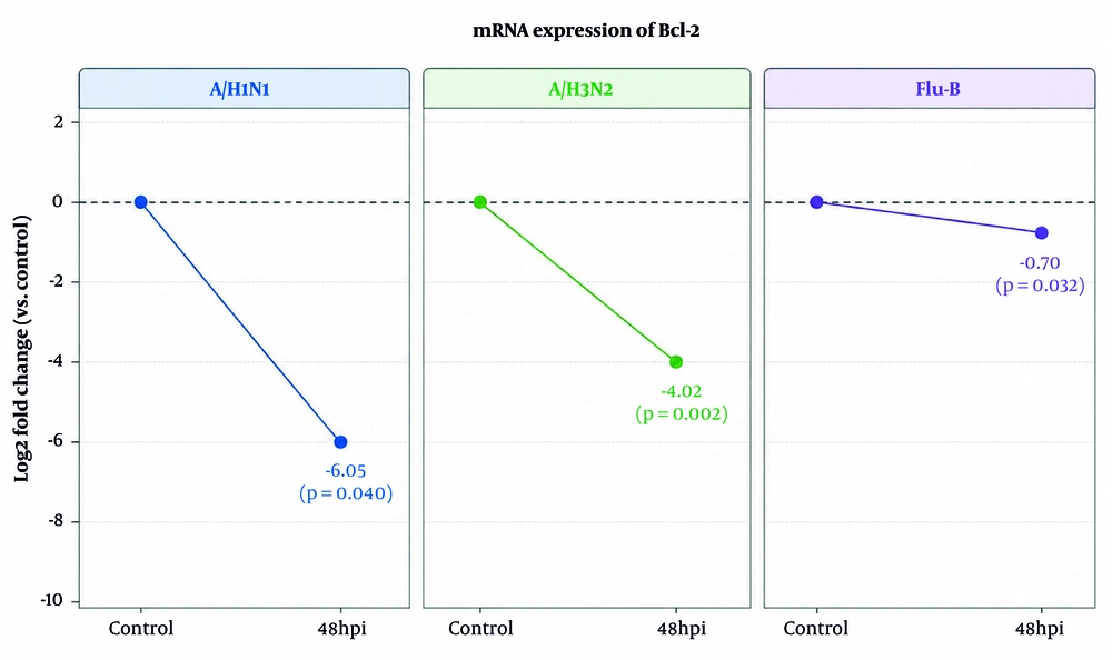
At 48 hours, Bcl-2 expression was reduced in influenza B-infected cells, with a mean difference of -0.702 ± 0.050 (95% CI, -1.149 to -0.255). Stronger downregulation was detected in A/H3N2 infection, with a mean difference of -4.023 ± 0.017 (95% CI, -4.172 to -3.874). Although A/H1N1 infection also showed decreased Bcl-2 expression (-6.015 ± 2.474), the wide confidence interval (-28.243 to 16.214) indicates considerable variability and limited statistical certainty (Figure 2).
At 48 hours post-infection, Bcl-2 mRNA expression was significantly downregulated in all virus-infected groups compared with control, with the greatest reduction observed in A/H1N1, followed by A/H3N2 and influenza B.
Figure 2.

At 48 hours post-infection, Bcl-2 mRNA expression was significantly downregulated in all virus-infected groups compared with control, with the greatest reduction observed in A/H1N1, followed by A/H3N2 and influenza B.

4.2. mRNA Expression of BAK1

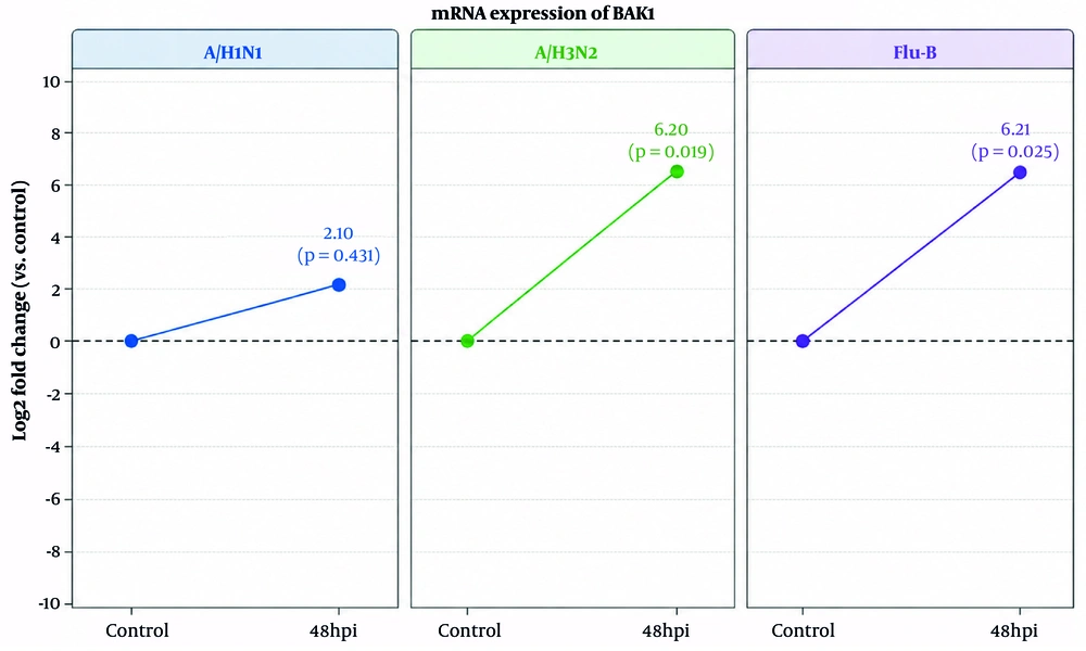
At 48 hours post-infection, A/H1N1 infection showed a smaller increase in BAK1 expression (2.099 ± 2.387), with a wide confidence interval (-19.344 to 23.542), indicating no statistically reliable change. In contrast, A/H3N2 infection showed significant upregulation (log fold change = 6.197 ± 0.266; 95% CI, 3.806 to 8.589; P = 0.019) after infection of A549 cells. Similar to A/H3N2, influenza B also showed significant upregulation of BAK1 expression (log fold change = 6.210 ± 0.339; 95% CI, 3.166 to 9.254; P = 0.025) (Figure 3).
At 48 hours post-infection, BAK1 mRNA expression was upregulated in all virus-infected groups compared with control; however, this increase was not significant in A/H1N1, while it was significant in both A/H3N2 and influenza B.
Figure 3.

At 48 hours post-infection, BAK1 mRNA expression was upregulated in all virus-infected groups compared with control; however, this increase was not significant in A/H1N1, while it was significant in both A/H3N2 and influenza B.

4.3. mRNA Expression of BAX

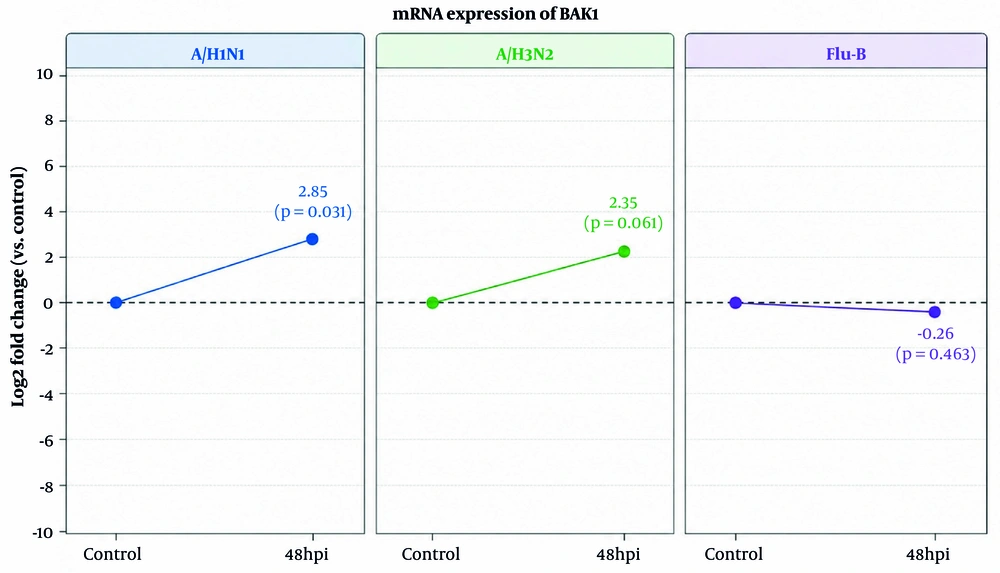
At 48 hours, influenza B infection resulted in a slight decrease in BAX expression, with a mean difference of -0.262 ± 0.329 (95% CI, -3.216 to 2.692), suggesting no significant alteration. A/H1N1 infection significantly increased BAX expression, with a mean difference of 2.847 ± 0.195 (95% CI, 1.093 to 4.602). A/H3N2 infection also showed elevated BAX expression (2.352 ± 0.322), although the confidence interval (-0.544 to 5.248) indicates that this increase was not statistically conclusive (Figure 4).
At 48 hours post-infection, A/H1N1 showed a significant increase in BAX expression (log fold change = 2.85, P = 0.031). Similarly, A/H3N2 showed an increase in BAX expression (log fold change = 2.35, P = 0.061), although this change was not statistically significant. In contrast, influenza B infection resulted in slight, nonsignificant downregulation of BAX expression (log fold change = -0.26, P = 0.463).
Figure 4.

At 48 hours post-infection, A/H1N1 showed a significant increase in BAX expression (log fold change = 2.85, P = 0.031). Similarly, A/H3N2 showed an increase in BAX expression (log fold change = 2.35, P = 0.061), although this change was not statistically significant. In contrast, influenza B infection resulted in slight, nonsignificant downregulation of BAX expression (log fold change = -0.26, P = 0.463).

4.4. Pairwise Comparison of Bcl-2, BAK1, and BAX Expression Between Influenza Virus Strains

Pairwise post-hoc comparisons of Bcl-2 expression in A/H1N1-, A/H3N2-, and influenza B-infected cells indicated strain-dependent differences. At 48 hours, a significant difference was observed between influenza B and A/H1N1 (P = 0.034), whereas the difference between influenza B and A/H3N2 (P = 0.103) was not significant. In addition, the difference between A/H1N1 and A/H3N2 (P = 0.258) was not significant.
Pairwise post-hoc comparisons of BAX expression in A/H1N1-, A/H3N2-, and influenza B-infected cells also indicated strain-dependent differences. At 48 hours, BAX expression was higher and significantly different in A/H1N1 and A/H3N2 compared with influenza B (influenza B vs A/H1N1: P = 0.002; influenza B vs A/H3N2: P = 0.003), while there was no significant difference between A/H1N1 and A/H3N2 (P = 0.185).
For BAK1 at 48 hours, no significant differences were observed between influenza B and A/H3N2 (P = 0.993), influenza B and A/H1N1 (P = 0.061), or A/H1N1 and A/H3N2 (P = 0.061).

5. Discussion

Apoptosis plays a critical role in the host response during viral infections, particularly those caused by influenza A virus and influenza B virus. Apoptosis can contribute to the cellular response to infection and limit viral spread; however, it may also exacerbate inflammation through cytokine release and immune activation. As a form of programmed cell death, apoptosis enables infected epithelial cells of the respiratory tract to undergo controlled self-destruction, thereby limiting viral spread within the body (13). The 48-hour post-infection time point was selected because it represents a biologically relevant window in which late viral replication events and host transcriptional responses, including apoptosis-related pathways, are robustly detectable. In this study, we observed marked upregulation of BAK1 after influenza virus inoculation of A549 cells, a well-established model of influenza virus infection. In addition, Bcl-2 downregulation and BAX upregulation, particularly in A/H1N1 compared with A/H3N2 and influenza B, suggest that these strains may use different strategies to affect cells and cellular responses.
The study by Fan et al. demonstrated that infection with influenza A virus promotes apoptosis partly through post-transcriptional regulation of BAX. Using A549 cells, they found that miR-34a was significantly downregulated following viral infection, which relieved its inhibitory effect on BAX mRNA and led to increased BAX protein expression. Functional assays further showed that overexpression of miR-34a reduced influenza-induced apoptosis, confirming that the miR-34a-BAX axis is an important mechanism in influenza-mediated cell death (16).
Our results align with previous reports. A/H1N1 significantly increased BAX expression, indicating activation of the intrinsic apoptotic pathway during infection, whereas A/H3N2 caused only a nonsignificant increase, suggesting subtype-dependent differences in apoptotic modulation. However, the changes were not large, and A/H3N2 may follow the same pattern as A/H1N1; this should be confirmed by flow cytometry. In contrast to the other influenza viruses in this study, influenza B slightly reduced BAX expression, implying that, compared with A/H1N1 and A/H3N2, this virus may use other mechanisms to regulate apoptosis after infection.
The study by Xu-Dong et al. investigated the effect of apoptosis after infection of mice with A/H1N1. Their results showed that A/H1N1 infection significantly reduced cell viability and induced apoptosis, as confirmed by flow cytometry with Annexin V. In addition, western blot analysis showed upregulation of caspase-3 expression and its cleaved active form at 24 and 32 hours post-infection. After infection, the BAX/Bcl-2 ratio was elevated, indicating activation of the intrinsic apoptotic pathway (17).
A study by McLean et al. provided important insights into the major role of BAX in influenza-induced apoptosis and viral replication. Their study showed that BAX activation and mitochondrial translocation were necessary for proper viral propagation (18).
Our findings are consistent with the mechanistic evidence reported by McLean et al. and Hossain et al. In our study, A/H1N1 infection led to a significant increase in BAX expression, supporting activation of BAX-mediated apoptosis during influenza virus infection, as described in previous studies. The results of Hossain et al. also suggest that such BAX upregulation may facilitate influenza virus replication in cells.
A study by Hossain et al. investigated the role of BAX inhibitor-1 as an antiviral factor during influenza A infection. Using MDCK cells overexpressing BAX inhibitor-1, they demonstrated that inhibition of BAX-mediated signaling significantly suppressed virus-induced cell death and reduced viral replication (19).
A study by Chen et al. investigated apoptotic signaling in lung epithelial cells infected with influenza A/H1N1 during coinfection with Porphyromonas gingivalis. Their results demonstrated that infection increased inflammatory cytokines and nitric oxide products within cells, thereby increasing the likelihood of apoptosis activation. BAX and caspase-3 protein expression were significantly increased, while Bcl-2 expression was significantly decreased. These findings indicated that influenza infection can promote apoptosis not only by increasing pro-apoptotic factors but also by suppressing anti-apoptotic regulators such as Bcl-2 (20).
Our results are consistent with these studies. In our study, decreased Bcl-2 expression was observed following infection with all three viruses (A/H1N1, A/H3N2, and influenza B), suggesting suppression of anti-apoptotic signaling by these three influenza virus strains. This suppression of anti-apoptotic signaling may occur because of viral effects that favor viral replication and spread within the body or as a delayed host-cell response to infection.
The study by Nencioni et al. investigated the role of Bcl-2 during infection with influenza A virus. They showed that the protective activity of Bcl-2 was impaired in influenza infection because of phosphorylation mediated by p38 mitogen-activated protein kinase signaling. This modification reduced the ability of Bcl-2 to inhibit apoptosis. Furthermore, Bcl-2 expression also influenced viral ribonucleoprotein export and viral replication, indicating an association between anti-apoptotic signaling and the influenza viral life cycle (21).
Our study is consistent with that report. In the present study, we observed significant downregulation of Bcl-2 following infection with A/H1N1, A/H3N2, and influenza B, implying strong suppression of Bcl-2-mediated anti-apoptotic signaling during infection. The research by Nencioni et al. supports the theory that influenza viruses may affect Bcl-2 function or expression, which can result in apoptosis after infection. While their study focused on post-translational modification of Bcl-2 through p38 mitogen-activated protein kinase, our mRNA expression data suggest that influenza infection may also downregulate Bcl-2 at the transcriptional level.
Regarding BAK1, few studies have investigated this protein after influenza virus infection and other respiratory viral infections. The study by Zhong et al. found that, in mammalian and avian cells, avian infectious bronchitis virus coronavirus upregulated BAK1 at the mRNA and protein levels. In that study, BAK1 knockdown delayed apoptosis activation and reduced viral release from cells. These findings suggest that BAK1 may be a key mediator of mitochondrial apoptosis that viruses exploit for more efficient replication and altered cell fate (22).
Similarly, we observed significant upregulation of BAK1 following A/H1N1 infection. A/H3N2 also showed upregulation following infection, whereas influenza B showed a minor decrease compared with A/H1N1 and A/H3N2. In both studies, viruses appear to disrupt BAK1-mediated mitochondrial apoptosis to balance cell survival and viral replication. Sufficient BAK1 activation promotes apoptosis at the appropriate stage, facilitating viral spread, while host anti-apoptotic factors, such as Bcl-2, Mcl-1, or Bcl-xL, modulate the timing and extent of cell death.
Another study by Pearson examined the effects of adenovirus-mediated wild-type p53 overexpression on apoptosis in human lung cancer cells, including H1299, H358, and H322 cells. They observed that BAX and BAK1 protein levels were markedly upregulated 18 to 36 hours after Adp53 transduction, preceding the morphological hallmarks of apoptosis, while Bcl-2 and Bcl-xL levels remained unchanged. This finding demonstrates that apoptosis induction can occur primarily through activation of pro-apoptotic mediators rather than suppression of anti-apoptotic proteins.
Compared with our influenza results, there is a clear parallel: we also observed upregulation of BAK1, significant for A/H1N1, and trends toward increased expression in A/H3N2, while influenza B showed minor upregulation. Similar to the p53 study, BAK1 upregulation in influenza infection likely promotes mitochondrial apoptosis independently of Bcl-2 modulation; however, in our data, Bcl-2 was decreased in all viral groups, further tipping the balance toward apoptosis.

5.1. Limitations

This study has several limitations. First, the results were obtained using an in vitro model. These expression patterns must be further confirmed in patients with active influenza infection, in whom host immune responses may affect gene expression. Strain differences may be time-point dependent and cannot be generalized across the infection course from a single endpoint. In addition, protein expression should be assessed by flow cytometry to confirm protein production. Measuring only a single late time point, 48 hours post-infection, limits the ability to determine whether the observed differences reflect early initiation events, peak responses, or downstream secondary effects. Measuring expression at other time points, including 4, 8, and 24 hours post-infection, is also recommended.

5.2. Conclusions

This study demonstrated that influenza virus infection changes the expression of pro-apoptotic genes in A549 human lung epithelial cells, with somewhat distinct patterns for each strain, including A/H1N1, A/H3N2, and influenza B. All influenza viruses caused downregulation of Bcl-2, indicating suppression of this anti-apoptotic gene and increased cellular susceptibility to programmed cell death. In addition, pro-apoptotic genes, including BAK1 and BAX, showed virus-specific regulation. A/H3N2 and influenza B sharply upregulated BAK1, whereas A/H1N1 also showed upregulation, although this was modest compared with A/H3N2 and influenza B. BAX expression was also significantly increased by A/H1N1 compared with A/H3N2 and influenza B.
Overall, our results suggest that influenza viruses may diminish cell survival signals by altering the regulation of pro-apoptotic genes, and that activation of pro-apoptotic pathways may vary across different strains. These findings suggest that different influenza viruses may employ distinct strategies to manipulate host-cell fate. Understanding these strain-specific effects can provide useful insights into influenza virus pathogenesis and possible targeted interventions in the future.

Acknowledgments

Footnotes

References

  • 1.
    Skelton RM, Huber VC. Comparing Influenza Virus Biology for Understanding Influenza D Virus. Viruses. 2022;14(5):1036. [PubMed ID: 35632777]. [PubMed Central ID: PMC9147167]. https://doi.org/10.3390/v14051036.
  • 2.
    Treanor JJ. Influenza Viruses. In: Kaslow, R.A., Stanberry, L.R., Powers, A.M. (eds) Viral Infections of Humans. Springer, New York. 2023;NY:1-57. https://doi.org/10.1007/978-1-4939-9544-8_19-2.
  • 3.
    Chauhan RP, Gordon ML. An overview of influenza A virus genes, protein functions, and replication cycle highlighting important updates. Virus Genes. 2022;58(4):255-269. [PubMed ID: 35471490]. https://doi.org/10.1007/s11262-022-01904-w.
  • 4.
    Kiseleva IV, Rudenko LG. Development of reassortant influenza vaccines: classical reassortment or reverse genetics? Russian Journal of Infection and Immunity. 2023;13(2):209-218. https://doi.org/10.15789/2220-7619-DOR-2449.
  • 5.
    Kang M, Wang LF, Sun BW, Wan WB, Ji X, Baele G, et al. Zoonotic infections by avian influenza virus: changing global epidemiology, investigation, and control. Lancet Infect Dis. 2024;24(8):e522-e531. [PubMed ID: 38878787]. https://doi.org/10.1016/S1473-3099(24)00234-2.
  • 6.
    Liang Y. Pathogenicity and virulence of influenza. Virulence. 2023;14(1). 2223057. [PubMed ID: 37339323]. [PubMed Central ID: PMC10283447]. https://doi.org/10.1080/21505594.2023.2223057.
  • 7.
    Zhu Y, Sun Y, Deng X, Cao P, Li S, Yu H, et al. Matrix protein 1 (M1) of influenza A virus: structural and functional insights. Emerg Microbes Infect. 2025;14(1). 2558881. [PubMed ID: 40925098]. [PubMed Central ID: PMC12451964]. https://doi.org/10.1080/22221751.2025.2558881.
  • 8.
    Pittman Ratterree DC, Dass SC, Ndeffo-Mbah ML. Mechanistic Models of Influenza Transmission in Commercial Swine Populations: A Systematic Review. Pathogens. 2024;13(9):746. [PubMed ID: 39338936]. [PubMed Central ID: PMC11434764]. https://doi.org/10.3390/pathogens13090746.
  • 9.
    Khanna M, Sharma K, Saxena SK, Sharma JG, Rajput R, Kumar B. Unravelling the interaction between Influenza virus and the nuclear pore complex: insights into viral replication and host immune response. Virusdisease. 2024;35(2):231-242. [PubMed ID: 39071870]. [PubMed Central ID: PMC11269558]. https://doi.org/10.1007/s13337-024-00879-6.
  • 10.
    Sun Y, Liu K. Mechanistic Insights into Influenza A Virus-Induced Cell Death and Emerging Treatment Strategies. Vet Sci. 2024;11(11):555. [PubMed ID: 39591329]. [PubMed Central ID: PMC11598850]. https://doi.org/10.3390/vetsci11110555.
  • 11.
    Liang Y. Pathogenicity and virulence of influenza. Virulence. 2023;14(1). 2223057.
  • 12.
    Blake ME, Kleinpeter AB, Jureka AS, Petit CM. Structural Investigations of Interactions between the Influenza a Virus NS1 and Host Cellular Proteins. Viruses. 2023;15(10):2063. [PubMed ID: 37896840]. [PubMed Central ID: PMC10612106]. https://doi.org/10.3390/v15102063.
  • 13.
    Soni S, Mebratu YA. B-cell lymphoma-2 family proteins-activated proteases as potential therapeutic targets for influenza A virus and severe acute respiratory syndrome coronavirus-2: Killing two birds with one stone? Rev Med Virol. 2023;33(2). e2411. [PubMed ID: 36451345]. [PubMed Central ID: PMC9877712]. https://doi.org/10.1002/rmv.2411.
  • 14.
    Sun Y, Liu K. Mechanistic Insights into Influenza A Virus-Induced Cell Death and Emerging Treatment Strategies. Vet Sci. 2024;11(11):555.
  • 15.
    Pacheco-Hernández LM, Ramírez-Noyola JA, Gómez-García IA, Ignacio-Cortés S, Zúñiga J, Choreño-Parra JA. Comparing the Cytokine Storms of COVID-19 and Pandemic Influenza. J Interferon Cytokine Res. 2022;42(8):369-392. [PubMed ID: 35674675]. [PubMed Central ID: PMC9422807]. https://doi.org/10.1089/jir.2022.0029.
  • 16.
    Gui R, Chen Q. Molecular Events Involved in Influenza A Virus-Induced Cell Death. Front Microbiol. 2022;12. 797789. [PubMed ID: 35069499]. [PubMed Central ID: PMC8777062]. https://doi.org/10.3389/fmicb.2021.797789.
  • 17.
    Fan N, Wang J. MicroRNA 34a contributes to virus-mediated apoptosis through binding to its target gene Bax in influenza A virus infection. Biomed Pharmacother. 2016;83:1464-1470. [PubMed ID: 27610823]. https://doi.org/10.1016/j.biopha.2016.08.049.
  • 18.
    Pei XD, Zhai YF, Zhang HH. Influenza virus H1N1 induced apoptosis of mouse astrocytes and the effect on protein expression. Asian Pac J Trop Med. 2014;7(7):572-575. [PubMed ID: 25063289]. https://doi.org/10.1016/S1995-7645(14)60096-1.
  • 19.
    McLean JE, Datan E, Matassov D, Zakeri ZF. Lack of Bax prevents influenza A virus-induced apoptosis and causes diminished viral replication. J Virol. 2009;83(16):8233-8246. [PubMed ID: 19494020]. [PubMed Central ID: PMC2715773]. https://doi.org/10.1128/JVI.02672-08.
  • 20.
    Hossain M, Saha S, Abdal Dayem A, Kim JH, Kim K, Yang GM, et al. Bax Inhibitor-1 Acts as an Anti-Influenza Factor by Inhibiting ROS Mediated Cell Death and Augmenting Heme-Oxygenase 1 Expression in Influenza Virus Infected Cells. Int J Mol Sci. 2018;19(3):712. [PubMed ID: 29498634]. [PubMed Central ID: PMC5877573]. https://doi.org/10.3390/ijms19030712.
  • 21.
    Chen Y, Zhou R, Yi Z, Li Y, Fu Y, Zhang Y, et al. Porphyromonas gingivalis induced inflammatory responses and promoted apoptosis in lung epithelial cells infected with H1N1 via the Bcl‑2/Bax/Caspase‑3 signaling pathway. Mol Med Rep. 2018. [PubMed ID: 29750299]. [PubMed Central ID: PMC6059728]. https://doi.org/10.3892/mmr.2018.8983.
  • 22.
    Nencioni L, De Chiara G, Sgarbanti R, Amatore D, Aquilano K, Marcocci ME, et al. Bcl-2 expression and p38MAPK activity in cells infected with influenza A virus: impact on virally induced apoptosis and viral replication. J Biol Chem. 2009;284(23):16004-16015. [PubMed ID: 19336399]. [PubMed Central ID: PMC2708894]. https://doi.org/10.1074/jbc.M900146200.

Crossmark
Crossmark
Checking
Share on
Cited by
Metrics

Ordering Reprints

Articles are published under the Creative Commons license stated on each article. No permission or royalty fee is required for uses permitted by that license. CCC handles optional bulk and customized reprint orders. Any quotation covers production and delivery services only, not copyright permission. > Request Reprints from CCC 

Search Relations

Author(s):

Related Articles