


1. Background
Influenza viruses are enveloped, negative-sense, single-stranded RNA viruses of the Orthomyxoviridae family and remain a leading cause of acute respiratory infections worldwide (1). Seasonal and pandemic strains, including A/H1N1, A/H3N2, and influenza B, are associated with substantial morbidity and mortality, primarily owing to their ability to induce cytopathic effects and dysregulated host immune responses (2).
Ferroptosis, a regulated form of cell death driven by iron-dependent lipid peroxidation, has recently been recognized as an important mechanism of virus-induced cytotoxicity (3). ACSL4 (acyl-CoA synthetase long-chain family member 4) is a central mediator of ferroptosis, promoting the accumulation of polyunsaturated fatty acids in membranes and sensitizing cells to lipid peroxidation. In addition, ferroptotic signaling intersects with inflammatory pathways, suggesting a potential link between virus-induced ferroptosis and host inflammatory responses (4, 5).
2. Objectives
Despite accumulating evidence implicating ferroptosis in viral infections, the temporal dynamics and magnitude of ACSL4 induction during influenza virus infection remain poorly characterized. Therefore, we assessed ACSL4 expression in A549 cells at 24 and 48 hpi after infection with A/H1N1, A/H3N2, and influenza B viruses. This study provides insight into the time-dependent activation of ferroptotic pathways in influenza-infected epithelial cells and highlights ACSL4 as a potential mediator of virus-induced cell death.
3. Methods
A549 human lung epithelial cells (RRID/IBRC: IBRC C10080) were cultured in high-glucose DMEM supplemented with 10% FBS and 1% penicillin/streptomycin at 37°C in 5% CO2. Cells were passaged at approximately 80% confluence. A549-adapted influenza A/H1N1, A/H3N2, and influenza B viruses were used to inoculate cells at 100 TCID50. Cells were incubated with the virus for 1 hour, followed by the addition of serum-free DMEM containing TPCK-treated trypsin (2 µg/mL). Non-infected cells served as controls.
Total RNA was extracted from the 10-4 dilution of infected cells, which showed the best TCID50 result, using the RNJIA kit (ROJE Technologies, Iran), according to the manufacturer’s protocol. RNA quality was verified using a NanoDrop spectrophotometer. cDNA was synthesized from 1 µg of RNA using the SMBIO cDNA synthesis kit with oligo(dT) primers for each infected cell group and the control group. cDNA dilutions of 1/1, 1/10, and 1/100 were prepared, and the 1/1 dilution was selected as the optimal dilution for all samples.
Real-time PCR was performed using SYBR Green Master Mix (Ampliqon) on a Rotor-Gene Q system (Qiagen). Each 25-µL reaction contained cDNA, primers (10 pmol each), and SYBR mix. Thermal cycling conditions were as follows: 95°C for 10 minutes; 40 cycles of 95°C for 15 seconds, 60°C for 30 seconds, and 72°C for 20 seconds. Melt curve analysis confirmed specificity. β-actin was used for normalization. Table 1 shows the primer sequences used in this study.
Table 1.Primer Sequences for ACSL4 and β-Actin a
| Targeted mRNA | Forward Primer | Reverse Primer | TM (F/R) |
|---|---|---|---|
| ACSL4 | TGACAACATCTGTGCTTTTGCC | CTACCCCTTTCTGTTGTGCCA | 60.22/60.20 |
| β-actin | CCACCATGTACCCTGGCATT | CGGACTCGTCATACTCCTGC | 60.03/59.97 |
a Abbreviation: ACSL4, acyl-CoA synthetase long-chain family member 4.
All statistical analyses were conducted using log2-transformed fold changes in gene expression levels relative to the control to enhance the detection of biologically meaningful changes. Parametric tests were also performed, and the results were similar to those of the nonparametric tests. Nonparametric tests were also used as supplementary analyses to provide comparative information.
To assess differential gene expression after exposure to different viruses at each time point, the Wilcoxon signed-rank test was used as a nonparametric method to evaluate significance against zero (control). In addition, the one-sample t-test was performed for further insight and comparison.
For comparisons of gene expression changes over time within each virus group, the nonparametric Kruskal-Wallis test and parametric one-way analysis of variance (ANOVA) were used. When significant differences were detected, pairwise post hoc comparisons were conducted. The pairwise Wilcoxon test was used as the post hoc test after the Kruskal-Wallis test, and the pairwise t-test was used after ANOVA. The Benjamini-Hochberg correction method was applied to control the type I error rate in multiple comparisons.
A significance level of less than 0.05 was considered statistically significant. All analyses were performed using R software version 4.5.1. Data visualization was conducted using the ggplot2 package in R software.
4. Results
At 24 hpi, ACSL4 expression was significantly increased in the influenza B group, with a mean log fold change of 1.972 ± 0.081 (95% CI, 1.247 to 2.696; P = 0.018). Similarly, the A/H1N1 group showed increased ACSL4 expression, with a mean log fold change of 1.524 ± 0.018 (95% CI, 1.364 to 1.683; P = 0.005). The A/H3N2 group showed moderate upregulation, with a mean log fold change of 0.989 ± 0.026 (95% CI, 0.758 to 1.220; P = 0.012).
At 48 hpi, ACSL4 expression was markedly decreased in the influenza B group, with a mean log fold change of -4.116 ± 0.212 (95% CI, -6.019 to -2.212; P = 0.023). In the A/H1N1 group, the mean log fold change was -1.637 ± 0.774 (95% CI, -8.594 to 5.321; P = 0.205), indicating no statistically significant change because of the wide confidence interval. The A/H3N2 group showed a mean increase of 0.908 ± 1.146 (95% CI, -9.387 to 11.203; P = 0.464), also without a statistically significant difference. These temporal changes are shown in Figure 1.
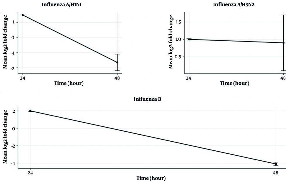
Figure 1.
ACSL4 upregulation was observed at 24 hpi. However, sharp downregulation of ACSL4 was observed at 48 hpi in the influenza B group, and this change was significant. All tests were performed in duplicate, and log fold changes were used for study analysis and comparison with control cells.
Post hoc analysis of ACSL4 gene expression changes showed that all pairwise comparisons were statistically significant at 24 hpi (P < 0.01), with influenza B showing the greatest increase relative to A/H1N1 and A/H3N2. This finding suggests virus-specific upregulation at the peak of early ferroptosis activation. At 48 hpi, differences between influenza B and A/H3N2 remained significant (P = 0.008), whereas comparisons involving A/H1N1 approached statistical significance (P = 0.05), reflecting divergence in ACSL4 regulation at later stages.
5. Discussion
Influenza virus infection can upregulate ACSL4, a pro-ferroptotic factor associated with viral replication, lipid peroxidation, and cell damage after infection. Our results demonstrated time-dependent induction of ACSL4, with the highest expression at 24 hpi for A/H1N1 and influenza B and the lowest expression at 48 hpi. These findings support a potential role for this pro-ferroptotic factor in influenza pathogenesis and the ferroptosis pathway.
Xia et al. found that influenza A/H9N2 can induce oxidative stress, mitochondrial dysfunction, and lipid peroxidation, ultimately leading to ferroptotic cell death through upregulation of ACSL4 (6).
In a study by Lv et al., influenza infection resulted in ACSL4 upregulation in lung tissue, with elevated cellular lipid peroxidation levels. This pattern highlighted the importance of ACSL4 as a key driver of ferroptosis because it channels polyunsaturated fatty acids (PUFA-CoA) into lipid peroxidation pathways, providing a direct substrate for oxidative damage within the cell (7).
In agreement with Lv et al. (7), our findings showed time-dependent upregulation of ACSL4 after influenza virus infection, with the highest induction at 24 hpi. This suggests that ACSL4-mediated ferroptosis is a conserved response across species and cell types.
Kung et al. showed that ACSL4 is more than a marker of ferroptosis; it is also an important component of viral replication. CRISPR-based genome-wide screens showed that ACSL4 is necessary for the development of viral replication organelles, which are membranous structures used by viruses, such as coronaviruses and SARS-CoV-2, to replicate their RNA. ACSL4 diverts certain polyunsaturated fatty acids to lipid peroxidation, thereby creating an environment rich in oxidized lipids. This environment may induce ferroptosis through its pro-ferroptotic activity and may also provide sufficient membrane permeability to organize viral replication complexes (8).
Chen et al. also studied the role of ferroptosis in lung injury caused by influenza (9). They reported that ACSL4 is upregulated and SLC7A11 is downregulated in the lungs of mice infected with A/H1N1. Pharmacological intervention in this pathway, as in the case of RuHaoDaShi granules, decreased ACSL4 and increased GPX4/SLC7A11/Nrf2 activity, confirming the potential of ferroptosis modulation to alleviate influenza-induced lung injury (9).
Multiple viral infections have been shown to affect ferroptotic pathways. Japanese encephalitis virus causes neuronal ferroptosis by acting on the GSH/GPX4 antioxidant system and increasing YAP1/ACSL4-supported lipid peroxidation. Newcastle disease virus inhibits system Xc- by downregulating SLC7A11 and SLC3A2 and stimulates the p53-SLC7A11-GPX4 axis, which promotes ferroptosis. Newcastle disease virus-induced YAP degradation further facilitates iron uptake and ferroptotic signaling. Epstein-Barr virus disrupts redox balance, increasing lipid reactive oxygen species and ferroptosis in B lymphocytes, although it may activate the p62-Keap1-Nrf2 axis in nasopharyngeal carcinoma cells and upregulate SLC7A11 and GPX4, conferring resistance to ferroptosis. Notably, influenza virus infection has been shown to trigger ferroptotic signaling. A/H1N1 infection impairs the system Xc-/GPX4 antioxidant system, resulting in diminished glutathione and lipid peroxidation. Inhibition of ferroptosis also reduces viral load and the virus-induced immune response (10).
We demonstrated time-dependent upregulation of ACSL4 in A549 cells after infection with influenza viruses, with the highest expression at 24 hpi, particularly for A/H1N1 and influenza B. This finding aligns with the report by Lv et al. (7), who observed significant ACSL4 upregulation in mouse lungs during influenza virus infection.
This study has several limitations. First, the experiments were performed in A549 cells, which are useful for studying influenza virus replication and pathogenesis but may not fully reflect the immune factors observed directly in patients. Second, the analysis was limited to mRNA expression, and protein-level validation of ACSL4 and functional ferroptosis assays were not performed. Functional ferroptosis assays and viral replication assays are recommended for future studies.
