
1. Background
Nontuberculous mycobacteria (NTM) are a diverse group of environmental bacteria within the Mycobacterium genus. Although numerous NTMs are present in nature, some act as opportunistic pathogens and cause serious illness, particularly in immunocompromised patients (1).
With approximately 200 known species, NTMs are primarily classified into 2 general groups based on growth rate: slow-growing mycobacteria (SGM) and rapidly growing mycobacteria (RGM). Mycobacterium simiae is among the most common NTMs. It is a slow-growing, photochromogenic species and an important opportunistic pathogen and public health threat in many regions worldwide (2).
M. simiae is the most important and well-defined member of the M. simiae complex, which consists of 17 known strains (3). It was first isolated in 1965 from a rhesus macaque (Macaca mulatta) and was later associated with human disease. Classical diagnostic approaches to pulmonary disease and lymphadenitis have predominantly focused on Mycobacterium tuberculosis. However, evidence indicates that M. simiae may present with clinical and radiologic findings indistinguishable from those of tuberculosis.
M. simiae infections are most frequently described in immunocompromised hosts, particularly in individuals with acquired immunodeficiency syndrome (AIDS) or chronic pulmonary diseases such as chronic obstructive pulmonary disease (COPD) (4). Biochemically, M. simiae is notable for being the only NTM that tests positive for niacin production, similar to M. tuberculosis (5). This biochemical similarity highlights the critical need for accurate differentiation between the 2 species, particularly given the potential of M. simiae to cause serious pulmonary disease in both immunocompetent individuals and those infected with HIV.
Epidemiologically, M. simiae is the most commonly isolated slow-growing NTM in Iran and the second most prevalent in other parts of the Middle East (6). Despite its clinical and epidemiological significance, the treatment of M. simiae infections remains a major challenge because of the lack of effective antimicrobial regimens. Most M. simiae strains are resistant to first-line antituberculosis drugs, often resulting in treatment failure. The American Thoracic Society (ATS) recommends a regimen including a macrolide, a fluoroquinolone, and trimethoprim/sulfamethoxazole for treating M. simiae infections (7). However, published studies on the drug susceptibility of clinical isolates are limited, largely because of the low prevalence of, and frequent misidentification of, this species.
To enable rapid detection and accurate differentiation between M. simiae and M. tuberculosis, various diagnostic methods have been employed. However, the lack of a unique and exclusive nucleotide sequence specific to M. simiae has hindered the development of efficient and user-friendly molecular diagnostic tools such as polymerase chain reaction (PCR) and loop-mediated isothermal amplification (LAMP).
Several molecular techniques have been developed for NTM detection, including restriction fragment length polymorphism (RFLP) (8), real-time PCR (9), matrix-assisted laser desorption/ionization time-of-flight mass spectrometry (MALDI-TOF MS) (10), line probe assays (11), whole-genome sequencing (WGS) (3), repetitive sequence-based PCR (rep-PCR) (12), and label-free electrochemical DNA nanobiosensors (13). However, these techniques can be technically demanding, time-consuming, and costly, particularly in resource-limited settings.
2. Objectives
Rapid and cost-effective molecular diagnostic tools are needed for accurate detection of these infections. This study aimed to introduce and evaluate a novel species-specific target for the identification of M. simiae. The proposed diagnostic target was identified using a modified genomic analysis approach based on previous research efforts (14, 15). The use of this novel target in a conventional PCR assay may address the current diagnostic gap and provide health care laboratories with a low-cost, reliable tool for rapid differentiation of M. simiae from M. tuberculosis and other NTMs.
3. Methods
3.1. Identification of Novel DNA Targets
To identify a novel and specific diagnostic target for M. simiae, a modified genome-comparison method was used. Initially, 2 complete genomes of M. simiae were retrieved from the NCBI database (Table 1). One genome was selected as the reference genome for further analyses, and the other was used to evaluate the specificity of the potential diagnostic target.
Table 1.
Summary of Genome Sequence Records for M. simiae Available in NCBI
| Accession Number | Database Source | Genome Assembly Sort | Strain | Genome Length |
|---|---|---|---|---|
| NZ_AP022568.1 | RefSeq | Complete Genome | JCM 12377 | 5788994 bp |
| NZ_HG315953.1 | GenBank | Whole genome shotgun | ATCC 25275 | 5782968 bp |
Given the total genome size of M. simiae (approximately 5,788,994 base pairs), the reference genome was computationally fragmented into 5,000-base-pair segments, yielding approximately 1,100 distinct genomic fragments. These fragments were analyzed individually using the BLASTN tool (Basic Local Alignment Search Tool for Nucleotides) provided by NCBI (https://blast.ncbi.nlm.nih.gov/Blast.cgi).
Fragments showing 100% identity and query coverage with the maximum alignment score in both complete M. simiae genomes were considered conserved sequences. These highly conserved fragments were subsequently subdivided into shorter nucleotide sequences. The resulting sequences were subjected to a more stringent similarity search using MegaBLAST against a genome sequence database containing M. simiae, M. tuberculosis, and other NTM species.
The aim of this comparative analysis was to identify fragments highly specific to M. simiae. Therefore, each candidate fragment was required to show less than 50% identity, query coverage, or alignment score with the genomes of nontarget microorganisms. This strict threshold was applied to ensure high specificity and to avoid cross-reactivity with other mycobacterial species that may coexist in clinical or environmental samples.
The study was designed to avoid selecting intergenic sequences as potential diagnostic targets. Intergenic regions can vary in length among strains of the same species without substantial genetic or functional diversity. Therefore, coding sequences (CDSs) encoding functional proteins were preferred. These genes can serve as key genomic markers that distinguish microorganisms, particularly among phylogenetically related species. Moreover, sequence variation within orthologous protein-coding genes among closely related mycobacterial species can provide valuable information for developing highly specific molecular diagnostics. The selected targets may also provide a basis for future molecular and evolutionary studies.
3.2. Primer Design
The most specific and relatively long gene fragment identified in the preceding analyses was selected for primer design. Primer Premier 5.0 software (Premier Biosoft International, CA, USA) was used to design primers for optimal amplification of the target region. The primers were assessed for potential secondary structures, including hairpins, self-dimers, and cross-dimers, using the OligoAnalyzer Tool (https://eu.idtdna.com/calc/analyzer). The designed primers were also evaluated using Primer-BLAST (NCBI) to confirm specificity against a broad range of genomic sequences.
To ensure compatibility with the human genome and prevent cross-reactivity with host DNA in clinical samples, an additional BLAST search restricted to the Homo sapiens genome was performed to identify and exclude potential alignments with human genomic sequences. This approach minimized the risk of false-positive results in clinical specimens containing human DNA.
3.3. Polymerase Chain Reaction
The PCR reaction mixture was prepared in a total volume of 25 μL and contained 1× reaction buffer with 50 mM KCl, 10 mM Tris-HCl (pH 9.0), and 1.5 mM MgCl2; 10 pmol of each primer; 200 μM of each deoxynucleotide triphosphate (dATP, dCTP, dGTP, and dTTP); and 1 U of Taq DNA polymerase (5 U/μL; Fermentas). In addition, 1 μL of genomic DNA was added as the amplification template.
PCR was performed using a thermal cycler (Techne TC-3000X) under the following cycling conditions: initial denaturation at 95 °C for 5 minutes, followed by 40 amplification cycles consisting of denaturation at 94 °C for 45 seconds, annealing at 61 °C for 30 seconds, and extension at 72 °C for 30 seconds. The annealing temperature was optimized within the range of 58 - 62 °C, with the most efficient amplification observed at 61 °C. A final extension was performed at 72 °C for 10 minutes.
After amplification, 5 μL of each PCR product was loaded onto a 1.5% agarose gel, stained with Green Viewer, and subjected to electrophoresis in 0.5× TBE buffer at 110 mV for 45 minutes. The expected amplicon size of 110 base pairs was visualized under UV illumination using a transilluminator (Spectroline).
Each PCR run included a positive control containing 1 ng of M. simiae DNA and a negative control in which sterile water was used instead of the DNA template to monitor for contamination.
3.4. Primer Specificity and Sensitivity
The analytical specificity of the designed primers was assessed using genomic DNA extracted from M. simiae and a panel of phylogenetically related mycobacterial species. These included M. tuberculosis, M. bovis BCG, M. smegmatis, M. kansasii, M. avium, M. chelonae, M. fortuitum, M. ulcerans, M. intracellulare, and M. marinum. Specificity was considered satisfactory when amplification was observed exclusively in reactions containing M. simiae DNA, with no detectable amplification products from DNA templates of nontarget mycobacterial species.
To evaluate the analytical sensitivity of the primers, a series of 10-fold serial dilutions of M. simiae genomic DNA was prepared in nuclease-free distilled water. The resulting concentrations were 100 pg/μL, 10 pg/μL, 1 pg/μL, 100 fg/μL, 10 fg/μL, 5 fg/μL, and 1 fg/μL. For each dilution, 1 μL was used as the template in separate PCR reactions to determine the lowest DNA concentration capable of yielding a detectable amplification product. This threshold represented the limit of detection (LOD) under the experimental conditions.
To ensure the reproducibility and robustness of the sensitivity results, each dilution series was tested in triplicate on 3 separate days.
3.5. Clinical Sensitivity of the Novel Diagnostic Target
To evaluate the sensitivity of the newly identified genomic target, DNA extracted from 10 clinical isolates of M. simiae was used. These isolates were collected from sputum samples of patients suspected of having tuberculosis and had previously been confirmed by molecular diagnostic techniques and sequencing in our laboratory.
All DNA samples derived from these clinical isolates were subjected to PCR amplification using the optimized protocol described above. To further validate the specificity of the novel-targeted PCR assay, amplicons from 5 clinical isolates were subjected to DNA sequencing. Sequencing was performed in both directions using the same forward and reverse primers employed in the PCR reactions. Each sequence was then compared against the GenBank database to confirm its identity and determine homology with reference sequences of M. simiae.
4. Results
4.1. Novel Specific Target and Designed Primers
The gene G6N33_RS00430 (Accession number: QB014449), spanning 978 base pairs and encoding an alpha/beta hydrolase (ABH) enzyme, was identified as a novel and specific molecular target for the detection of M. simiae based on bioinformatic analysis of the reference strain M. simiae JCM 12377 (Figure 1). This gene was selected through a comprehensive bioinformatics pipeline designed to identify unique genomic sequences suitable for species-specific detection.
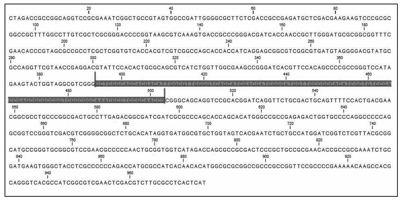
Figure 1.
Alpha/beta hydrolase gene sequence of M. simiae (978 bp). The location of the amplicon is illustrated in purple.
A MegaBLAST search against the NCBI database of complete bacterial genomes indicated that the G6N33_RS00430 gene sequence is uniquely present in M. simiae and absent from the genomes of other bacterial species, including closely related members of the Mycobacterium genus. To further evaluate potential homology, a BLASTN analysis was performed. Although the ABH-encoding gene is also present in a few closely related species within the M. simiae complex, specifically M. saskatchewanense and M. kubicae, sequence similarity was substantially lower. The BLAST maximum alignment score for M. simiae was 1764, whereas the scores decreased to 296 and 269 for M. saskatchewanense and M. kubicae, respectively. These reductions in alignment scores indicate sequence divergence of this gene among these species and support its discriminatory potential for identifying M. simiae.
Following identification of this unique gene, a pair of primers was designed to target the most conserved and diagnostically relevant region of the sequence. Primer design focused on minimizing undesirable secondary structures, including hairpins, self-dimers, and cross-dimers, which can interfere with amplification efficiency and specificity.
The primer sequences and their thermodynamic and structural properties, including melting temperature (Tm), GC content, nucleotide length, and predicted amplicon size, are presented in Table 2. Primer-BLAST analysis confirmed that the designed primers exclusively produced an amplicon in M. simiae, with no cross-reactivity detected in other mycobacterial species.
Table 2.
Sequences and Characteristics of Primers Used in This Study
| Primer pair | Sequence (5'→3') | Length (bp) | Tm (°C) | GC% | Product Length |
|---|---|---|---|---|---|
| Forward primer | GCTCGGCAGCACGACATTG | 19 | 62.37 | 63.16 | 110 bp |
| Reverse primer | CGGTTGCGGAGGCGTTT | 17 | 61.06 | 64.71 | 110 bp |
4.2. Analytical Sensitivity and Specificity of the ABH-PCR Assay
The analytical sensitivity of the ABH-PCR assay was evaluated to determine the minimum quantity of genomic DNA that could be reliably detected. The assay detected low concentrations of M. simiae genomic DNA, with a detection limit of 5 fg of extracted DNA (Figure 2). Amplification products were clearly observed across DNA concentrations ranging from 100 pg to 5 fg/μL, as demonstrated by distinct and reproducible amplicon bands of the expected size on agarose gel electrophoresis. In contrast, no amplification was observed with 1 fg of template DNA, indicating that this concentration was below the assay’s reliable detection threshold.
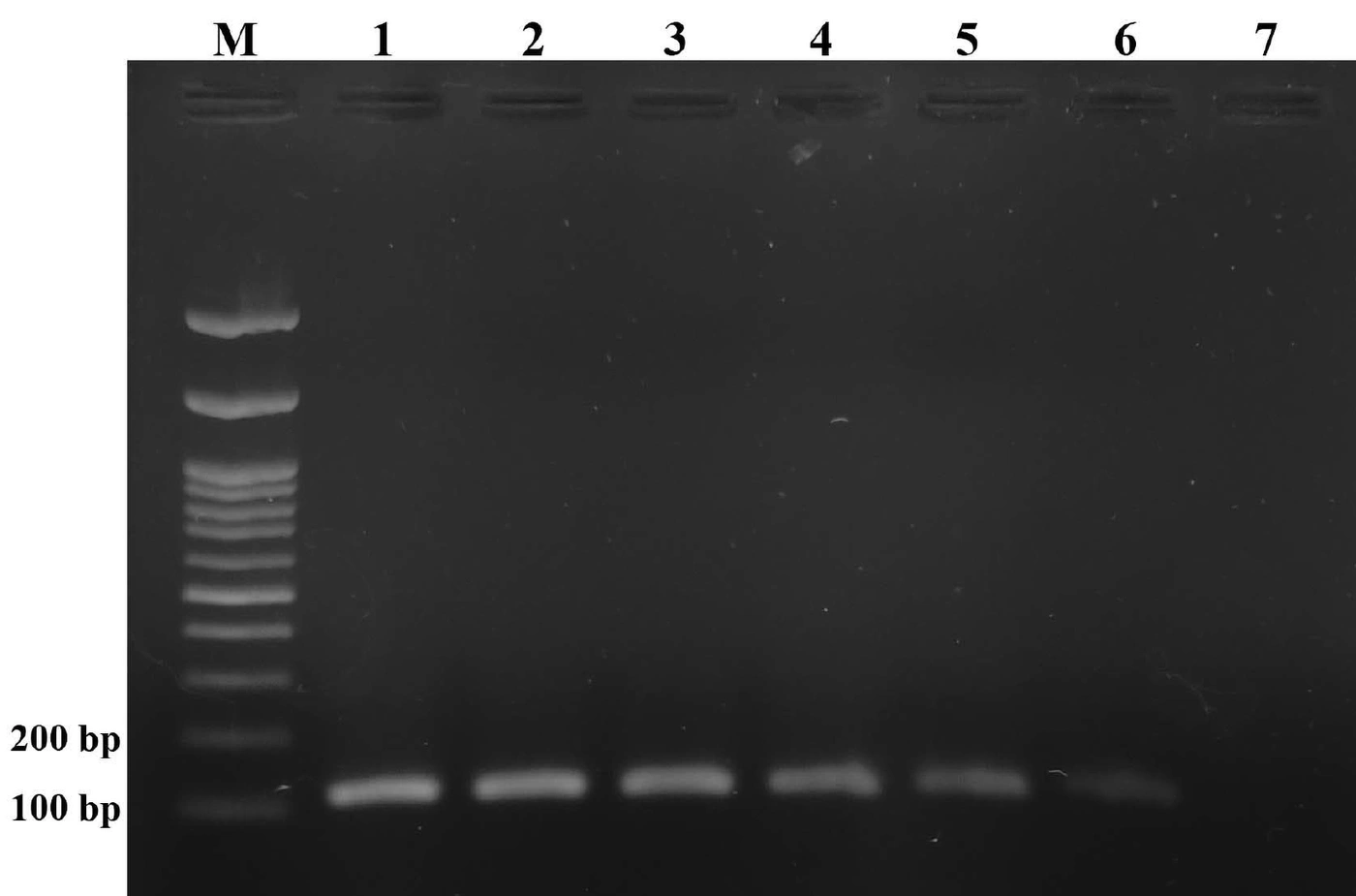
Figure 2.
Agarose gel electrophoresis of PCR products using serial dilutions of M. simiae genomic DNA. M: 100-bp DNA marker; lane 1: 100 pg; lane 2: 10 pg; lane 3: 1 pg; lane 4: 100 fg; lane 5: 10 fg; lane 6: 5 fg; lane 7: 1 fg.
The analytical specificity of the ABH primers was assessed using genomic DNA from a panel of 11 mycobacterial strains. This panel included 1 M. simiae strain, 2 members of the M. tuberculosis complex (M. tuberculosis and M. bovis), and 8 non-simiae mycobacterial species (M. smegmatis, M. kansasii, M. avium, M. chelonae, M. fortuitum, M. ulcerans, M. intracellulare, and M. marinum). Among all tested strains, only DNA extracted from M. simiae yielded a detectable amplicon, as evidenced by a clear and specific PCR band on the agarose gel (Figure 3). No amplification products were detected from the other Mycobacterium species, demonstrating a high degree of specificity of the ABH primers for M. simiae.
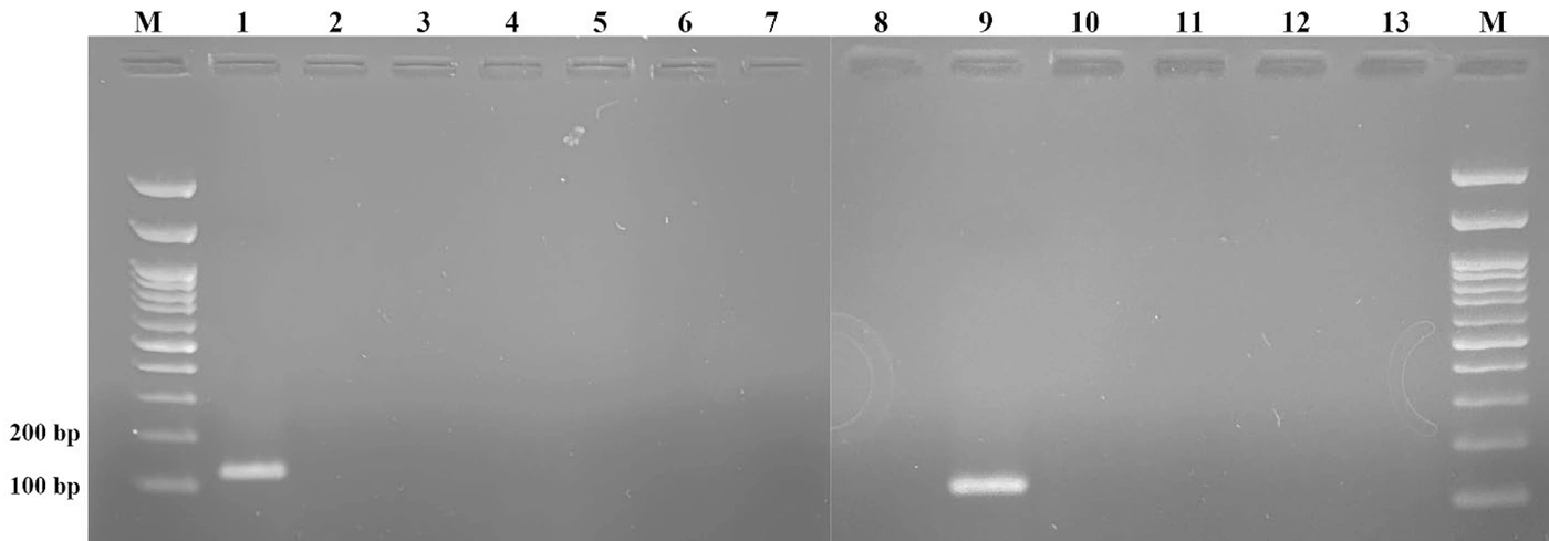
Figure 3.
Specificity of PCR assessed using genomic DNA from various mycobacterial species. M: 100-bp DNA marker; lane 1: positive control; lane 2: M. bovis; lane 3: M. smegmatis; lane 4: M. kansasii; lane 5: M. avium; lane 6: M. chelonae; lane 7: M. intracellulare; lane 8: M. fortuitum; lane 9: M. simiae; lane 10: M. ulcerans; lane 11: M. marinum; lane 12: M. tuberculosis; lane 13: negative control.
4.3. Evaluation of the ABH-PCR Assay in Clinical Isolates of M. simiae
Following extraction of genomic DNA from 10 clinical specimens previously confirmed to be positive for M. simiae, a minimal volume of 1 μL was used as the template for each PCR reaction. This limited template volume was used to reduce the potential inhibitory effects of residual substances commonly present in clinical samples, which can adversely affect PCR amplification efficiency and reliability.
The ABH-PCR assay successfully amplified the target DNA sequence in all 10 clinical isolates. Agarose gel electrophoresis revealed a distinct 110-bp amplicon corresponding to the expected target region in every sample tested. No amplification was observed in the negative-control reactions, ruling out contamination and supporting assay specificity (Figure 4).
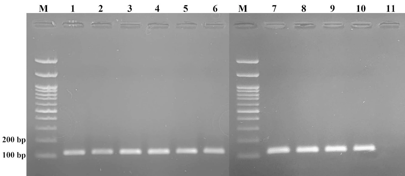
Figure 4.
Agarose gel electrophoresis showing amplification of the 110-bp target in 10 clinical isolates of M. simiae. Lane M: 100-bp DNA marker; lanes 1 - 10: M. simiae isolates; lane 11: negative control.
To further validate assay specificity and the identity of the amplified products, 5 of the 10 positive PCR samples were randomly selected for sequence analysis. Sequencing confirmed that the amplicons matched the target region of M. simiae, reinforcing the molecular accuracy and diagnostic value of the ABH-PCR assay.
5. Discussion
Given the increasing clinical significance of M. simiae, particularly among immunocompromised patients, the development of a rapid and reliable molecular diagnostic assay is important. Early and accurate detection of M. simiae is critical for the timely initiation of treatment and may improve patient outcomes.
A major challenge in the molecular diagnosis of M. simiae is the lack of a species-specific diagnostic target. This limitation has often necessitated expensive materials, complex procedures, or specialized equipment, thereby hindering widespread implementation. Although several molecular methods are available for the detection and differentiation of NTMs, their integration into routine diagnostic workflows remains challenging.
PCR-RFLP is one of the more accessible molecular techniques for the rapid identification of M. simiae (8). This method relies on genus-specific amplification of gene regions such as hsp65 or ITS, followed by digestion with restriction enzymes. Although PCR-RFLP is relatively inexpensive and suitable for basic laboratory settings, its discriminatory power is limited for differentiating closely related species, and it often requires multiple enzymes.
More advanced methods, such as real-time PCR and line probe assays, use species-specific probes for the simultaneous detection of multiple NTMs with high accuracy (9, 11). However, these assays typically target conserved regions such as rpoBC, hsp65, 16S rRNA, or ITS, which can result in cross-hybridization or failure to detect certain species. For example, M. genevanse and M. simiae are indistinguishable using rpoB-based probes (9), highlighting the limitations of these approaches. Moreover, the high cost of hybridization-based methods makes them impractical for routine use in many clinical laboratories.
Whole-genome sequencing is regarded as the gold standard for definitive species identification (3). Despite its high resolution and accuracy, WGS is impractical for routine diagnostics because of its cost, technical complexity, and requirement for bioinformatics expertise. Similarly, label-free electrochemical DNA biosensors targeting conserved regions such as ITS offer high sensitivity, portability, and rapid results (13). However, this technology remains at an early stage of development and requires further standardization before widespread implementation.
To address these limitations, we developed and validated a novel PCR-based diagnostic assay targeting a newly identified species-specific genomic sequence in M. simiae, designated G6N33_RS00430, which encodes an alpha/beta hydrolase (ABH). This study used modified comparative genomic analysis, optimized primer design, and analytical and clinical validation to establish a sensitive and specific detection platform for M. simiae.
Identification of the G6N33_RS00430 gene as a unique genetic target was a key outcome of this study. Comparative genomic analysis supported the specificity of this gene for M. simiae, with limited homology to other mycobacteria, including members of the M. simiae complex. Although limited similarity to M. saskatchewanense and M. kubicae was observed, alignment scores were markedly lower, supporting the discriminatory capacity of the target. This finding underscores the potential of refined comparative genomics to identify reliable diagnostic targets, particularly for NTMs, which often show substantial phenotypic and genotypic similarities. In addition, these 2 species belong to the M. simiae complex; therefore, the possibility of cross-reaction does not interfere with the objectives of the study.
Analytical evaluation demonstrated that the ABH-PCR assay was highly sensitive, with a detection limit of 5 fg of genomic DNA. This sensitivity may be particularly relevant for clinical samples with low bacterial loads, especially in the early stages of infection.
Specificity was validated using a panel of 11 clinically relevant mycobacterial species, including members of the M. tuberculosis complex and various NTMs. The assay amplified only M. simiae DNA, with no observed cross-reactivity.
Clinical validation further supported the performance of the ABH-PCR assay. All 10 M. simiae-positive clinical samples yielded specific amplification products, even with a minimal DNA input of 1 μL, which was selected to reduce the impact of potential PCR inhibitors commonly found in clinical samples.
5.1. Conclusions
The ABH-PCR assay offers a sensitive, specific, and cost-effective tool for the rapid identification of M. simiae. Its analytical performance may offer advantages over conventional culture-based and biochemical identification methods, which are often time-consuming, labor-intensive, and prone to misidentification. In addition, the low cost of the assay and its compatibility with standard laboratory equipment may make conventional PCR a practical molecular tool for routine diagnostic laboratories. This assay could also be adapted for epidemiological surveillance and environmental monitoring, contributing to a better understanding of M. simiae transmission dynamics and public health impact.
5.2. Study Limitations
This study has several limitations. First, the assay was evaluated using a limited number of isolates and samples; therefore, larger multicenter studies are needed to validate its performance. Second, we did not directly compare the assay with an established gold standard or alternative molecular diagnostic methods. Future studies should therefore address comparative diagnostic accuracy in clinical specimens.