


1. Context
Poliomyelitis is a highly contagious and transmissible infectious disease which is mainly transmitted via contaminated food and drinking water by human feces (fecal-oral route) (1). The clinical spectrum outcomes are as follows:
1) Asymptomatic form (90% of cases), 2) Mild illness characterized by fever, malaise, headache, nausea, vomiting, diarrhea or constipation (9% of cases), 3) Central nervous involvement such as aseptic meningitis, 4) Paralytic form (less than 1% of cases), 5) Post - polio syndrome characterized by cold intolerance in the affected limbs, muscle pain and weakness, 30 - 40 years after exposure to the wild virus in childhood (2, 3).
This disease is caused by three types of polio virus (P1, P2 and P3). The most neurovirulent types are P1 and P2 known as vaccine derived poliovirus type. Immunity against one type does not protect Individuals against the other types. Several doses of oral polio vaccine (OPV) are needed to induce immunity and it provides long-term protection against poliomyelitis (4).
Humans are the main reservoir of the virus. This point provides the possibility of eradication of disease by vaccination. Thus, in 1988, the world health assembly (WHA) adopted the goal of global polio eradication by the year 2000. The objectives of this strategy were as follows: a) Immunize people with OPV, b) Achieve and maintain high coverage vaccination levels among children under five, c) Develop an appropriate epidemiologic and laboratory acute flaccid paralysis (AFP) surveillance system, d) Hold the national polio days (NPDs) (2, 5).
The current study aimed to report the status of polio eradication program in Iran and attract the attention of the authorities seriously to the possibility of emerging the disease in the country.
-WPV1, 2 & 3: wild poliovirus type 1, 2 & 3
-GPEI: global poliomyelitis eradication initiative
-WHA: world health assembly
-NIDs: national immunization days
-SNIDs: sub-national immunization days
-NITAG: national immunization technical advisory group
-OPV: oral polio vaccine
-IPV: inactivated (injectable) polio virus
-tOPV: trivalent oral polio virus
-bOPV: bivalent oral polio virus
-mOPV1: monovalent type-1 oral poliovirus vaccine
-VDP: vaccine derived paralysis
-VDPVs: vaccine derived polio viruses
-VAAFP: vaccine associated acute flaccid paralysis
-iVDPVs: immunodeficiency-associated vaccine derived polio viruses
-cVDPVs: circulatory-associated vaccine derived polio viruses
-aVDPVs: ambiguous vaccine derived polio viruses
-PID: primary immune deficient
2. Evidence Acquisition
PubMed was used as an international databank and IranMedex as a national motor search to assess the subject. The key words were as follows: polio, poliomyelitis and polio eradication, OPV, IPV, AFP, VDP and Iran. One hundred and eighty-nine assessed abstracts included: polio and Iran (48), poliomyelitis and Iran (34), polio eradication and Iran (7), OPV and Iran (10), oral polio vaccine and Iran (18), IPV and Iran (12), inactivated polio vaccine and Iran (3), AFP and Iran (40), acute flaccid paralysis and Iran (15), VDP and Iran (0), and vaccine derived paralysis and Iran (2). Of these abstracts, 33 articles which were entirely relevant to the study goals were evaluated. There were no deterrent ethical considerations in the study.
3. Results
3.1. Polio Vaccines
For the first time, trivalent inactivated (injectable) polio vaccine (IPV) was prepared by Jonas Edward Salk in 1952, and then live attenuated OPV was introduced by Albert Sabine in 1961 (2, 5, 7). Oral polio vaccine is formed by a combination of live and attenuated types 1, 2 and 3 of the polio viruses and is called trivalent oral polio vaccine (tOPV) (8, 9).
Since the attenuated OPV has many benefits, its consumption is accepted worldwide. Some of these advantages are as follows: ease of use, induction of efficient intestinal and durable humeral immunity and low cost. Nevertheless, the most important disadvantage of OPV is genetic instability, resulting in vaccine-associated paralytic poliomyelitis (VAPP) (10).
Mir et al. evaluated the monovalent type-1 oral poliovirus vaccine (mOPV1) immunogenicity, given with an interval of one week in Karachi, Pakistan. This study was carried out on 1009 healthy newborn babies with a birth weight of at least 2.5 kg. The authors recommended that, shortening the interval between mOPV doses to seven days could be beneficial (11).
3.2. Immunogenicity
The oral polio vaccine creates an appropriate intestinal and humeral immunity against all three biotypes of the virus. The immunity against the biotypes is not the same.
Izadi et al. in a cross-sectional study conducted on 365 children aged 20 (± 2) months who had received at least five doses of trivalent oral polio vaccine, evaluated serological status against three serotypes of polioviruses. In this study, seropositive children against poliovirus serotypes 1, 2 and 3 were 94.1%, 96.7% and 78.3%, respectively. They concluded that, since the immune response against polio virus type 3 (PV3) was lower than those of other serotypes, improving the population immunity against this serotype is an urgent priority (12).
In many developing countries, the immunogenicity of the three doses of OPV is lower than that of industrialized countries (13).
Habib et al. conducted a double-blind, randomized placebo-controlled trial on 404 newborns aged 0-14 days. They assessed the impact of zinc supplementation on immune response to OPV and concluded that zinc supplementation had no effect on OPV immunogenicity (14).
3.3. Vaccine-Associated Poliovirus Paralysis (VAPP)
Nowadays, the wild polio virus is eradicated in most countries, but acute flaccid paralysis (AFP) caused by the vaccine is reported in some countries. This is called vaccine derived polioviruses paralysis (VDPP) (15).
Three categories of VDPVs are recognized: 1) Circulating VDPVs (cVDPVs) which occurs in the settings with low coverage of immunization by OPV, 2) Immunodeficiency-associated VDPVs (iVDPVs) which occurs in individuals with primary immunodeficiency, and 3) Ambiguous VDPVs (aVDPVs), which cannot be definitively categorized. In other words, OPV may cause paralysis in non-immune vaccine recipients. Immunodeficiency-associated vaccine derived polio viruses can replicate and be excreted for years from the body of the patients with primary immunodeficiency (4, 8, 10).
In patients with primary immune disorders (PID), especially those with B-cell system disorders, chronic excretion of polio virus rises and the risk of paralytic poliomyelitis increases (16). The first presentations of VAPP may be neurologic abnormalities in some patients with primary immunodeficiency (17).
The risk of VAPP in normal population is 1 case per 750000 and this rate in patients with immunodeficiency particularly for persons with agammaglobulinemia and hypogammaglobinemia is 1 per 7000. However, there is no appropriate estimation for patients with impaired cellular immunity (17).
Vaccine derived paralysis is caused by OPV; thus, eliminating this risk requires stopping the OPV use (8).
The majority of VDPPs are associated with type 2 circulating VDPVs (cVDPV2). Therefore, it is suggested to remove type 2 virus from trivalent vaccines (tOPV) and change this vaccine to bivalent vaccines (bOPV) consisting only types 1 and 3 polioviruses.
3.4. WHA New Strategy for Polio Eradication
Since most of the vaccine induced paralytic forms are caused by polio virus type 2, according to the world health organization (WHO) suggestion, this type is removed from the oral vaccine and bOPV is used Instead of tOPV in some countries since April 2016.
At least one dose of IPV is introduced prior to bOPV in routine immunization schedule (4).
Six hundred and eighty-six cases of paralytic polio were detected due to cVDPVs since 2006. More than 97% of the paralytic cases were caused by type 2 cVDPVs. Therefore, to eliminate the risk of paralytic forms by cVDPV2s, OPV serotype 2 is withdrawn from all immunization programs throughout the world. Therefore, trivalent OPV (tOPV) should be replaced with bivalent OPV (bOPV) which contains only types 1 and 3 polioviruses using IPV and switching from tOPV to bOPV (8).
In 2013, the world health assembly (WHA) endorsed a plan to withdraw OPV from immunization programs in the world. This program started with the removal of type 2 component of OPV in 2016. According to this program, before the above date (by the end of 2015), at least one dose of IPV vaccine is included in routine vaccination programs of all 126 countries that use OPV (10, 18).
3.5. Polio Eradication Status in the World
In 1988, the WHA predicted that poliomyelitis should be eradicated by the year 2000 (19). Up to 2014, the wild polio virus (WPV) transmission in all countries except Afghanistan, Pakistan and Nigeria was disrupted. (8, 9). However, reports indicate that, in 2015, Nigeria reaches one year without polio (20), promising news.
3.6. Polio Eradication Status in Iran
In 1982, an advisory committee was formed and named national immunization technical advisory group (NITAG) in Iran. It provided numerous scientific and technical recommendations to eradicate polio in the country (21). Since 1994, the Islamic Republic of Iran adopted strategies and programs to eradicate polio (19).
Mass vaccination of 10 million children under five in a national immunization day program started in 1994 and continued twice every year up to 1998. After that, the regional supplementary immunization began in the form of, sub-national immunization days (SNIDs) and still continues twice a year (19).
The last case was an imported case from Afghanistan and the circulation of wild poliovirus was stopped in December 2000 in the country and it was confirmed in 2001 by WHO and at present Iran is known as a polio free country for about 15 years (19). However, since the disease is active in the two neighboring countries Afghanistan and Pakistan (15), the risk of re-emergence of wild polio virus due to importation is high. Figure 1 shows Iran’s geographical location.
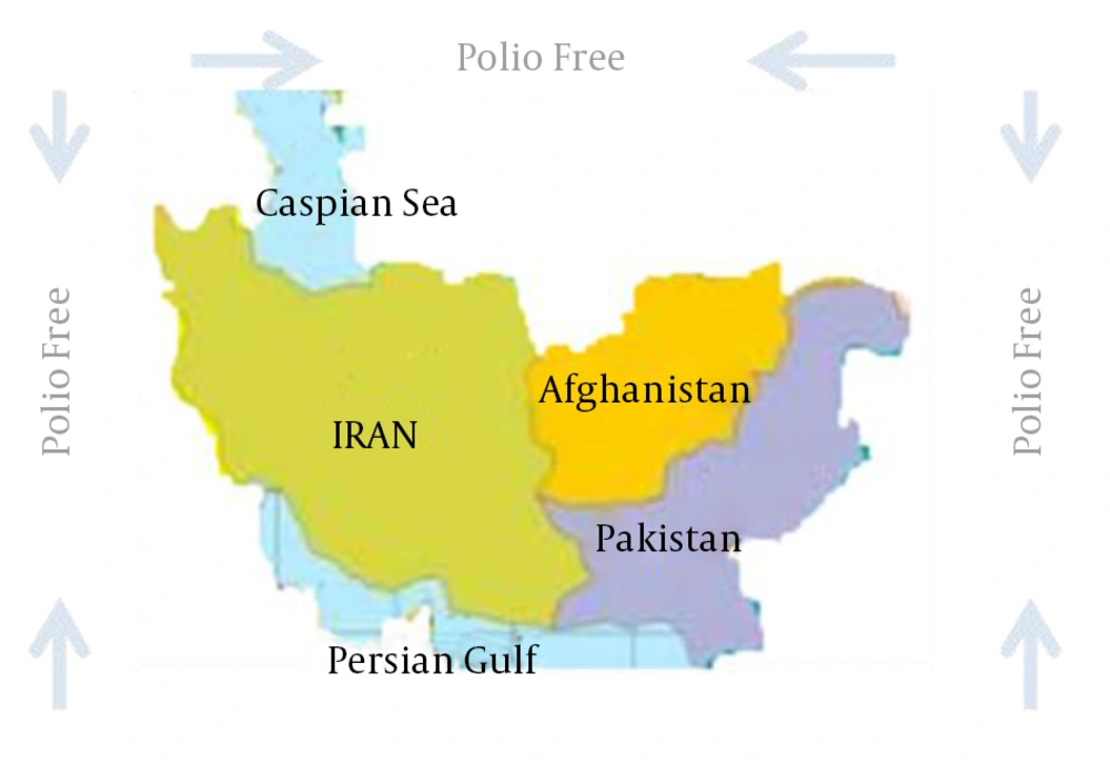
Figure 1.
Iran’s Geographical Location
3.7. AFP and VAPP Reports in Iran
Acute flaccid paralysis (AFP) is a clinical manifestation characterized by reduced muscle tone (weakness) or paralysis. The causes of AFP are as follows:
Guillain‐Barre syndrome, acute axonal neuropathy, neuropathies of infectious diseases such as diphtheria and Lyme disease, acute toxic neuropathies by heavy metals, arthropod bites, focal mono neuropathy, dermatomyositis, periodic paralyses, corticosteroids and blocking agents, Post viral myositis, myasthenia gravis, botulism, poisoning by organophosphoric insecticides, tick bite paralysis, snake bites, acute porphyrias, critical illness neuropathy, acute myopathy in patients attended to the intensive care unit (ICU), cord compression, multiple sclerosis, transverse myelitis, acute disseminated encephalomyelitis (ADEM), ischemic cord damage and vaccine-associated paralytic poliomyelitis (23).
Vaccine-associated paralytic poliomyelitis (VAPP) is an uncommon side effect of oral polio vaccine. Moussavi et al., in their report entitled “polio eradication in Iran” stated the main causes of AFP as follows:
Guillain-Barre syndrome (the most common cause), cerebral nervous infarctions, transverse myelitis, myelopathy, cerebral palsy, peripheral neuropathy, ADEM, metabolic disorders, synovitis, ischemic encephalopathy, acute lymphocytic leukemia (ALL), myositis, hereditary neuropathy and mediastinal teratoma (5).
There is an appropriate surveillance system in Iran’s health network. In this system, all the target diseases, including AFP are monitored (5).
According to Moussavi et al., the lowest rate of non-polio AFP was observed in Semnan province, Iran, with 1.4% and the highest one in Mazandaran province with 6.3%. The total reported cases in Iran were 622 with a total rate of 3.3% in 2010.
The following table shows flaccid paralysis cases caused by wild polio virus and oral polio vaccine in Iran and the three mentioned countries compared to Iran in 2014 and 2015 (9-15).
Table 1. Poliomyelitis and AFP Cases in Iran and Non-Eradicated Polio Countries in 2014 and 2015a
| Country | AFP surveillance, 2015 | Poliomyelitis Cases | |||
|---|---|---|---|---|---|
| AFP Cases Reported | 2014 | 2015 | |||
| WPV1 | cVDPV2* | WPV1 | cVDPV1 | ||
| Iran | 324 | 0 | 0 | 0 | 0 |
| Afghanistan | 1242 | 28 | 0 | 3 | 0 |
| Pakistan | 1964 | 306 | 22 | 25 | 0 |
| Nigeria | 5341 | 6 | 30 | 0 | 0 |
| Global total | 37258 | 359 | 55 | 28 | 0 |
aAFP, acute flaccid paralysis; WPV1, wild poliovirus type 1; cVDPV 1&2, circulatory-associated vaccine derived polio virus type 1 and 2.
The total number of reported cases of non-polio was 708 in 2013 (4 per 100000, among under 15 years population) (19).
Soltani et al. conducted an observational study on 139 children aged < 15 years with AFP from January 2000 to December 2010 in Kurdistan, Iran. In this study, in 138 (99%) stool samples no poliovirus was isolated and none of them were diagnosed with polio. They concluded that to monitor AFP, the key requirement is an appropriate surveillance system (24).
Naeini et al. in a cross-sectional survey, reviewed the records of youth under 15-year-old with AFP in Isfahan province, Iran, from 2007 to 2013. All the cases were visited and three stool samples were collected from each of them and sent to the national polio laboratory for poliovirus isolation. In this study, 85 cases were analyzed, 54 males (63.5%) and 31 females (36.5%). The mean age of the patients was 5.7 ± 3.9 years. The most common cause (83.5%) of paralysis among these patients was Guillian-Barre syndrome. No poliomyelitis was found in this study except one case of vaccine associated poliomyelitis (VAPV) (22). Guillain-Barré syndrome was also the most frequent final diagnosis in the report by Soltani et al. (24). Poorolajal et al. in an eight-year surveillance of 88 children with non-polio AFP, aged < 15 in Hamadan, Iran, reported 74 patients (84.0%) with complete paralysis. Guillain-Barré syndrome was the major leading cause of AFP in these children and in none of them wild polioviruses were detected (25). Salehiomran et al. showed high incidence of Guillain-Barré syndrome among AFP cases (26).
Shahmahmoodi et al., isolated type 3 immunodeficiency-associated vaccine-derived polioviruses (iVDPVs) in a 15-month-old Iranian boy with AFP in 2008. All of the seven contacts, who had been tested, were negative for polio and no secondary AFP cases were found (27).
Dehghani et al. reported that 80% of the liver transplant recipients had protective antibody titers for poliomyelitis (28).
Li et al. investigated the polio virus excretion in a total of 562 PID cases from 2008 to 2013. In this study, 17 patients (3%) shed poliovirus from the stool, but none of them were developed paralysis during the study period. They concluded that, surveillance for polioviruses among the patients with immune disorders should be established (16).
Rahimi et al. reported three AFP cases with paralysis in which Sabin-like type 1 polioviruses (neurovirulent) were isolated in Iran in 2001 (29).
Parvaneh et al. in a case report described a fatal case of VAPP in an eight–month-old infant with major histocompatibility (MHC) class II deficiency (30).
Shahmahmoodi et al. in a study conducted on infants with immunodeficiency, determined the prevalence of VAPP from 1995 to 2008 in Iran. They suggested that, screening the neonates for immunodeficiency could reduce the risk for VAPP; thus, infants should be screened for immune disorders and inactivated polio vaccine should be used for them (31).
4. Discussion
To eradicate poliomyelitis from an area, using the vaccine alone is not enough. The other factors that coincide with the vaccination should be considered. Some of these factors include: geographical location (32, 33), water and food contaminated by human feces, non-healthy sewages, abundance of the insects in the environment (1, 34), low literacy, poor personal hygiene, being afraid of vaccine complications (35, 36), attitudinal factors, religion (32), political and security problems, lack of an appropriate and comprehensive health system, population immunity level, low rate of vaccination coverage, Inappropriate management of holding national immunization days (NIDs), economic poverty (37) including absence of an appropriate budgeting system and low per capita for health, genetic factors (33), malnutrition, etc.
Hereunder, the status of each effective factor in non- eradicated polio countries is compared with that of Iran.
4.1. Geographical Location
Traditionally, it is known that the response of children in the tropical and developing countries to the oral polio vaccine is low. Iran is not located in a tropical zone (24, 32, 33); in the other words, it is known that children in the tropical and developing countries respond poorly to OPV (33). None of the two countries that are still contaminated by wild poliovirus, and also Iran, is classified among the tropical regions (38).
4.2. Contamination of Water by Human Feces and Non-Healthy Sewages
Poliomyelitis is a highly contagious disease mainly transmitted via the fecal-oral route. Studies show that drinking water in Pakistan and Nigeria is not safe (1, 35). Total population access to improved drinking water sources in 2015 is reported 22%, 47%, 90% and 94% in Afghanistan, Nigeria, Pakistan and Islamic Republic of Iran, respectively (39). Iran’s health officials should continuously monitor the health of the Helmand River water flowing from Afghanistan to Iran (40).
4.3. Economic Poverty
Economic poverty (absence of an appropriate budgeting system and low per capita for health) is an important factor.
4.4. Low Literacy
In a study in 2014, female literacy rate in the world was 79.7%, in Afghanistan 12.6%, in Nigeria 50.4%, in Pakistan is 42% and in Iran 82.5% (41). Therefore, the literacy rate of the mothers in the addressed countries is lower than that of Iran.
4.5. Fear of Mothers from Vaccine Complications
The attenuated virus in OPV reverts into a form that can paralyze (36). Thus, the fear of vaccination is not a non-reasonable reaction.
4.6. Religious Factors
Religious extremists with wrong impressions from the religious orders are an important factor against using polio vaccine and they are a major factor in the failure of immunization programs against polio in Nigeria (41), Pakistan (42) and Afghanistan (43). In these countries, focusing on the communication strategies including dialogues with religious leaders, holding some individual meetings, seminars, workshops, campaigns and media programs is believed to be necessary to eliminate such misconceptions, which can in turn contribute to raising public awareness about the urgency of the need for polio vaccination.In Iran, more than 20 years ago when the national vaccination days were held, religious (Shia or Sunni) leaders encouraged people to participate in the vaccination programs.
4.7. Political and Security Issues
The world news repeatedly expresses the military attacks on Afghanistan and Pakistan. These attacks may lead to regional instability and migration of the people into Iran.
Based on the latest statistics, about 2.5 million Afghan refugees live in Iran (44). Therefore, it is very important to control and vaccinate the immigrants.
4.8. Lack of an Appropriate and Comprehensive Health System
Holding the national polio days requires a coordinated and powerful health care delivery system, and it seems that, in the countries where wild virus is still reported, this system is not powerful. Appropriate management of holding national immunization days (NIDs) is directly associated with health delivery system in the countries. Some factors in this domain are weaknesses of policy making, planning, preparation and provision of a safe and effective vaccine, correct cold chain monitoring, organizing, mobilizing of manpower and implementing the mass vaccination on NIDs and continuing the vaccination monitoring. Iran’s primary health care system is approved by the worldwide health authorities (45).
4.9. The Rate of Vaccination Coverage
Once the polio vaccine coverage is low, polioviruses vaccines may circulate among insufficiently immunized people and become pathogenic [46].
Estimated coverage of polio vaccination among infants aged < 1 year with three doses of OPV3, in 2013 was 90% in Afghanistan, 67% in Nigeria, and 66% in Pakistan (37). Current immunization coverage with three doses of OPV in Iran is more than 95% percent (19), and this coverage rate is sufficient to eradicate polio.
4.10. Genetic Factors
The role of genetics in polio is known (33), but, information is not available about the role of genetics in Iran, Afghanistan and Pakistan.
4.11. Recommended Vaccine for Iran
Despite using IPV in most of the developed countries, in Iran, OPV is still used.
As already mentioned, Iran is considered as a polio-free country since 2001; however its two neighboring countries, Afghanistan and Pakistan are still as two endemic countries for the wild virus. Therefore, especially in the areas around the geographical borders, using the OPV vaccine is preferred. In addition, using IPV or combined schedules (OPV + IPV) is not cost-effective (19).
4.12. Conclusions
Trivalent oral polio vaccine (tOPV) is a live and attenuated vaccine, consisting types 1, 2 and 3 of the polio viruses. The most important disadvantage of OPV is genetic instability, resulting in vaccine-associated paralytic poliomyelitis. The immune response against polio virus type 3 (PV3) was lower than those of other serotypes. Therefore, improving the population immunity against this serotype is an urgent priority. The wild polio virus is disrupted in most countries, except Afghanistan and Pakistan. The majority of VDPPs are associated with type 2 circulating VDPVs (cVDPV2). Therefore, it is suggested to remove type 2 virus from trivalent vaccines (tOPV) and change this vaccine to bivalent vaccines (bOPV), consisting only types 1 and 3 polioviruses.
From 2014 to 2015, 324 non-polio paralyses were reported in Iran. Iran’s health managers should continuously monitor the vaccination coverage with three doses of OPV in the level over 95%, monitor the health of the Helmand River water which flows from Afghanistan to Iran and vaccinate the immigrants.
