Asian J Sports Med

Image Credit:Asian J Sports Med

Comparative Effects of Vibration and Mechanical Massage on Exercise-Induced Delayed-Onset Muscle Soreness in Sedentary Students: A Randomized Controlled Trial

Author(s):
Saixi SongSaixi SongSaixi Song ORCID1, Wichai EungpinichpongWichai EungpinichpongWichai Eungpinichpong ORCID2,*
1Department of Exercise and Sport Sciences, Faculty of Graduate School, Khon Kaen University, Khon Kaen, Thailand
2School of Physical Therapy, Faculty of Associated Medical Sciences, Khon Kaen University, Khon Kaen, Thailand

Asian Journal of Sports Medicine:Vol. 16, issue 3; e169855
Published online:May 12, 2026
Article type:Research Article
Received:Jan 20, 2026
Accepted:Mar 26, 2026
How to Cite:Song S, Eungpinichpong W. Comparative Effects of Vibration and Mechanical Massage on Exercise-Induced Delayed-Onset Muscle Soreness in Sedentary Students: A Randomized Controlled Trial. Asian J Sports Med. 2025;16(3):e169855. doi: https://doi.org/10.5812/asjsm-169855

Abstract

Background:

Delayed-onset muscle soreness (DOMS) significantly impairs functional capacity and exercise tolerance in sedentary college students. Although massage interventions show promise in promoting recovery from DOMS, comparative evidence between mechanical and vibration modalities remains limited.

Objectives:

To compare the efficacy of mechanical massage (leg massager) versus vibration massage (massage gun) in promoting acute recovery from exercise-induced DOMS in the lower-limb muscles of collegiate athletes.

Methods:

This single-blind (assessor-blinded) randomised controlled trial enrolled 34 sedentary college students (n = 17 per group), based on an a priori power analysis (G*Power 3.1.9.7; f = 0.25, α = 0.05, power = 0.80), aged 18 - 23 years with a BMI of 18 - 24 kg/m², who underwent exercise-induced gastrocnemius DOMS modelling. Participants were randomly allocated (using a random number table stratified by baseline VAS scores by an independent researcher) to receive either mechanical massage (leg massager, 50 - 80 mmHg, five minutes per limb) or vibration massage (massage gun, 50 Hz, five minutes per limb) at 24 hours post-exercise. The pre-registered primary outcome was pain intensity, measured using the visual analogue scale (VAS) at 24 hours post-intervention. Secondary outcome measures included pain intensity (VAS), pressure pain threshold (PPT), muscle strength (hand-held dynamometer, HHD), range of motion (ROM), and calf circumference, assessed at baseline, immediately post-exercise, and at 24, 48, and 72 hours post-exercise. Data were analysed using a two-factor repeated measures ANOVA (time × group).

Results:

Both groups showed significant time-dependent improvements across all outcomes (P < 0.05). VAS decreased from 24 to 72 hours (mechanical: 4.24 ± 2.04 to 1.49 ± 1.61; vibration: 3.98 ± 2.06 to 0.57 ± 0.68; F = 30.819, P < 0.001), with no between-group difference (F = 0.598, P = 0.445); the group × time interaction showed a trend toward significance (F = 2.267, P = 0.086). At the prespecified primary endpoint (VAS at T2, 24 hours post-intervention), the between-group mean difference was 0.26 points (95% CI: −1.12 to 1.64; Cohen’s d = 0.13), indicating negligible between-group separation. The time effect was large (partial η² = 0.491). At 72 hours, the between-group difference was 0.92 points (95% CI: 0.09 to 1.75; Cohen’s d = 0.74), which did not exceed the MCID of 1.5 - 2.0 points for acute musculoskeletal pain. PPT, muscle strength, ROM, and calf circumference demonstrated comparable recovery patterns between groups (all P > 0.05). No adverse events occurred.

Conclusions:

Both mechanical and vibration massage interventions were associated with similar patterns of acute recovery from DOMS, with no statistically or clinically meaningful between-group differences observed in any outcome measure. Because neither modality demonstrated superiority and the absence of a no-treatment control group precludes definitive attribution of improvements to the interventions, these findings support the feasibility and tolerability of both devices as non-pharmacological options for sedentary young adults, pending confirmation in adequately controlled trials.

1. Background

Delayed-onset muscle soreness (DOMS) is a common condition characterised by muscle discomfort, stiffness, tenderness, and functional impairment that typically emerges 24 to 48 hours after unaccustomed or intense eccentric exercise, peaks between 24 and 72 hours post-exercise, and gradually resolves within five to seven days (1). The pathophysiology of DOMS is multifactorial, involving mechanical microtrauma to muscle fibres, inflammatory cascades, oxidative stress, and disrupted calcium homeostasis (2). Eccentric contractions generate excessive tensile forces, leading to sarcomere disruption and myofibrillar protein degradation, particularly in Type II muscle fibres (3). This damage triggers secondary inflammatory responses that sensitise nociceptors and contribute to perceived muscle soreness (4). Beyond subjective discomfort, DOMS impairs neuromuscular function, reducing maximal voluntary contraction force by up to 50% and compromising proprioceptive acuity (5). While these consequences are well recognised in athletic contexts, they are particularly relevant for sedentary individuals, who lack the repeated-bout conditioning that attenuates DOMS severity (6). For sedentary young adults attempting to initiate or maintain an active lifestyle, DOMS-associated discomfort and functional limitation may discourage continued exercise participation, with potential implications for long-term physical activity levels and chronic disease prevention. Moreover, engaging in physical activity while DOMS remains unresolved substantially increases the risk of secondary musculoskeletal injuries (7-9), further hindering regular participation.
Various recovery interventions have been used to manage DOMS, including cryotherapy, contrast water immersion, active recovery, nutritional supplementation, and massage-based therapies (10-12). Among these, massage therapy has gained considerable attention because of its purported mechanisms, including enhancing local blood flow, facilitating metabolic waste removal, reducing muscle stiffness, and modulating pain perception (13, 14). More recently, mechanical massage devices—including pneumatic compression systems and percussive vibration therapy tools—have emerged as accessible, cost-effective, user-administered alternatives to traditional manual massage (15). These modalities deliver rhythmic mechanical stimulation to superficial and deep myofascial tissues, potentially modulating pain pathways (16) and facilitating muscle fibre recruitment (13). Despite their widespread adoption, the comparative efficacy of these two device categories for DOMS recovery remains poorly established. Two systematic reviews addressing massage-based interventions for DOMS — Nahon et al. and Torres et al. — identified considerable methodological heterogeneity across existing studies (17-19), including the absence of standardised DOMS induction protocols, reliance on subjective pain ratings without objective functional outcomes, insufficient follow-up periods beyond 48 hours, and substantial variability in intervention parameters. Critically, direct head-to-head RCT evidence comparing pneumatic compression and percussive vibration devices for DOMS remains extremely limited, precluding evidence-based device selection for clinicians and consumers alike. To our knowledge, no published RCT has directly compared a pneumatic compression leg massager with a percussive vibration massage gun for DOMS recovery. The two systematic reviews identified do not include such a head-to-head comparison, and no meta-analysis specifically addressing this device pairing could be identified. The present study therefore addresses a specific and unmet gap in the comparative evidence base.

2. Objectives

Therefore, this randomised controlled trial aims to systematically compare the acute recovery effects of mechanical massage (a pneumatic compression leg massager) and vibration massage (a percussive massage gun) following exercise-induced gastrocnemius DOMS (a muscle frequently affected by eccentric exercise owing to its predominant Type II fibre composition) in a sedentary college student population. By employing standardised DOMS induction protocols and comprehensive, multidimensional outcome assessments, this study seeks to generate robust comparative efficacy data to inform evidence-based recovery strategies. We hypothesised that vibration massage, through high-frequency mechanoreceptor activation consistent with gate-control pain modulation (16), would produce significantly greater reductions in pain intensity, as measured by the visual analogue scale (VAS) at 24 hours post-intervention, compared with pneumatic compression massage.

3. Methods

3.1. Study Design

This study was approved by the Ethics Committee of Khon Kaen University, Thailand (Approval No. HE682109). The trial registration number is [TCTR20251218001]. The registered primary outcome of the trial was visual analog scale (VAS) pain intensity.
This parallel-group, single-blind (assessor-blinded) randomised controlled trial was conducted at the Exercise Laboratory of Guangxi University of Finance and Economics. The study employed a repeated-measures design with five assessment time points over 72 hours following exercise-induced DOMS.

3.2. Participants

A total of 42 healthy adults were recruited from Guangxi University of Finance and Economics (Nanning, China) via poster advertising. After screening, eight individuals were excluded because they did not meet the criteria. Inclusion criteria required participants to be free of musculoskeletal injury or lower-limb pathology within the previous six months, not regularly engaged in structured lower-limb resistance training, and classified as having low physical activity levels (Physical Activity Rating Scale [PARS-3] score ≤19, indicating a small amount of exercise (20). Exclusion criteria included pregnancy, cardiovascular or neurological disorders, current use of anti-inflammatory medication, and prior adverse reactions to massage therapy. Thirty-four volunteers enrolled and signed consent forms to participate in the study.

3.3. DOMS Induction Protocol

DOMS was induced using standardised eccentric calf raises. After warm-up and 1RM determination, participants performed 7 sets of 15 repetitions at 60% 1RM per leg, with controlled 4-s eccentric lowering to 15° dorsiflexion, a 1-s hold, and a 1-s concentric return, with 90-s rest between sets.

3.4. Randomisation and Blinding

Following DOMS induction, participants were randomly allocated to either the mechanical massage group (MMG, N = 17) or the vibration massage group (VMG, N = 17) using a random number table, stratified by baseline VAS pain scores (<5 vs. ≥5) to ensure balanced pain levels between groups. An independent researcher not involved in outcome assessment generated the allocation sequence. Allocation concealment was achieved through centralised third-party allocation. The independent researcher exclusively held the allocation sequence and disclosed individual assignments only at the moment of intervention delivery, after DOMS induction was completed. The recruiting investigator remained unaware of assignments throughout enrolment, and the treating therapist was informed only immediately prior to each session. Baseline equivalence between groups was confirmed using independent t-tests for all continuous variables (all P > 0.05). Due to the nature of the interventions, participants and treatment administrators could not be blinded. However, outcome assessors were fully blinded to intervention delivery. All outcome measures were collected by trained evaluators who remained blinded to group allocation throughout data collection and analysis. Participants were instructed not to discuss their intervention with assessors to maintain blinding integrity. Given that the primary outcome (VAS) is subjective in nature, potential susceptibility to expectancy and placebo effects was acknowledged. To mitigate this risk, objective outcome measures—including PPT, HHD, ROM, and calf circumference—were incorporated alongside VAS. The convergent patterns across subjective and objective outcomes support the internal validity of the findings.

3.5. Intervention Protocols

Mechanical Massage: Participants sat with their calves positioned in a pneumatic leg massager (50 - 80 mmHg, second intensity level), which delivered sequential compression for 5 minutes per limb at 24h post-exercise.
Vibration Massage: Participants lay prone with relaxed calves. A percussion gun (50 Hz, 3000 rpm) with a circular flat head (3.8 cm) was applied at 45° with consistent pressure (2 - 3 kg/cm²), progressing from proximal to distal. Each limb received 2.5 minutes of medial and 2.5 minutes of lateral gastrocnemius treatment (5 minutes total per limb) at 24h post-exercise.

3.6. Outcome Measures

All outcome measures were assessed at baseline (T0, pre-exercise), immediately post-exercise (T1), and at 24 hours (T2, post-intervention), 48 hours (T3), and 72 hours (T4) post-exercise. The intervention was administered at 24 hours post-exercise, with T2 measurements conducted immediately following the intervention, consistent with established DOMS time courses (21). Trained evaluators conducted assessments blinded to group allocation. To minimise fatigue effects and maintain measurement reliability, assessments followed a standardised sequence: 1. calf circumference, 2. pain intensity (VAS during half-squat), 3. pressure pain threshold (PPT), 4. range of motion (ROM), and 5. muscle strength (hand-held dynamometer, HHD). This order progressed from the least to the most physically demanding measurements. Participants were instructed not to disclose their intervention assignment to evaluators, and assessments were conducted in a separate room to maintain blinding. Standardised protocols were followed to minimise inter-rater variability.
Subjective muscle soreness was quantified using a 100-mm visual analogue scale (VAS), anchored at 0 mm (no pain) and 100 mm (worst pain imaginable). A review indicates that the VAS has ρ values ranging from 0.60 to 0.77 in reliability studies and from 0.76 to 0.84 in validity studies (22), demonstrating adequate measurement reliability and validity. VAS was assessed during a standardised half-squat position (thigh parallel to the ground) to capture movement-evoked pain representative of DOMS symptoms during daily activities.
Maximal isometric plantarflexion strength was assessed using a hand-held dynamometer with established reliability (ICC = 0.91 - 0.98) (23). Participants assumed a prone position with the knee fully extended and the ankle in neutral dorsiflexion. The dynamometer was positioned perpendicular to the plantar surface of the foot at the level of the metatarsal heads. Three maximal isometric contractions (5s each, 60s rest) were performed. The highest force output (recorded in Newtons) across the three trials was used for analysis. Consistent verbal encouragement was provided to ensure maximal effort.
Mechanical pain sensitivity was quantified using a hand-held pressure algometer with a 1-cm² probe tip, demonstrating excellent reliability (ICC = 0.91) (24). The pressure pain threshold (PPT) was defined as the minimum pressure intensity required to elicit a pain sensation. The algometer was applied perpendicular to the skin at the midpoint of the gastrocnemius muscle belly, with pressure increasing at a constant rate of 1 kg/cm²/second. Participants indicated when pressure first became painful, and the value (kg/cm²) was recorded. Three measurements were taken at each time point with 30-second intervals, and the mean value was calculated.
Active ankle plantarflexion ROM was measured using a standard plastic goniometer following established reliability protocols (ICC = 0.89 - 0.94) (25, 26). Participants lay prone with the knee extended and the foot hanging freely over the edge of the examination table. The goniometer axis was aligned with the lateral malleolus, the stationary arm parallel to the fibular shaft, and the moving arm parallel to the fifth metatarsal. Participants actively plantarflexed the ankle to the maximal comfortable range without compensatory movements, and the angle was recorded in degrees. Three measurements were taken and averaged (27).
Muscle swelling was assessed by measuring calf circumference at the point of maximal girth using a flexible anthropometric tape measure. Participants stood with equal weight distribution, and the measurement was taken with the tape positioned perpendicular to the long axis of the lower leg. Three measurements were recorded and averaged, with the measurement site marked to ensure consistency across time points. This method of assessing calf circumference has been shown to have high reliability [SEM = 0.5 - 0.6 cm; intraclass correlation coefficient (ICC) = 0.97] (28) and to correlate with muscle volume (R2 = 0.42) (29).

3.7. Statistical Analysis

Sample size was calculated a priori using G*Power 3.1.9.7 (Franz Faul, University of Kiel, Germany) for ANOVA: Repeated measures, within-between interaction. Based on previous studies, a medium effect size of f = 0.25 was assumed, with α = 0.05, power (1−β) = 0.80, two groups, five measurement time points, a correlation among repeated measures of 0.50, and a nonsphericity correction of ε = 0.71, yielding a minimum total sample size of 26 participants (actual power = 0.808). Accounting for a 20% anticipated dropout rate, the required final sample was 34 participants (17 per group). All statistical analyses were performed using SPSS version 29.0 (SPSS Inc., Chicago, IL, USA). Baseline characteristics were compared between groups using independent t-tests for continuous variables. A two-factor repeated-measures ANOVA (time × group) was conducted to examine the effects of time, group, and their interaction on each outcome measure. The significance level was set at α = 0.05 for all analyses. Partial eta-squared (η²) values were calculated as measures of effect size. Post-hoc pairwise comparisons were performed when significant main effects or interactions were detected. Bonferroni correction was applied to all post-hoc pairwise comparisons to control the familywise Type I error rate across multiple time-point comparisons. Prior to analysis, normality was assessed using the Shapiro-Wilk test, and homogeneity of variance was confirmed (P > 0.05). Sphericity was assessed using Mauchly’s test; when the assumption of sphericity was violated, Greenhouse-Geisser corrections were applied to degrees of freedom and associated p-values. Data are presented as mean ± standard deviation (SD).

4. Results

4.1. Participant Characteristics and Study Completion

A total of 34 participants were enrolled and randomly allocated to two groups. All participants completed the study protocol, with no dropouts or protocol violations. The mechanical massage group (MMG) comprised seven males and 10 females (mean age 19.76 ± 1.09 years, BMI 21.22 ± 1.79 kg/m²), whereas the vibration massage group (VMG) comprised five males and 12 females (mean age 20.12 ± 1.58 years, BMI 20.83 ± 1.84 kg/m²). Independent t-tests showed no statistically significant between-group differences in baseline characteristics (all P > 0.05), confirming successful randomisation. No adverse events were reported during the study period.

4.2. Visual Analogue Scale (VAS) Pain Intensity

The results of the two-way repeated measures ANOVA for VAS pain intensity are summarised in Table 1. A significant main effect of time was observed (F = 30.819, P < 0.001, partial η² = 0.491), whereas the main effect of group was not statistically significant (F = 0.598, P = 0.445, partial η² = 0.019). The time × group interaction effect was also not significant (F = 2.267, P = 0.086, partial η² = 0.066).
Table 1.Visual Analog Scale Pain Intensity at Different Time Points a, b
TimeVibration Massage Group (VMG)Mechanical Massage Group (MMG)
T00.06 ± 0.220.25 ± 0.68
T12.17 ± 1.92 c1.19 ± 1.10 c
T23.98 ± 2.06 c4.24 ± 2.04 c
T31.22 ± 0.63 c1.78 ± 1.0 c
T40.57 ± 0.68 c1.49 ± 1.61 c

a Values are expressed as mean ± SD (N = 34, scores 0 - 10).

b Group × time interaction: P = 0.086.

c P < 0.001 compared to baseline (T0) within the same group.

Although the between-group difference at T4 reached a medium effect size (d = 0.74), the absolute mean difference (0.92 points) remained below the commonly accepted MCID threshold of 1.5 - 2.0 points for acute musculoskeletal pain, suggesting that the observed separation is unlikely to represent clinically meaningful superiority of either intervention.

4.3. Pressure Pain Threshold (PPT)

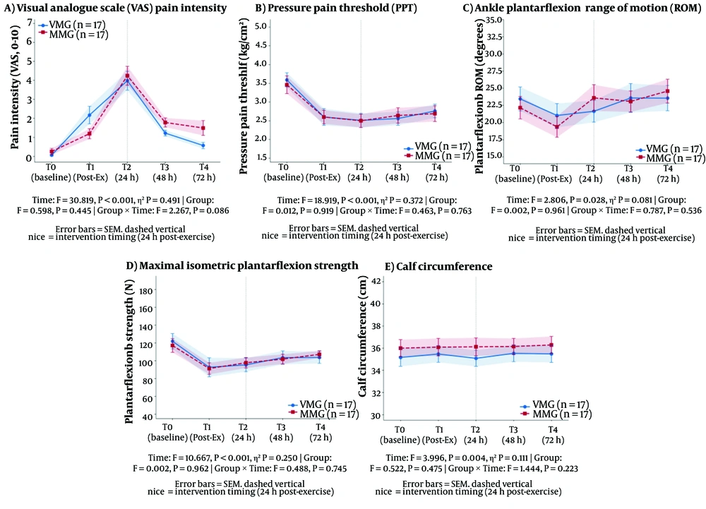
As shown in Table 2, PPT demonstrated a significant main effect of time (F = 18.919, P < 0.001, partial η² = 0.372), with no significant main effect of group (F = 0.012, P = 0.919, partial η² = 0.000) or time × group interaction (F = 0.463, P = 0.763, partial η² = 0.014) (Figure 1).
Table 2.Pressure Pain Threshold at Different Time Points a
TimeVibration Massage Group (VMG)Mechanical Massage Group (MMG)
T03.59 ± 0.793.46 ± 0.97
T12.60 ± 0.92 b2.60 ± 0.75 b
T22.51 ± 0.78 b2.50 ± 0.73 b
T32.56 ± 0.74 b2.64 ± 0.85 b
T42.76 ± 0.75 b2.69 ± 0.87 b

a Values are are expressed as mean ± SD (N = 34, kg/cm²).

b P < 0.001 compared to baseline (T0) within the same group.

Recovery trajectories across all outcome measures following mechanical and vibration massage interventions for exercise-induced DOMS. Panels show (A) Visual Analogue Scale (VAS) pain intensity, (B) Pressure Pain Threshold (PPT), (C) Ankle Plantarflexion Range of Motion (ROM), (D) Maximal Isometric Plantarflexion Strength, and (E) Calf Circumference. Data are presented as mean ± SEM (n = 17 per group). The dashed vertical line indicates the timing of the intervention (24h post-exercise). Abbreviations: VMG, vibration massage group; MMG, mechanical massage group.
Figure 1.

Recovery trajectories across all outcome measures following mechanical and vibration massage interventions for exercise-induced DOMS. Panels show (A) Visual Analogue Scale (VAS) pain intensity, (B) Pressure Pain Threshold (PPT), (C) Ankle Plantarflexion Range of Motion (ROM), (D) Maximal Isometric Plantarflexion Strength, and (E) Calf Circumference. Data are presented as mean ± SEM (n = 17 per group). The dashed vertical line indicates the timing of the intervention (24h post-exercise). Abbreviations: VMG, vibration massage group; MMG, mechanical massage group.

At the post-intervention time point (T2), the between-group mean difference in PPT was −0.01 kg/cm² (95% CI: −0.52 to 0.50; Cohen’s d = −0.01), confirming negligible between-group separation at the primary assessment point.

4.4. Range of Motion (ROM)

Active ankle plantarflexion ROM showed a significant main effect of time (F = 2.806, P = 0.028, partial η² = 0.081), with no significant main effect of group (F = 0.002, P = 0.961, partial η² = 0.000) or time × group interaction (F = 0.787, P = 0.536, partial η² = 0.024) (Table 3).
Table 3.Range of Motion at Different Time Points a
TimeVibration Massage Group (VMG)Mechanical Massage Group (MMG)
T023.44 ± 7.1522.15 ± 6.76
T121.02 ± 7.20 b19.36 ± 6.29 b
T221.63 ± 6.5423.59 ± 7.86
T323.57 ± 8.6823.07 ± 6.27
T423.55 ± 7.5924.58 ± 7.14

a Values are expressed as mean ± SD (N=34, degrees).

b P < 0.001 compared to baseline (T0) within the same group.

At T2, the between-group mean difference in ROM was 1.96° (95% CI: −2.90 to 6.82; Cohen’s d = 0.27), indicating a small, non-significant between-group effect.

4.5. Muscle Strength (Hand-Held Dynamometer)

Hand-held dynamometry revealed a significant main effect of time (F = 10.667, P < 0.001, partial η² = 0.250), with no significant main effect of group (F = 0.002, P = 0.962, partial η² = 0.000) or time × group interaction (F = 0.488, P = 0.745, partial η² = 0.015) (Table 4).
Table 4.Muscle Strength at Different Time Points a
TimeVibration Massage Group (VMG)Mechanical Massage Group (MMG)
T0121.83 ± 35.43117.00 ± 31.75
T192.56 ± 44.06 b91.13 ± 26.62 b
T295.25 ± 31.22 b97.61 ± 24.21 b
T3103.39 ± 30.98 b101.72 ± 22.41 b
T4103.61 ± 27.46 b107.06 ± 16.68 b

a Values are expressed as mean ± SD (N = 34, Newtons).

b P < 0.001 compared to baseline (T0) within the same group.

Strength had not returned to the ≥95% baseline threshold commonly considered indicative of full functional recovery, suggesting that the 72-hour observation window may be insufficient to capture complete strength restoration following DOMS. At T2, the between-group mean difference in plantarflexion strength was 2.36 N (95% CI: −16.42 to 21.14; Cohen’s d = 0.08), confirming a negligible between-group effect at the post-intervention assessment.

4.6. Calf Circumference

Calf circumference exhibited a significant main effect of time (F = 3.996, P = 0.004, partial η² = 0.111), with no significant main effect of group (F = 0.522, P = 0.475, partial η² = 0.016) or time × group interaction (F = 1.444, P = 0.223, partial η² = 0.043) (Table 5).
Table 5.Calf Circumference at Different Time Points a
TimeVibration Massage Group (VMG)Mechanical Massage Group (MMG)
T035.16 ± 3.2836.00 ± 3.15
T135.47 ± 3.0736.09 ± 3.28
T235.08 ± 2.9836.12 ± 3.37
T335.53 ± 3.11 b36.15 ± 3.01 b
T435.48 ± 3.15 c36.29 ± 3.19 c

a Values are expressed as mean ± SD (N = 34, cm).

b P < 0.05.

c P < 0.001 compared to baseline (T0) within the same group.

At T2, the between-group mean difference in calf circumference was 1.04 cm (95% CI: −1.10 to 3.18; Cohen’s d = 0.33), which remains within the published measurement error range for this technique (SEM = 0.5 - 0.6 cm) and is therefore unlikely to represent a clinically meaningful difference.

5. Discussion

We documented immediate post-exercise changes across all outcomes, an aspect that has received relatively little attention in previous DOMS research (21, 30). Participants reported mild to moderate pain immediately after exercise, consistent with acute muscle damage, before the delayed pain peak typically observed at 24 hours (11, 21). Concurrent immediate reductions in muscle strength, pain pressure threshold, and ankle ROM confirmed acute muscle dysfunction induced by the eccentric protocol.
The group × time interaction for VAS approached, but did not reach, statistical significance (F = 2.267, P = 0.086), indicating a possible trend toward greater pain reduction with vibration massage that requires confirmation in larger trials.
Similar patterns were observed for muscle strength, pain pressure threshold, ankle ROM, and calf circumference. All variables showed significant recovery over time, with no statistically significant group differences (all P > 0.05), indicating that both mechanical and vibration massage produced comparable trajectories of functional and symptomatic improvement over the 72-hour follow-up. No adverse events were reported, supporting the short-term safety and tolerability of both devices in sedentary young adults.
These findings are generally consistent with previous literature (17, 18, 31) indicating that massage can attenuate DOMS symptoms and facilitate the recovery of muscle function, although the magnitude and duration of effects vary across studies and modalities (17-19). Our results extend existing evidence by directly comparing a mechanical leg massager with a hand-held vibration massage gun under controlled conditions. Notably, the between-group difference at T4 (0.92 points) did not exceed the commonly accepted MCID of 1.5 - 2.0 points for acute musculoskeletal pain on a 0 - 10 VAS, indicating that the absence of between-group superiority is not only statistically but also clinically meaningful.
The near-significant group × time interaction for VAS suggests a possible advantage of vibration massage for pain reduction, with a larger absolute decrease in VAS scores from 24 to 72 hours in the vibration group. However, given the P-value of 0.086 and modest sample size, this pattern should be interpreted cautiously and viewed as hypothesis-generating rather than conclusive. Future studies with larger samples and greater statistical power are needed to clarify whether vibration massage confers clinically meaningful incremental benefits over mechanical massage for DOMS-related pain.
It should be noted that the following mechanistic explanations are speculative, as this trial was not designed to directly assess physiological mechanisms. Several mechanisms may underlie the observed improvements in both groups. First, both modalities are thought to activate cutaneous and muscle mechanoreceptors, modulating afferent nociceptive signalling consistent with gate-control theory (16, 32). This mechanism may account for the immediate post-intervention reductions in VAS observed in both groups. Second, mechanical pressure applied to soft tissues may facilitate local circulation and clearance of pro-inflammatory metabolites such as prostaglandins (13, 14), thereby attenuating peripheral sensitisation associated with DOMS. Third, vibration-specific effects (32-34), including activation of muscle spindles and Golgi tendon organs at 50 Hz, may additionally promote neuromuscular relaxation and alter viscoelastic muscle properties, potentially contributing to the trend toward greater pain reduction observed in the VMG (P = 0.086). However, this interpretation should be considered exploratory and requires confirmation in adequately powered trials.
This study has several limitations. Most importantly, the absence of a no-intervention or sham control group prevents definitive attribution of improvements to the massage interventions rather than natural recovery (12, 19). Previous studies (11, 17, 21, 30) have reported natural DOMS recovery rates of approximately 60 - 70% over 72 hours, similar to the reductions observed in both groups in the present study. Therefore, while our findings indicate that both massage modalities were well tolerated and coincided with improvements in symptoms and function, they do not establish that either modality accelerates recovery beyond the natural course. The inclusion of control or sham conditions in future trials is essential to isolate intervention-specific effects.
The sample consisted of sedentary college students aged 18 - 23 years, which limits generalisability to athletes, older adults, or individuals with underlying musculoskeletal or metabolic conditions. We evaluated the effects of a single massage session administered at 24 hours post-exercise; thus, our results may not apply to different dosing regimens, earlier or repeated applications, or other muscle groups. In addition, we focused on subjective pain, muscle strength, ROM, and calf circumference; we did not include biochemical markers of muscle damage or inflammation (3, 14), which could provide insight into potential mechanistic differences between modalities.
Despite these limitations, our findings have practical implications. For sedentary or novice exercisers experiencing gastrocnemius DOMS, both mechanical and vibration massage appear to be feasible and well-tolerated strategies that coincide with reductions in pain and improvements in muscle function over 72 hours. Given the absence of clear superiority of one modality over the other in our data, clinicians and practitioners may reasonably base their choice on device availability, cost, usability, and patient preference. However, given the lack of a no-intervention control and the exploratory nature of this trial, these findings should not be interpreted as definitive evidence of therapeutic efficacy.
Future research should include adequately powered randomised controlled trials incorporating no-treatment and/or sham controls (11, 19), diverse populations (including recreational and competitive athletes), and varying massage doses and timings. Integrating objective biomarkers (14) and longer-term follow-up would help clarify mechanisms and determine whether repeated exposure to specific massage modalities modifies DOMS susceptibility or longer-term training adaptations.

5.1. Conclusions

Both mechanical massage (leg massager) and vibration massage (massage gun) produced comparable effects on recovery from exercise-induced gastrocnemius DOMS in sedentary college students. Significant time-dependent improvements were observed across all outcome measures over 72 hours, with no statistically significant between-group differences. Both modalities were safe and well tolerated, supporting their feasibility and tolerability as non-pharmacological options in this population, although efficacy beyond natural recovery cannot be established from the present design.

Footnotes

References

  • 1.
    Afonso José, Clemente Filipe Manuel, Nakamura Fábio Yuzo, Morouço Pedro, Sarmento Hugo, Inman Richard A, et al. The Effectiveness of Post-exercise Stretching in Short-Term and Delayed Recovery of Strength, Range of Motion and Delayed Onset Muscle Soreness: A Systematic Review and Meta-Analysis of Randomized Controlled Trials. Frontiers in physiology. 2021;12:677581. 677581. [PubMed ID: 34025459]. [PubMed Central ID: PMC8133317]. https://doi.org/10.3389/fphys.2021.677581.
  • 2.
    Armstrong RB. Mechanisms of exercise-induced delayed onset muscular soreness: a brief review. Medicine & Science in Sports & Exercise. 1984;16(6):529-38. [PubMed ID: 6392811].
  • 3.
    Fridén J, Sjöström M, Ekblom B. Myofibrillar damage following intense eccentric exercise in man. International journal of sports medicine. 1983;4(3):170-6. [PubMed ID: 6629599]. https://doi.org/10.1055/s-2008-1026030.
  • 4.
    Vila-Chã Carolina, Bovolini Antonio, Francisco Cristiana, Costa-Brito Ana R, Vaz Cláudia, Rua-Alonso María, et al. Acute effects of isotonic eccentric exercise on the neuromuscular function of knee extensors vary according to the motor task: impact on muscle strength profiles, proprioception and balance. Frontiers in Sports and Active Living. 2023;5:1273152. 1273152. https://doi.org/10.3389/fspor.2023.1273152.
  • 5.
    Hamill Joseph, Freedson Patty S, Clarkson Priscilla M, Braun Barry. Muscle soreness during running: biomechanical and physiological considerations. Journal of Applied Biomechanics. 1991;7(2):125-37. https://doi.org/10.1123/ijsb.7.2.125.
  • 6.
    Clarkson Priscilla M, Hubal Monica J. Exercise-induced muscle damage in humans. American journal of physical medicine & rehabilitation. 2002;81(11):S52-S69. [PubMed ID: 12409811]. https://doi.org/10.1097/00002060-200211001-00007.
  • 7.
    Orchard John, Marsden John, Lord Stephen, Garlick David. Preseason hamstring muscle weakness associated with hamstring muscle injury in Australian footballers. The American journal of sports medicine. 1997;25(1):81-5. [PubMed ID: 9006698]. https://doi.org/10.1177/036354659702500116.
  • 8.
    Gulick DT, Kimura IF. Delayed onset muscle soreness: what is it and how do we treat it? Journal of Sport Rehabilitation. 1996;5(3):234-43.
  • 9.
    Proske U, Morgan D. L. Muscle damage from eccentric exercise: mechanism, mechanical signs, adaptation and clinical applications. The Journal of physiology. 2001;537(2):333-45. [PubMed ID: 11731568]. [PubMed Central ID: PMC2278966]. https://doi.org/10.1111/j.1469-7793.2001.00333.x.
  • 10.
    Dupuy Olivier, Douzi Wafa, Theurot Dimitri, Bosquet Laurent, Dugué Benoit. An evidence-based approach for choosing post-exercise recovery techniques to reduce markers of muscle damage, soreness, fatigue, and inflammation: a systematic review with meta-analysis. Frontiers in physiology. 2018;9:312968. 403. [PubMed ID: 29755363]. [PubMed Central ID: PMC5932411]. https://doi.org/10.3389/fphys.2018.00403.
  • 11.
    Cheung Karoline, Hume Patria A, Maxwell Linda. Delayed onset muscle soreness. Sports medicine. 2003;33(2):145-64. https://doi.org/10.2165/00007256-200333020-00005.
  • 12.
    Howatson Glyn, van Someren Ken A. The prevention and treatment of exercise-induced muscle damage. Sports medicine. 2008;38(6):483-503. [PubMed ID: 18489195]. https://doi.org/10.2165/00007256-200838060-00004.
  • 13.
    Weerapong Pornratshanee, Hume Patria A, Kolt Gregory S. The mechanisms of massage and effects on performance, muscle recovery and injury prevention. Sports medicine. 2005;35(3):235-56. [PubMed ID: 15730338]. https://doi.org/10.2165/00007256-200535030-00004.
  • 14.
    Crane Justin D, Ogborn Daniel I, Cupido Colleen, Melov Simon, Hubbard Alan, Bourgeois Jacqueline M, et al. Massage therapy attenuates inflammatory signaling after exercise-induced muscle damage. Science translational medicine. 2012;4(119):119ra13-ra13. [PubMed ID: 22301554]. https://doi.org/10.1126/scitranslmed.3002882.
  • 15.
    Cheatham SW, Kolber MJ, Cain M, Lee M. The effects of self‐myofascial release using a foam roll or roller massager on joint range of motion, muscle recovery, and performance: a systematic review. International journal of sports physical therapy. 2015;10(6):827.
  • 16.
    Melzack R, Wall PD. Pain Mechanisms: A New Theory: A gate control system modulates sensory input from the skin before it evokes pain perception and response. Science. 1965;150(3699):971-9.
  • 17.
    Wei Ming, Liu Xin, Wang Siyu. The impact of various post-exercise interventions on the relief of delayed-onset muscle soreness: a randomized controlled trial. Frontiers in physiology. 2025;16:1622377. 1622377. [PubMed ID: 40832134]. [PubMed Central ID: PMC12358485]. https://doi.org/10.3389/fphys.2025.1622377.
  • 18.
    Nahon Roberto Lohn, Silva Lopes Jaqueline Santos, Monteiro de Magalhães Neto Aníbal. Physical therapy interventions for the treatment of delayed onset muscle soreness (DOMS): Systematic review and meta-analysis. Physical Therapy in Sport. 2021;52:1-12. [PubMed ID: 34365084]. https://doi.org/10.1016/j.ptsp.2021.07.005.
  • 19.
    Torres Rui, Ribeiro Fernando, Alberto Duarte José, Cabri Jan M.H. Evidence of the physiotherapeutic interventions used currently after exercise-induced muscle damage: systematic review and meta-analysis. Physical Therapy in Sport. 2012;13(2):101-14. [PubMed ID: 22498151]. https://doi.org/10.1016/j.ptsp.2011.07.005.
  • 20.
    Liang D. Stress level of college students and its relationship with physical exercise. Chin Ment Health J. 1994;8(2).
  • 21.
    Hotfiel Thilo, Freiwald Jürgen, Hoppe Matthias, Lutter Christoph, Forst Raimund, Grim Casper, et al. Advances in delayed-onset muscle soreness (DOMS): Part I: Pathogenesis and diagnostics. Sportverletzung· Sportschaden. 2018;32(4):243-50. [PubMed ID: 30537791]. https://doi.org/10.1055/a-0753-1884.
  • 22.
    Boonstra Anne M, Schiphorst Preuper Henrica R, Reneman Michiel F, Posthumus Jitze B, Stewart Roy E. Reliability and validity of the visual analogue scale for disability in patients with chronic musculoskeletal pain. International journal of rehabilitation research. 2008;31(2):165-9. [PubMed ID: 18467932]. https://doi.org/10.1097/mrr.0b013e3282fc0f93.
  • 23.
    Hawker Gillian A, Mian Samra, Kendzerska Tetyana, French Melissa. Measures of adult pain: Visual analog scale for pain (vas pain), numeric rating scale for pain (nrs pain), mcgill pain questionnaire (mpq), short‐form mcgill pain questionnaire (sf‐mpq), chronic pain grade scale (cpgs), short form‐36 bodily pain scale (sf‐36 bps), and measure of intermittent and constant osteoarthritis pain (icoap). Arthritis care & research. 2011;63(S11):S240-S52. [PubMed ID: 22588748]. https://doi.org/10.1002/acr.20543.
  • 24.
    Maffiuletti Nicola A, Aagaard Per, Blazevich Anthony J, Folland Jonathan, Tillin Neale, Duchateau Jacques. Rate of force development: physiological and methodological considerations. European journal of applied physiology. 2016;116(6):1091-116. [PubMed ID: 26941023]. [PubMed Central ID: PMC4875063]. https://doi.org/10.1007/s00421-016-3346-6.
  • 25.
    Konor MM, Morton S, Eckerson JM, Grindstaff TL. Reliability of three measures of ankle dorsiflexion range of motion. International journal of sports physical therapy. 2012;7(3):279-87. [PubMed ID: 22666642]. [PubMed Central ID: PMC3362988].
  • 26.
    Wilken Jason, Rao Smita, Estin Miriam, Saltzman Charles, Yack H. John. A new device for assessing ankle dorsiflexion motion: reliability and validity. journal of orthopaedic and sports physical therapy. 2011;41(4):274-80. [PubMed ID: 21460462]. https://doi.org/10.2519/jospt.2011.3397.
  • 27.
    Norkin CC, White DJ. Measurement of joint motion: a guide to goniometry. 2016.
  • 28.
    Carmont M.R, Silbernagel K. Grävare, Mathy A, Mulji Y, Karlsson J, Maffulli N. Reliability of Achilles tendon resting angle and calf circumference measurement techniques. Foot and Ankle Surgery. 2013;19(4):245-9. [PubMed ID: 24095232]. https://doi.org/10.1016/j.fas.2013.06.007.
  • 29.
    Rosso Claudio, Vavken Patrick, Polzer Caroline, Buckland Daniel M, Studler Ueli, Weisskopf Lukas, et al. Long-term outcomes of muscle volume and Achilles tendon length after Achilles tendon ruptures. Knee Surgery, Sports Traumatology, Arthroscopy. 2013;21(6):1369-77. [PubMed ID: 23370984]. https://doi.org/10.1007/s00167-013-2407-1.
  • 30.
    Cleak M.J, Eston R.G. Delayed onset muscle soreness: mechanisms and management. Journal of sports sciences. 1992;10(4):325-41. [PubMed ID: 1518094]. https://doi.org/10.1080/02640419208729932.
  • 31.
    Zainuddin Z, Newton M, Sacco P, Nosaka K. Effects of massage on delayed-onset muscle soreness, swelling, and recovery of muscle function. Journal of athletic training. 2005;40(3):174-80. [PubMed ID: 16284637]. [PubMed Central ID: PMC1250256].
  • 32.
    Lundeberg T, Nordemar R, Ottoson D. Pain alleviation by vibratory stimulation. Pain. 1984;20(1):25-44. [PubMed ID: 6333660]. https://doi.org/10.1016/0304-3959(84)90808-x.
  • 33.
    Broadbent S, Rousseau J J, Thorp R M, Choate S L, Jackson F S, Rowlands D S. Vibration therapy reduces plasma IL6 and muscle soreness after downhill running. British journal of sports medicine. 2010;44(12):888-94. [PubMed ID: 18812416]. https://doi.org/10.1136/bjsm.2008.052100.
  • 34.
    Cochrane Darryl J. Effectiveness of using wearable vibration therapy to alleviate muscle soreness. European journal of applied physiology. 2017;117(3):501-9. [PubMed ID: 28168554]. https://doi.org/10.1007/s00421-017-3551-y.

Crossmark
Crossmark
Checking
Share on
Cited by
Metrics

Ordering Reprints

Articles are published under the Creative Commons license stated on each article. No permission or royalty fee is required for uses permitted by that license. CCC handles optional bulk and customized reprint orders. Any quotation covers production and delivery services only, not copyright permission. > Request Reprints from CCC 

Search Relations

Author(s):

Related Articles