


1. Background
2. Objectives
3. Methods
3.1. Study Design and Animals
3.2. Coronary Artery Ligation Procedure
3.3. Echocardiographic Measurements


3.4. Training Protocol
3.5. Quantitative RT PCR (qRT-PCR)
3.6. Statistical Analysis
4. Results
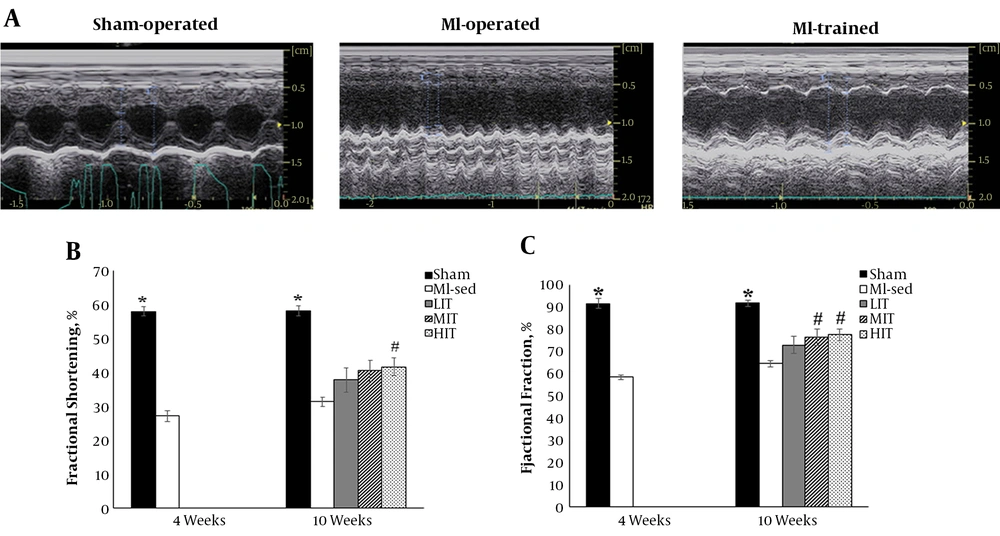
Left ventricular M-mode echocardiographic recordings at four and 10 weeks after surgery. A, Samples of m mode echocardiography in Sham-operated and MI-operated (MI-sed, LIIT, MIIT) animals. B, Left ventricular fractional shortening at 4 and 10 weeks after surgery. C, Left ventricular ejection fraction at 4 and 10 weeks after surgery. * P < 0.05 vs. MI-sed and MI-trained, # P < 0.05 vs. MI-sed.
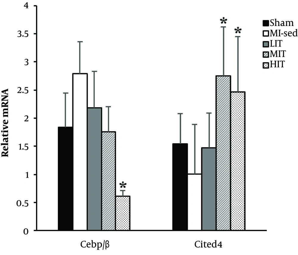
mRNA level of indicated genes from left ventricular at 10 weeks after surgery in Sham-operated and MI-operated (MI-sed, LIIT, MIIT and HIIT) animals. * P < 0.05 vs MI-sed. n = 6 animals per group.
| Sham, N = 6 | MI-Operated, N = 24 | ||||
|---|---|---|---|---|---|
| Four Weeks After Surgery | |||||
| IVSD (cm) | 0.17 ± 0.04 | 0.14 ± 0.03b | |||
| IVSS (cm) | 0.27 ±. 0.04 | 0.20 ± 0.04 | |||
| LVIDd (cm) | 0.50 ± 0.11 | 0.55 ± 0.09 | |||
| LVIDs (cm) | 0.22 ± 0.07 | 0.40 ± 0.07b | |||
| LVPWd (cm) | 0.15 ± 0.05 | 0.17 ± 0.17 | |||
| LVPWs (cm) | 0.23 ± 0.04 | 0.19 ± 0.03b | |||
| EF (%) | 90.49 ± 2.44 | 58.30 ± 10.79b | |||
| FS (%) | 55.40 ± 3.94 | 27.06 ± 6.58b | |||
| Sham, N = 6 | MI-Sed, N = 6 | LIIT, N = 5 | MIIT, N = 6 | HIIT, N = 6 | |
| 10 Weeks After Surgery | |||||
| IVSD (cm) | 0.17 ± 0.02 | 0.16 ± 0.02 | 0.15 ± 0.02 | 0.18 ± 0.03 | 0.15 ± 0.02 |
| IVSS (cm) | 0.27 ± 0.03 | 0.22 ± 0.02 | 0.26 ± 0.05 | 0.21 ± 0.02 | 0.26 ± 0.05 |
| LVIDd (cm) | 0.51 ± 0.11 | 0.57 ± 0.08 | 0.63 ± 0.07 | 0.53 ± 0.10 | 0.67 ± 0.08b |
| LVIDs (cm) | 0.22 ± 0.08 | 0.39 ± 0.05b | 0.39 ± 0.08b | 0.32 ± 0.09 | 0.39 ± 0.07b |
| LVPWd (cm) | 0.15 ± 0.04 | 0.17 ± 0.02 | 0.14 ± 0.02 | 0.14 ± 0.04 | 0.16 ± 0.02 |
| LVPWs (cm) | 0.22 ± 0.04 | 0.22 ± 0.03 | 0.21 ± 0.01 | 0.19 ± 0.07 | 0.27 ± 0.04 |
| EF (%) | 91.59 ± 3.30 | 64.48 ± 3.69b | 72.87 ± 8.95b | 76.41 ± 8.85b,c | 77.46 ± 7.02b,c |
| FS (%) | 56.44 ± 3.74 | 31.32 ± 3.46b | 37.72 ± 8.11b | 40.45 ± 7.60b | 41.62 ± 6.84b,c |
Abbreviations: EF, left ventricular ejection fraction; FS, left ventricular fractional shortening; IVSD and IVSS, interventricular septal end diastole and end systole; LVIDd and LVIDs, left ventricular internal diameter end diastole and end systole; LVPWd and LVPWs, left ventricular posterior wall end diastole and end systole.
a Values are expressed as mean ± SD.
b P < 0.05 vs. Sham.
c P < 0.05 vs. MI-Sed.


