
1. Background
Breast cancer remains one of the most prevalent and deadly malignancies in women, constituting a major global public health concern (1). With an approximate annual incidence of 2.3 million new cases, it is the second leading cause of cancer-related mortality in the female population (2). The complexity of this cancer stems from its significant molecular and phenotypic heterogeneity, influenced by genetic, epigenetic, and transcriptomic alterations, which profoundly impact diagnosis strategies, treatment response, and patient prognosis (3).
Current standard treatments include surgery, radiotherapy, chemotherapy, and hormone therapy. However, despite significant advancements, the efficacy of these approaches is compromised by major limitations, including considerable side effects, the non-selective targeting of many anticancer drugs, systemic toxicity, the development of drug resistance, and inadequate bioavailability (4, 5). This highlights the necessity for the search for novel and safer therapeutic agents. In this context, the MCF-7 cell line is widely employed as a standard and validated in vitro laboratory model for the initial assessment of cytotoxicity and antiproliferative effects (4).
A broad spectrum of anticancer drugs has been derived from natural sources (6, 7). Among promising medicinal plants, Nigella sativa has attracted modern research interest due to its rich historical background in traditional medicine and a wide range of bioactive compounds (8). Pharmacological studies have confirmed its anti-inflammatory, antioxidant, and antimicrobial properties. Notably, its significant anticancer potential against various neoplasms, including breast cancer, has been extensively reported (9-11).
The most important and abundant bioactive compound of N. sativa is thymoquinone (TQ), which constitutes 30–50% of its essential oil content (12, 13). Other important compounds include monoterpenes such as p-cymene, thymol, 4-terpineol, and carvacrol. Furthermore, N. sativa contains valuable amounts of non-volatile compounds, including alkaloids, saponins, flavonoids, and unsaturated fatty acids, which may exert synergistic effects. Extracts of N. sativa and its key compounds, particularly thymoquinone, have demonstrated potent antiproliferative effects in various in vitro models of breast cancer, including the MCF-7 cell line (11, 14).
2. Objectives
Therefore, considering the aforementioned background and the importance of discovering more selective and safer therapeutic agents, the primary objective of this study is the preliminary evaluation of the cytotoxic effects of aqueous and hydroalcoholic extracts of N. sativa on the MCF-7 breast cancer cell line, as well as the assessment of their selective toxicity on normal human dermal fibroblasts. It does not investigate molecular mechanisms but aims to provide essential baseline data for more in-depth future research.
3. Methods
3.1. Chemicals
All chemicals were purchased from Sigma-Aldrich (St. Louis, MO, USA), and all plasticware was obtained from Corning (USA).
3.2. Initial Preparation
Raw N. sativa seeds were purchased and assessed for purity. After washing with distilled water, they were immersed in a 20% ethanol solution for surface sterilization. The seeds were thoroughly dried in the shade at room temperature. Subsequently, the samples were placed in an oven at 50°C for 24 h to achieve complete dryness. To increase the surface area and facilitate better extraction, the dried seeds were ground into a fine powder using a mill. The powder was stored in an airtight container, protected from light and moisture, until further use.
3.3. Preparation of Hydroalcoholic Extract
For the hydroalcoholic extraction, 40 g of the seed powder was mixed with 400 mL of a 50% ethanol solution (v/v, diluted with distilled water). This mixture was stirred on a shaker at 130 revolutions per minute (RPM) for 24 hours at room temperature. Upon completion of the extraction, the contents of the container were vacuum-filtered using filter paper to obtain a clear extract.
3.4. Preparation of Aqueous Extract
The aqueous extract was obtained using a Soxhlet apparatus. For this purpose, 40 g of the seed powder was placed into filter paper thimbles, and the soluble components were extracted with distilled water. The resulting dilute extracts were then transferred to a rotary evaporator for concentration and removal of excess solvents, whereby the water and alcohol were evaporated off.
3.5. Extract Concentration and Dry Matter Determination
The obtained dilute extracts (both types) were placed in a rotary evaporator under vacuum to remove the solvent and achieve concentration, allowing the excess water and ethanol to be gently evaporated off, resulting in a concentrated extract. To determine the dry matter content of the final extracts, a precise quantity of each concentrated extract (approximately 2 milliliters) was poured into pre-weighed Petri dishes. These dishes were then dried in an oven at 50°C until a constant weight was achieved. The dry matter of each extract was calculated by subtracting the weight of the empty Petri dish from the weight of the dish containing the dried extract and was reported in grams per gram (g/g).
3.6. Qualitative High-performance Liquid Chromatography Profiling
The analysis of phenolic compounds in the N. sativa seed extract was performed using high-performance liquid chromatography (HPLC). This analysis was conducted solely for qualitative identification. The analyses were carried out using a Knauer (Germany) HPLC system, consisting of a K-2700 binary pump, a K-3700 diode array detector, and an RP-C18 column (model Perfectsil Target ODS-3, dimensions: 4.6 × 250 mm, particle size 5 µm). The mobile phase used was a mixture of acetonitrile and distilled water (containing 0.1% acetic acid) in a volume ratio of 60:40, delivered isocratically at a flow rate of 1 mL/min. The injection volume was 20 µL, and the detector wavelength was set to 360 nm for the measurement of phenolic compounds.
3.7. Cell Lines
The cancerous and normal cell lines used in this study were obtained from the Iranian Biological Resource Center (IBRC). The cell lines used were human dermal fibroblasts (HDF; IBRC code: C10506) and breast cancer cells (MCF-7; IBRC code: C10682). DMEM culture medium containing penicillin, streptomycin, and 10% FBS was used for cell culture. The cells were cultured in an incubator at 37°C, with 5% CO₂ and saturated humidity.
3.8. Evaluation of Cell Viability Using the MTT Assay
3-Methyl-2,5-diphenyltetrazolium bromide (MTT), a yellow, water-soluble tetrazolium salt, is reduced by the mitochondrial enzyme succinate dehydrogenase in living cells. The reduction of this compound leads to the cleavage of the tetrazolium ring and the formation of insoluble purple formazan crystals. These crystals are dissolved using the organic solvent dimethyl sulfoxide (DMSO), and the resulting color intensity is measured at a wavelength of 570 nm. The amount of formazan produced is directly proportional to the number of cells with active metabolism. To perform the assay, 100 µL of cells were seeded per well in a 96-well plate. Cells were treated with concentrations of 6.25, 12.5, 25, 50, 100, 200, 400, 800, 1600, 3200, and 6400 µg/mL of both N. sativa seed extract types (aqueous and hydroalcoholic) for 24 h. After the treatment period, MTT solution was added to the wells, and the plate was incubated for 4 hours in a 5% CO₂ incubator at 37°C. Subsequently, the supernatant containing MTT was removed, and 100 µL of DMSO was added to each well to dissolve the formazan crystals. The plate was shaken in an incubator shaker at 250 rpm for 20 min, and finally, the optical density at 570 nm was measured using an ELISA reader.
3.9. Statistical Analysis
Data analysis was performed using SPSS (version 16, IBM Corporation, USA). Data are presented as Mean ± SEM from three independent replicates (n = 3). After confirming normality (Shapiro-Wilk test) and homogeneity of variances (Levene's test), a one-way ANOVA followed by Duncan's post hoc test was used for group comparisons. Differences were considered significant at P < 0.05.
4. Results
4.1. Analysis of the Active Compound Profile in Nigella sativa
Based on HPLC analysis, the predominant peak in the aqueous extract of N. sativa corresponded to p-coumaric acid, followed by peaks for ferulic acid, quercetin, and hydroxybenzoic acid, respectively (Figure 1).
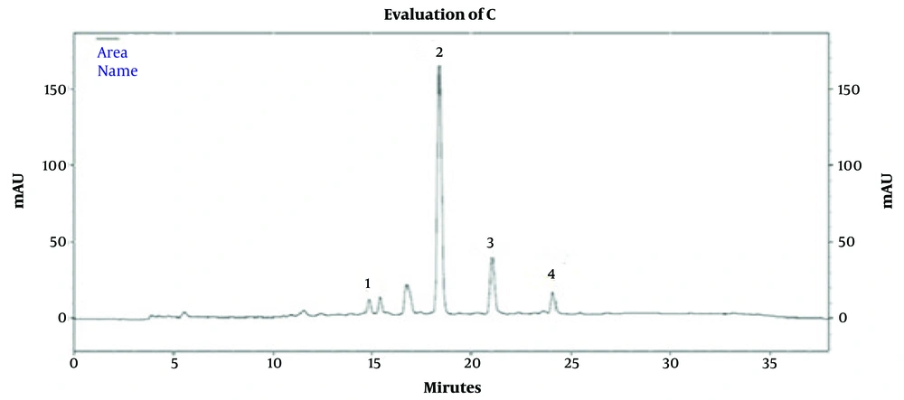
Figure 1.
High-performance liquid chromatography (HPLC) chromatogram of compounds in the aqueous extract of Nigella sativa; the identified peaks correspond to the following: (1) Hydroxybenzoic acid; (2) P-Coumaric acid; (3) Ferulic acid; (4) Quercetin
The HPLC profile of the hydroalcoholic extract of N. sativa also indicated that hydroxybenzoic acid, syringic acid, and ferulic acid showed the highest peak areas among the identified active compounds, respectively (Figure 2).
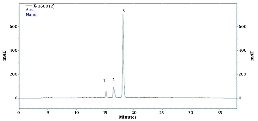
Figure 2.
High-performance liquid chromatography (HPLC) chromatogram of the hydroalcoholic extract of Nigella sativa; the identified peaks are: (1) Hydroxybenzoic acid; (2) Syringic acid; (3) Ferulic acid
4.2. Evaluation of the Cytotoxic Effect of the Aqueous Extract of Nigella sativa on the Viability of Human Dermal Fibroblasts Cells
The evaluation of the cytotoxic effects of different concentrations of aqueous and hydroalcoholic extracts of N. sativa seeds on the viability of HDF cells and MCF-7 cells demonstrated that in this study, concentration ranges of 6.25, 12.5, 25, 50, 100, 200, 400, 800, 1600, 3200, and 6400 µg/mL from both extract types were assessed, and the data obtained from cell treatment with the aqueous extract are presented in Figures 3. and 4.
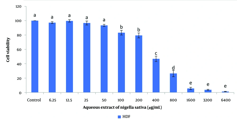
Figure 3.
The effect of different concentrations of the aqueous extract of Nigella sativa on the viability of human dermal fibroblasts (HDF) cells (P < 0.05) (different small letters above each bar indicate statistically significant differences between groups at the level of P < 0.05. Groups sharing common letters do not differ significantly).
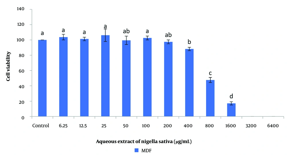
Figure 4.
Effect of different concentrations of aqueous Nigella sativa extract on the viability of MCF-7 cells (P < 0.05) (different small letters above each bar indicate statistically significant differences between groups at the level of P < 0.05. Groups sharing common letters do not differ significantly).
The results of the statistical analysis indicated a significant and concentration-dependent decrease in the viability of HDF cells following exposure to aqueous extract concentrations of 100 µg/mL and higher, compared to the control group (P < 0.05). Accordingly, at concentrations of 100, 200, and 400 µg/mL, HDF cell viability decreased significantly, reaching 83.3 ± 3.3%, 79.4 ± 4.3%, and 46.7 ± 8.3% respectively, while the viability in the control group was 100% (Figure 3).
4.3. Evaluation of the Cytotoxic Effect of the Aqueous Extract of Nigella sativa on the Viability of MCF-7 Cells
In an investigation of the effect of different concentrations of aqueous N. sativa extract on MCF-7, the results showed that at concentrations of 100 and 200 µg/mL, no statistically significant difference in cell survival was observed compared to the control group. However, at concentrations of 400 µg/mL and higher, the aqueous N. sativa extract induced a significant, concentration-dependent reduction in the viability of MCF-7 cells compared to the control group (P < 0.05).
Accordingly, the percentage survival of MCF-7 cells at concentrations of 100, 200, and 400 µg/mL was measured as 102.5 ± 2.5%, 97.5 ± 2.5%, and 88.2 ± 2.2%, respectively, whereas this value was considered 100% in the control group (Figure 4).
4.4. Evaluation of the Cytotoxic Effect of the Hydroalcoholic Extract of Nigella sativa on the Viability of Human Dermal Fibroblasts Cells
Data regarding the effect of different concentrations of hydroalcoholic N. sativa extract on normal (HDF) and cancerous (MCF-7) cells are presented in Figures 5. and 6. The results indicated that this extract, in the concentration range of 12.5 to 50 µg/mL, caused a significant increase in the viability of normal HDF cells. In contrast, at concentrations of 200 µg/mL and higher, a significant decrease in the survival of these cells compared to the control group was observed (P < 0.05).
Based on the findings, the percentage viability of HDF cells at concentrations of 12.5, 25, 50, 100, 200, 400, and 800 µg/mL was measured as 114.1 ± 4.0%, 112.0 ± 1.0%, 112.6 ± 3.6%, 93.4 ± 3.4%, 64.9 ± 5.0%, 42.9 ± 3.0%, and 31.5 ± 2.5%, respectively, while this value was considered 100% in the control group (Figure 5).
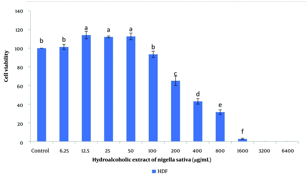
Figure 5.
Effect of different concentrations of hydroalcoholic Nigella sativa extract on the viability of human dermal fibroblasts (HDF) cells (P < 0.05) (different small letters above each bar indicate statistically significant differences between groups at the level of P < 0.05. Groups sharing common letters do not differ significantly).
4.5. Evaluation of the Cytotoxic Effect of the Hydroalcoholic Extract of Nigella sativa on the Viability of MCF-7 Cells
The results showed that only at concentrations of 800 µg/mL and higher (toxic but non-selective concentrations), the hydroalcoholic extract of N. sativa caused a significant reduction in the viability of MCF-7 cancer cells compared to the control group (P < 0.05). In contrast, at lower concentrations (6.25 and 12.5 µg/mL), this extract led to a significant increase in the survival of MCF-7 cells (P < 0.05). Based on this, the percentage viability of MCF-7 cells at concentrations of 6.25, 12.5, and 800 µg/mL was measured as 119.1 ± 4.1%, 131.7 ± 7.7%, and 59.7 ± 2.6%, respectively, while the corresponding value for the control group was considered 100% (Figure 6).
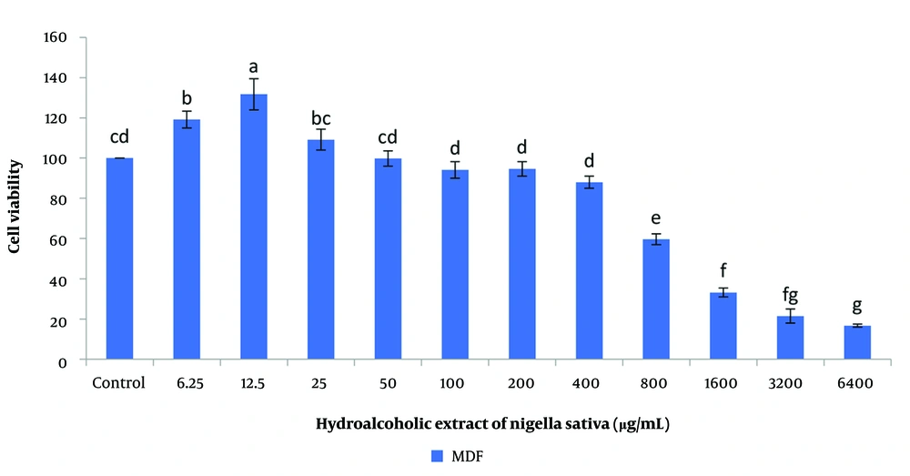
Figure 6.
Effect of different concentrations of hydroalcoholic Nigella sativa extract on the viability of MCF-7 cells (P < 0.05) (different small letters above each bar indicate statistically significant differences between groups at the level of P < 0.05. Groups sharing common letters do not differ significantly).
The present study evaluated the efficacy, which is dependent on dose and type, of aqueous and hydroalcoholic extracts of N. sativa on MCF-7 and HDF cell lines. The findings indicate that although both extracts show concentration-dependent inhibitory effects on the growth of MCF-7 cells, they lack the necessary selective property for targeted anticancer application. This is because, within the concentration range effective against cancer cells, they also exert significant toxicity on normal HDF cells, and thus no significant difference is observed in the selective death of cancer cells compared to the preservation of healthy cell viability. These results emphasize the complexity of the cellular response and its dependence on factors such as the type of extract, concentration, and cell nature, and reveal the need for more in-depth pharmacological studies to determine an optimal dose that can realize the anticancer potential of N. sativa with minimal side effects on healthy cells.
5. Discussion
This study reveals complex and concentration-dependent outcomes of N. sativa aqueous and hydroalcoholic extracts on normal and cancerous cells. Although both extracts exhibited anti-proliferative effects on MCF-7 cells at high concentrations, they lacked the requisite therapeutic selectivity, as they simultaneously induced significant toxicity in healthy HDF cells. Furthermore, dual responses (growth stimulation or inhibition) were observed depending on the extract type and concentration used, underscoring the pharmacological complexity of these extracts. These findings highlight the need for a deeper understanding of the structure-activity relationship and the determination of an optimal dose.
A growing body of scientific evidence supports the potent anticancer potential of N. sativa and its bioactive compounds, particularly TQ. Studies indicate that these agents exhibit multifaceted and promising anticancer effects against a broad spectrum of cancers, including breast cancer. Numerous in vitro studies have investigated the molecular mechanisms of this activity on various breast cancer cell lines, such as MDA-MB-231 and MCF-7 (2, 14, 15).
For instance, studies on MDA-MB-231 breast cancer cells have shown that a methanolic extract of N. sativa at low concentrations (2.5 and 5 µg/mL) induces dose-dependent apoptosis by increasing pro-apoptotic gene expression and decreasing anti-apoptotic gene expression (14). This finding is consistent with a study on SiHa cervical cancer cells, where a methanolic extract of N. sativa (125 µL/mL) led to apoptosis through the activation of caspase- and p53-dependent pathways (16). Furthermore, methanolic, hexane, and chloroform extracts of N. sativa demonstrated strong antiproliferative effects on HeLa (cervical) cells with very low IC50 values (ranging from ng/mL to μg/mL) (17).
This apoptotic mechanism has also been confirmed for the pure compound TQ (13, 15). Specifically, TQ exerts its effects by inhibiting cell proliferation, inducing oxidative stress (increasing ROS), and inhibiting inflammatory pathways (9, 13). Additionally, thymol, another derivative of N. sativa, disrupts the growth of MCF-7 cells at an IC50 of 200 µM by inhibiting the expression of key cell cycle genes (Cyclin D1 and PCNA) (18).
This study clearly demonstrates that both aqueous and hydroalcoholic extracts of N. sativa exert distinct and significant effects on HDF and MCF-7 cell lines. These effects are highly dependent on the extract type, concentration, and specific cell line. N. sativa and its compounds also influence tumor progression processes. Studies indicate that these compounds can inhibit cancer cell invasion and metastasis by suppressing the expression of matrix metalloproteinase enzymes (MMP-2 and MMP-9) and the angiogenic factor (VEGF), while simultaneously increasing the expression of the adhesion molecule E-cadherin (19, 20). For example, N. sativa oil at a concentration of 200 µg/mL significantly reduced MMP2-9 gene expression in MCF-7 and AGS cell lines. Furthermore, these compounds can modulate the cellular stress response by regulating the expression of heat shock proteins (HSPs). The same study showed that N. sativa oil decreased HSP60-70 expression (9).
One of the challenges in using natural compounds like N. sativa is their limited therapeutic selectivity and concurrent toxicity to healthy cells at effective concentrations, as also observed in the present study. To overcome this limitation, novel formulations such as nano emulsions and nanoparticles have been developed. These formulations significantly enhance anticancer efficacy by improving the bioavailability and penetration of active compounds (such as TQ) (21). For example, a nanoemulsion formulation of N. sativa oil extract demonstrated potent cytotoxicity against MCF-7 cells (22). Additionally, silver nanoparticles synthesized with N. sativa extract (N-AgNPs) at concentrations of 10 - 100 µg/mL affected MCF-7 cells by inducing apoptosis and regulating inflammatory markers (such as COX-2) (4).
Another promising approach is combination therapy with common chemotherapeutic drugs. Evidence suggests that the concurrent use of N. sativa extracts or its nano formulations with drugs such as doxorubicin can have a synergistic (enhancing) effect, increasing treatment efficacy (22). This highlights the importance of developing combined formulations (e.g., nano emulsions containing doxorubicin and N. sativa extract) as a potential therapeutic strategy.
In summary, the findings of this study indicate that the concentrations required to inhibit MCF-7 cancer cells simultaneously cause significant cytotoxic effects on normal HDF cells. These observations underscore the necessity for future research to focus on identifying and optimizing specific active compounds, precisely determining the therapeutic dose, and developing novel formulations to achieve selective efficacy. Considering the statistical power limitation of the present study, confirmation of these findings in future studies with a more robust statistical design is essential.
5.1. Conclusions
In summary, this study demonstrates that N. sativa extracts, particularly the aqueous form, exhibit anticancer potential against MCF-7 cells at intermediate concentrations. A key finding is the strongly concentration-dependent response pattern: Very low concentrations are ineffective or even stimulate cell growth, intermediate concentrations suggest potential selective activity, while high concentrations are generally cytotoxic. Furthermore, the extraction solvent (aqueous versus hydroalcoholic) significantly alters the biological activity profile of the extract. Given these results, precise therapeutic dose determination and formulation optimization are essential prerequisites for developing any therapeutic application of N. sativa to achieve maximum anticancer efficacy with minimal toxicity to healthy cells. As this research aimed at preliminary screening and delineating the effective concentration range, future studies should focus on lower, physiologically more relevant concentrations, likely achievable through advanced drug delivery technologies.