
1. Background
Stem cells are undifferentiated cells that retain the ability of proliferation and produce progenitor cells in themselves and can be classified into three main categories including embryonic, adult, and cancerous stem cells. A variety of adult stem cells are found in differentiated cells of the body tissues and organs including hematopoietic, mesenchymal, and neural stem cells (1, 2).
Mesenchymal stem cells, which encompass fibroblast cells, were first discovered in 1996 (3). Mesenchymal stem cells are, by definition, cells with high self-ability that under suitable conditions can be distinguished from a variety of specialized cells such as osteoblasts, chondrocytes, and adipocytes (4). Almost all adult tissues contain mesenchymal stem cells including bone marrow, peripheral blood, blood vessels, skeletal muscles, skin, liver, and adipose tissues. Regardless of tissue sources, these cells have some common properties such as proliferation ability, long-term self-renewal under controlled cell culture conditions, spindle morphology, and the potential for differentiation into mesodermal and even non-mesodermal types (5).
Today, mesenchymal cells have many applications and can be used to repair tissues such as in wound healing, spinal cord injury, Parkinson’s disease, and cerebrovascular accident (CVA). In the past, it was believed that if specialized cells were hardly damaged, they could not be replaced with healthy cells, but this problem was largely solved by extracting stem cells and using them as therapeutic cells (6, 7). Mouse embryonic fibroblasts (MEFs) are considered as fibroblast cells that are extracted from mouse embryos. Cultured in suitable conditions, MEFs have a spindle morphology which is the usual form of fibroblasts. MEFs are confined cell lines and can proliferate up to 4 - 5 passages in vitro. MEFs are often used for virus infection and wound healing and as feeder layers (7). Cell therapy is an interesting and effective strategy in the treatment of injuries and diseases for the replacement of stem cells or tissues made of stem cells. So far, various stem cells have been used in the treatment of diseases in various studies.
Cell therapy as a non-invasive approach is a good alternative for old-fashioned treatments including autograft and allograft methods. The use of stem cells in cell therapy involves using them as allogeneic, syngeneic, and autologous stem cells (8). One of the goals of cell therapy research is to track the fate of cells used in cell therapy. In cell therapy, the used cells should be marked and visualized. The marking method has been extensively developed in biology as a method of analyzing the fate of a cell and its descendants at different growth stages (9). Most of these markers are fluorescent or they can create a process that produces color reactions. Fluorescent markers are stimulated with certain wavelengths and reflect light with a longer wavelength. Some of these compounds are used for tracing including permanent fluorescent molecules such as Lucifer yellow and Carboxyfluorescein.
Enzymes such as horseradish peroxidase, biocytin, heavy metals such as cobalt and nickel, and compounds containing radioactive labels can be detected using the autoradiography method (10). Colors that are injected to determine the overall structure of cells should have specific properties such as being visible, either immediately or after chemical reactions. Also, they must be durable in the injected cell, should not be toxic, must not break down into products that have different characteristics, and survive under the pressure of histological processes (11). Some of these dyes are Dil and oxycarbonyl cyanine which are highly fluorescent lipophilic compounds, which are easily soluble in lipids of the plasma membrane and nontoxic and remain in the membrane for a long time. They are also published in fixed tissues. Dil can be detected using a fluorescence microscope. This dye has the same characteristics as rhodamine, and if irradiated with green light, it emits a red fluorescence light (12, 13).
2. Objectives
In the present study, we aimed to investigate the effects of Dil dye on fetal fibroblastic cells and examine the cell survival and durability of this fluorescence detector over a specific period of time in stem cells.
3. Methods
The present study is based on the results of a master’s thesis conducted in the cell culture laboratory of Shahid Chamran University of Ahvaz, Iran in the winter of 2017. In this study, pregnant mice (NMRI strain) were procured from animals’ house in Ahvaz Jundishapur University of Medical Sciences. The animals were kept in 12 light/12 dark cycle. After the purchase, the animals were moved to the animals’ house of Shahid Chamran University of Ahvaz and were kept in cages until the 12th day of pregnancy. Access to water and food was unlimited. To isolate MEFs on the 13th day of pregnancy, according to bioethical rules, the animals were sacrificed by chloroform (Merck, Germany). The abdominal surface was completely sterilized with alcohol. After the abdomen was exposed, embryos were removed from the uterus and in a petri dish, a sterile glass containing phosphate buffer saline (PBS, Sigma, USA) (with 1% antibiotic <Pen Strep.gibco. America>). Embryonic organs were removed along with internal organs. Mechanical digestion was performed by scalpel blade. For enzyme digestion, the carcasses of the embryos were immersed in trypsin and incubated for 15 - 20 minutes (Gibco 0.25%, United Kingdom). Then, centrifugation was performed and the contents of the tube were washed with PBS and pipetted several times with the culture medium (DMEM, Gibco. England) to separate the tissue cells. Then, the contents of the tube were passed through a sterile filter. The embryo soup was poured into a flask containing Dulbecco’s modification of Eagle medium (DMEM) and 10% fetal bovine serum (FBS, Gibco, America), and the flask was transferred to the incubator. After 24 h incubation, the supernatant of each flask with non-adhesion cells was removed. When a cell line reached about 80% confluence, the cells were subcultured by trypsin solution and transferred to T25 flasks for further passage (2, 5).
For staining with Dil, the sticky MEFs inside the flasks were isolated by trypsin and after counting by Neobar Lam, 1 × 10⁴ DMEM containing 10% FBS was placed in each well of a 96-well plate. After 24 hours of incubation, the culture medium was removed from the wells and 100 µL of medium containing Dil dye (Sigma, America) with concentrations of 0.5 µg/mL, 1 µg/mL, and 2 µg/mL were added to each well.
For the control group, only the culture medium was changed (10). Then, the plate was transferred to the incubator again, and after 20 to 30 minutes of incubation, it was removed from the incubator and the solutions in the wells were evacuated by the sampler. Three washes were performed with PBS, and again 100 µL of culture medium containing colorless serum was added to each well. The above operations were performed for the four groups that were treated for 1, 5, 7 and 14 days, respectively. 3-(4, 5-Dimethylthiazol-2-yl)-2, 5-diphenyltetrazolium bromide (MTT) test was performed for the treated and control groups on days 1, 5, 7, and 14. To perform this test, the MTT solution containing 5 mg/mL was prepared as the main stock and stored in darkness. Then, the solution was combined with a DMEM medium at a ratio of 9:1 (nine parts culture medium and one part MTT solution), and in each well, 100 µL of the above solution was added and the plate was incubated for 3 - 4 hours. After that, the wells were evacuated again, and in each well, 100 µL of DMSO (Merck, Germany) was added to dissolve the sediment and incubated for 15 minutes. Then, the contents of each well were slowly pipetted, and absorbance was measured at 570 nm by ELISA reader (Fax Stat, USA). For staining the nucleus with 4’, 6-diamidino-2-phenylindole (DAPI, Sigma), and the contents of the wells were drained and the wells were washed with PBS. Then, 100 µL of para-formaldehyde was poured into each well and incubated for 20 - 30 minutes until the cells were fixed. Then, each well was stained with DAPI at a concentration of 300 nano molar and photographed with a fluorescence microscope, and the images were examined.
The data were analyzed by two-way analysis of variance (ANOVA) in SPSS. The curves were drawn by Microsoft Excel (2016), and P-value less than 0.05 were considered significant.
4. Results
The morphology of the MEFs was evaluated using an inverted microscope. The results indicated that the treated cells receiving 0.5 µg/mL of Dil dye were not significantly different from the control group cells. It showed that Dil dye does not have any adverse effects on cell viability after entering the cell membrane and cell marking. Therefore, it is suitable for marking and tracing MEFs (Figure 1).
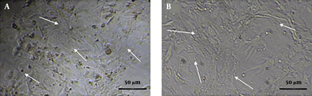
Figure 1.
Morphology of fibroblast cells treated with 0.5 µg/mL concentration of Dil dye by invert microscopy after seven days of treatment. A, treated sample; B, control sample.
Therefore, Dil concentrations lower than 0.5 µg/mL were suitable and non-toxic for MEFs, whereas concentrations higher than 0.5 µg/mL had a comparable toxic effect on these cells. In this study, MEFs treated with Dil dye were studied after 1, 5, 7 and 14 days using fluorescence microscopy. As shown in Figure 2, at concentration of 0.5 µg/mL, this detector retained its fluorescence properties after 14 days, such that no morphologic difference was observed between the images of the first to fourteenth days. Therefore, Dil with concentrations lower than 0.5 µg/mL was ideal for MEFs staining.
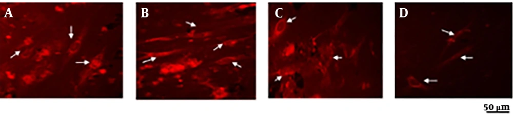
Figure 2.
Evaluation of stability of Dil dye in fibroblast cells treated with 0.5 µg/mL concentration of DiI dye by fluorescent microscopy A, One day after treatment; B, five days after treatment; C, seven days after treatment; and D, 14 days after treatment.
In the cell survival analysis method using MTT test, cell survival was found to be about 98% in the treatment group at a concentration of 0.5 µg/mL, indicating non-toxicity of the concentration, whereas with increasing the concentration, toxic effects also increased (Figure 3).
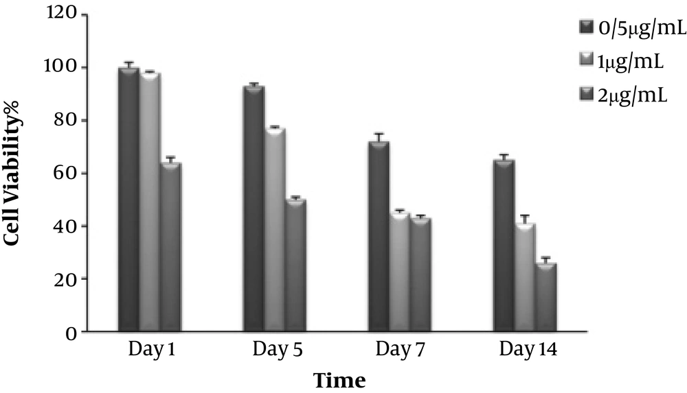
Figure 3.
MTT assay for comparing cell viability of fibroblast cells treated with 0.5, 1 and 2 µg/mL concentrations of DiI dye at 1, 5, 7 and 14 days after treatment (n = 3). Non-consecutive letters indicate a significant difference at the confidence level of P < 0.05.
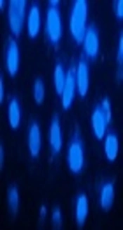
As shown in Figure 4, in the nucleus study by using the DAPI method, it was determined that the nuclei were not apoptotic and Dil dye at the concentration of 0.5 µg/mL had no negative effects on the MEFs nuclei. In the control samples, the nuclei were round, complete, and not fractionated, but at concentrations higher than 0.5 µg/mL, the signs of cell death were evident in the treated samples (the relevant images are not shown in this article). Therefore, the detector used at concentrations above 0.5 µg/mL had a toxic effect on cell nucleus.

Figure 4.
DAPI staining for the evaluation of morphology and nucleus properties of fibroblast cells treated with 0.5 µg/mL concentration of Dil dye A, one day after treatment; B, five days after treatment; C, seven days after treatment; D, 14 days after treatment; and E, control samples.
5. Discussion
In this study, the compatibility and durability of fluorescent Dil dye on MEFs were investigated. The marking of MEFs was performed with the aim of evaluating the durability and toxicity of Dil dye and its effect on cell survival. The results showed that cell survival was different between MEFs that received Dil dye at different concentrations, such that there was no significant difference between the group receiving Dil dye at a concentration of 0.5 µg/mL and the control group; however, there was a significant difference between the control group and the recipients of dye at concentrations higher than 0.5 µg/mL. Thus, Dil with different concentrations showed different levels of toxicity for MEFs. DAPI-colored nucleus staining studies also confirmed that the 0.5 µg/mL concentration of Dil dye does not any have toxic effects on MEFs. Also, color durability studies via fluorescence microscopy imaging showed that during the test period, this dye remained stable in MEFs with an excellent reflection. As a result, Dil dye at a concentration of 0.5 µg/mL can be used as a safe and suitable fluorescent detector to detect MEFs in vitro and in vivo.
Choi et al. stained and detected neurons using Dil dye, and their study revealed that the detector was still present in the membrane after eight weeks (14). In a study by Dai et al., which was conducted for cell differentiation study, mesenchymal stem cells of rats were labeled with Dil, which showed that after six months, this dye remained in the cell membrane, confirming the durability and stability of the dye in in vivo conditions (15). In a study by Hemmrich et al. on the use of Dil, PKH26 and CFSE markers were used to detect fat-precursor cells. It was found that CFSE has been used to mark the cytoplasm, and two other colors were suitable for staining cell membrane. It was also found that marking of the membrane using Dil dye was appropriate for immunohistochemical analysis, and this dye remained throughout the cell membrane representing a very useful property during analyzing fat in tissue engineering (16). In addition, Weir et al. studied the differentiation and division of mesenchymal stem cells using the Dil dye and CFSE. Marking of the cells was performed in vivo and in vitro and finally, it became clear that Dil dye was a better marker than the rest of the tested dyes. The results showed that CFSE quickly lost its fluorescence property, and the loss of the fluorescence signal was observed after eight days at all the staining concentrations (17).
In a study by Li et al., blood vessels were marked through heart perfusion using Dil solution. Due to lateral diffusion, Dil dye was used to stain membrane structures for angiogenesis and pseudopodia processes. Then, the tissue was examined immediately by a conventional confocal fluorescence microscope. It was proved that in contrast with the similar methods for vascular visualization, such as dextran conjugate color and Indian ink, the staining method using Dil was superior, and vascular marking was performed in less than an hour and high-quality optical serials were obtained from the tissue sections. Eventually, it was observed that blood vessels under the microscope strongly emitted red light using the rhodamine filter (18).
For cell tracking, other compounds and methods have also been discussed. For example, radioactive detectors can be seen using the autoradiography method. For example, large proteins can be traced using the I125 atom (19). One of the disadvantages of these detectors is that they only last a fixation when combined with one of the cell components and they can only be used for cut tissues. It is also possible to obtain ambiguous results due to the use of autoradiography. In the absence of these restrictions and many of the constraints of other compounds for the Dil dye, it can be considered as an ideal dye for cell tracking surveys (20). According to the results of this study, we recommend Dil fluorescence dye as an appropriate and fully concentration-dependent dye to trace MEFs, but it is noteworthy that it is necessary to determine the appropriate concentration based on the type of cells before using this dye, since in spite of its advantages and high durability, this dye can have significant toxic effects, and at very low concentrations, the efficiency and durability of the dye reduce.