Effects of a Yoga-Based Facial Massage Intervention on Reducing Eye Strain Among Young Adults with Prolonged Screen Time: A Randomized Pilot Trial

Author(s):
Hongxiu ChenHongxiu ChenHongxiu Chen ORCID1, Tanapat RatanapakornTanapat RatanapakornTanapat Ratanapakorn ORCID2, Manichaean SukonpatipManichaean Sukonpatip2, Somkiat AsawaphureekornSomkiat Asawaphureekorn2, Xingze WangXingze Wang3, Beibei WangBeibei WangBeibei Wang ORCID1, Wichai EungpinichpongWichai EungpinichpongWichai Eungpinichpong ORCID4,*
1Department of Exercise and Sport Sciences, Faculty of Graduate School, Kohn Kaen University, Khon Kaen, Thailand
2Department of Ophthalmology, Faculty of Medicine, Khon Kaen University, Khon Kaen, Thailand
3School of Physical Education, Huzhou University, Zhejiang, China
4School of Physical Therapy, Faculty of Associated Medical Sciences, Khon Kaen University, Khon Kaen, Thailand

Health Scope:Vol. 15, issue 2; e169874
Published online:Mar 30, 2026
Article type:Research Article
Received:Jan 28, 2026
Accepted:Feb 24, 2026
How to Cite:Chen H, Ratanapakorn T, Sukonpatip M, Asawaphureekorn S, Wang X, et al. Effects of a Yoga-Based Facial Massage Intervention on Reducing Eye Strain Among Young Adults with Prolonged Screen Time: A Randomized Pilot Trial. Health Scope. 2026;15(2):e169874. doi: https://doi.org/10.5812/healthscope-169874

Abstract

Background:

This study investigates the effects of specialised Thai Yoga facial massage (STYFM) on visual, musculoskeletal, cardiovascular, and autonomic functions in young adults with computer vision syndrome (CVS).

Methods:

A randomized controlled trial was conducted with 30 participants with visual fatigue. Participants were randomly assigned to either an STYFM group (FMG, n = 15) or a control group (CTG, n = 15). The FMG received 10 - 15 minutes of STYFM, while the CTG rested with eyes closed for 15 minutes to control for time and removal of visual load. The primary outcome was visual fatigue assessed by the Computer Vision Syndrome Questionnaire (CVS-Q). Secondary outcomes included near point of convergence (NPC), amplitude of accommodation (AMP), visual acuity (VA), spherical equivalent refraction (SE), handgrip strength (HS), cervical range of motion (CROM), and cardiovascular/autonomic indicators (heart rate, heart rate variability [HRV], and peripheral oxygen saturation [SpO₂]). Outcomes were assessed at baseline and immediately post-intervention.

Results:

The FMG demonstrated significant improvements in CVS-Q, NPC, AMP, VA (P < 0.001), HS (P < 0.05), CROM (P < 0.001), SpO₂, HRV, high-frequency power (HF), RMSSD, and PNN50 (P < 0.05). The CTG showed modest improvements in CVS-Q and NPC (P < 0.05), a decline in HS (P < 0.05), and increases in HRV, HR, RMSSD, and PNN50 (P < 0.05). Significant between-group differences were found in CVS-Q, NPC, AMP, VA, HS, CROM subcomponents (P < 0.001), SpO₂, and HR (P < 0.05).

Conclusions:

STYFM, as a noninvasive and easily self-implementable yoga-based mind–body intervention, appears to reduce visual fatigue and improve musculoskeletal and autonomic-related indicators in young adults. Given yoga’s role in stress regulation, STYFM may also help mitigate stress-related and extraocular symptoms associated with prolonged screen use, including headache. Larger, adequately powered trials are warranted. No intervention-related adverse events were reported.

1. Background

Computer vision syndrome (CVS), also referred to as digital eye strain, has become an important public health concern with the widespread use of smartphones, tablets, and computers (1). It affects approximately 75% - 90% of computer users worldwide (2), presenting with ocular symptoms such as visual fatigue, blurred vision, dry eyes, photophobia, and conjunctival hyperemia, as well as extraocular symptoms including headache, neck, and shoulder discomfort (3, 4). These symptoms often persist beyond working hours and worsen with prolonged screen exposure (5, 6), severely impairing academic and occupational performance and increasing the risk of chronic ocular conditions such as dry eye disease (7). Moreover, large population-based eye cohort studies have demonstrated a substantial prevalence of refractive errors and their associations with demographic factors, underscoring the broad public health burden of visual disorders (8).
Current approaches to managing CVS and visual fatigue primarily focus on optimizing ergonomic conditions and promoting practices such as the 20–20–20 rule to reduce accommodative load (9). In addition to these behavioral strategies, optical corrections such as blue light-filtering lenses (10) and progressive lenses (11) aim to improve visual comfort by enhancing refractive support for near and intermediate tasks (12, 13). Moreover, pharmacological and nutritional interventions, such as oral antioxidants (4), omega-3 fatty acid supplementation (14), and artificial tears (15), have been explored for their potential to mitigate ocular surface inflammation and improve tear stability. Despite these benefits, most existing treatments are primarily symptomatic, failing to address deeper deficits in accommodation, convergence, musculoskeletal performance, and autonomic regulation. These limitations highlight the need for innovative, multifactorial, non-pharmacological strategies capable of targeting the complex pathophysiology underlying CVS and visual fatigue.
Specialised Thai yoga facial massage (STYFM) is an integrative intervention that combines diaphragmatic breathing with targeted facial stimulation techniques, resembling a low-intensity form of yoga practice. Yoga, as a mind–body approach, has been widely applied in stress regulation and autonomic modulation, both of which are implicated in the development and persistence of computer vision syndrome and its extraocular symptoms (16). Preliminary evidence indicates that STYFM, by stimulating the facial, cranial, and cervical regions, may alleviate symptoms such as visual fatigue, blurred vision, ocular irritation, and burning sensations (17). Further research has also shown that massage interventions (18), acupressure techniques (19), and facial massage therapies (20) can activate the parasympathetic nervous system, improve autonomic nervous system balance, and enhance ocular and lymphatic circulation, thereby alleviating symptoms of visual fatigue and ocular discomfort. Notably, facial massage has also been shown to improve dry eye symptoms (21), which are closely associated with visual fatigue (22), suggesting that the alleviation of dry eye may indirectly reduce visual discomfort. Moreover, facial massage has been reported to improve the cervical range of motion (CROM) (23), further relieving the visual symptoms associated with cervical tension. Additionally, recent studies indicate that facial massage, neck massage, and acupoint massage may promote the relaxation of the periorbital muscles and modulate neural function, facilitating improvements in accommodation and convergence functions (24). These findings suggest that targeted massage interventions could offer novel strategies for relieving visual fatigue and enhancing visual performance. Near point of convergence (NPC) testing is considered an important clinical method for evaluating the convergence function and visual fatigue symptoms associated with convergence insufficiency (25, 26). From a physiological perspective, the therapeutic effects of STYFM are thought to result from synergistic mechanisms, including the regulation of visual accommodation and convergence functions (27, 28), modulation of autonomic nervous system activity (29), activation of lymphatic circulation (20, 30), and enhancement of musculoskeletal performance (31). These converging pathways may potentially explain the improvements in multiple physiological domains reported in prior studies and support the rationale for further investigation.
Despite preliminary evidence suggesting the potential benefits of STYFM, existing studies have been limited by narrow outcome scopes and insufficient methodological rigor. To date, no systematic investigations have comprehensively evaluated the multidimensional effects of STYFM on visual, musculoskeletal, cardiovascular, and autonomic parameters in individuals with CVS. The absence of high-quality randomized controlled trials (RCTs) in this domain underscores a critical gap in the evidence base for its therapeutic utility.

2. Objectives

Therefore, this RCT aims to evaluate the efficacy of STYFM as a non-pharmacological intervention for CVS by assessing the visual parameters (CVS-Q, NPC, AMP, VA, SE), musculoskeletal outcomes (HS, CROM), and cardiovascular/autonomic indices (SpO₂, HR, HRV).

3. Methods

The study was approved by the Ethics Committee of Khon Kaen University, Thailand (approval number: HE672161) and registered in the Thai Clinical Trials Registry (TCTR) on 19/11/2024 (registration number: TCTR20241119007).

3.1. Participants

Participants were recruited through an announcement on the Khon Kaen University platform and comprised young adults aged 18 - 35 years. Visual fatigue was screened using the Computer Vision Syndrome Questionnaire (CVS-Q). Inclusion criteria were a CVS-Q score ≥ 6 and an NPC > 10 cm. Participants were screened for ocular disorders, autonomic dysfunction, cardiovascular diseases, and current medication use. Caffeine consumption and dietary intake were restricted for at least two hours prior to the trial. The study was conducted in accordance with the principles of the Declaration of Helsinki. Exclusion criteria were: (1) Ocular and/or systemic conditions that directly induce visual fatigue (e.g., cataracts, glaucoma, cranial nerve palsy); (2) history of refractive or ocular surgery; (3) moderate-to-severe refractive errors (SE > ± 3.00 D, astigmatism > ± 2.00 D, or anisometropia > ± 1.00 D); (4) pregnancy or lactation; (5) use of medications or supplements that may affect visual fatigue; and (6) engagement in alternative therapies for visual fatigue within the preceding six months.

3.2. Randomization and Allocation Concealment
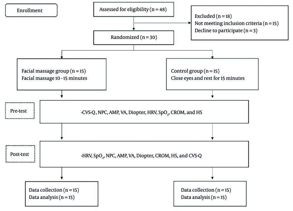
An independent researcher who was not involved in recruitment, outcome assessment, or intervention delivery generated the random allocation sequence (1:1 ratio) using Randomization.com (Figure 1). No stratification was applied, and the allocation sequence was generated without restrictions (simple randomization). Allocation concealment was ensured using a password-protected allocation file that was accessible only to the independent researcher. After eligibility confirmation and completion of baseline assessments, the enrolling researcher contacted the independent researcher to obtain the group assignment. Participants were allocated either to the STYFM group (FMG), which received the intervention, or to the control group (CTG), which rested quietly with eyes closed for 15 minutes to control for time and removal of visual load.
Participant recruitment flowchart
Figure 1.

Participant recruitment flowchart

3.3. Experimental Methodology and Measurements
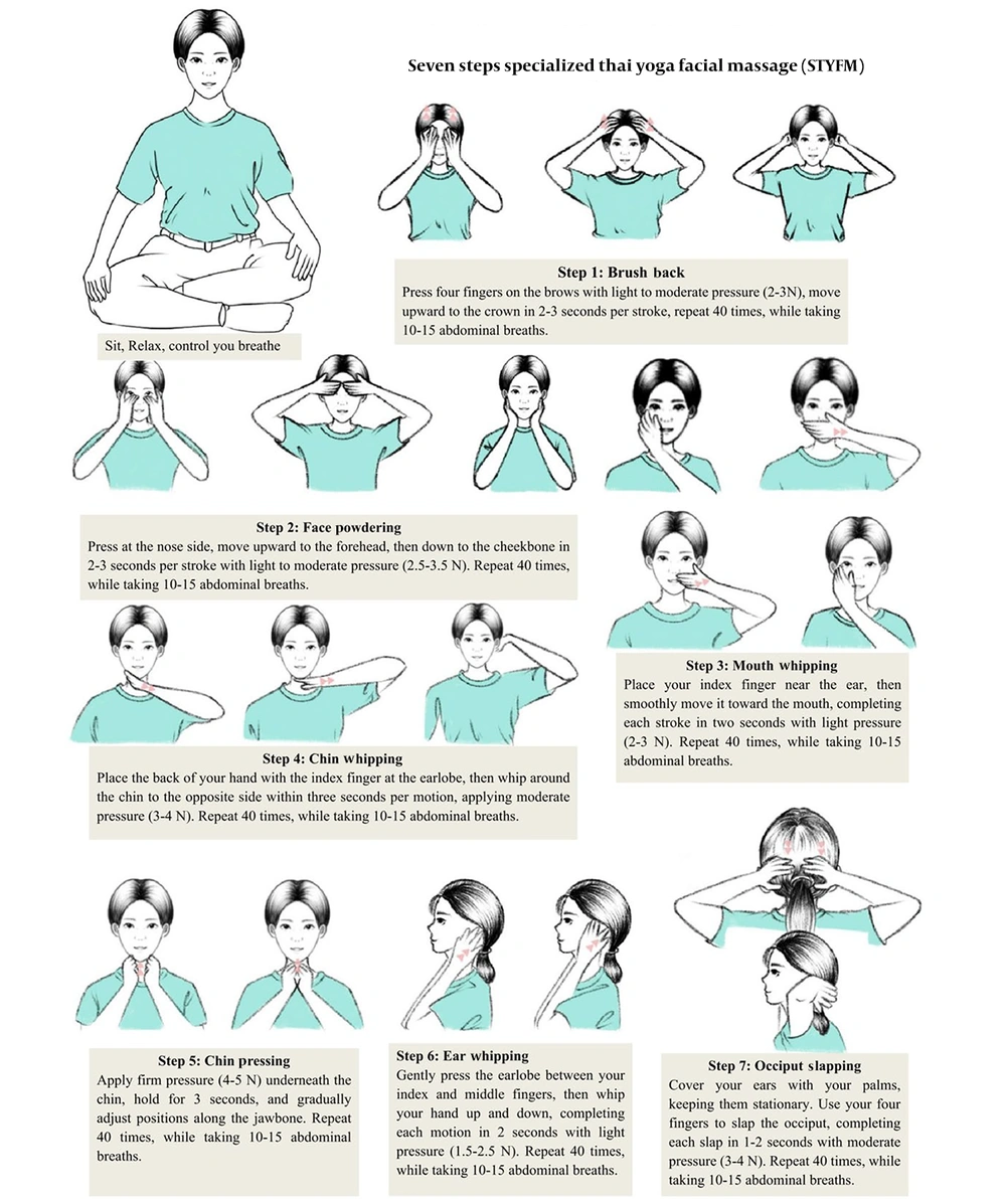
Prior to the measurement process, participants were seated for 20 minutes in a climate-controlled environment (24°C) to ensure thermal acclimatization. Sequential baseline assessments included the CVS-Q, NPC, AMP, VA, diopter, HRV, SpO₂, CROM, and HS. Participants in the FMG received the STYFM intervention for 10 - 15 minutes (Figure 2), while those in the CTG rested quietly with eyes closed for 15 minutes. Post-intervention assessments were conducted in the following order: HRV, SpO₂, NPC, AMP, VA, diopter, CROM, HS, and CVS-Q.
Steps of the STYFM
Figure 2.

Steps of the STYFM

3.4. Computer Vision Syndrome Questionnaire

The validated CVS-Q (ICC = 0.802, AUC = 0.826), developed by the University of Alicante (32) and translated in accordance with copyright regulations (33), was used. It comprised a self-assessment and a researcher-administered evaluation, followed by a summary of both. The questionnaire was completed prior to objective measurements. The CVS-Q consists of 16 items, with a total possible score of 64. Based on the score, the severity of visual fatigue is classified as follows: 0 - 5 points indicate no visual fatigue, 6 - 20 points indicate mild visual fatigue, 21 - 40 points indicate moderate visual fatigue, and a score greater than 41 points indicates severe visual fatigue.

3.5. Near Point of Convergence and Amplitude of Accommodation

The NPC was measured using the pen tip method, gradually advancing the sight marker until binocular vision could no longer be maintained, and the corresponding distance was recorded. The near point of accommodation (NPA) was measured using an accommodation card and ruler, recording the distance to the sight marker. To enhance accuracy, the Royal Air Force Near Point Rule was applied for NPA measurement, requiring participants to focus on a target with one eye. The measurement was repeated three times, and the average of the recorded distances was used to represent the NPA, ensuring the reliability of the results (34). Subsequently, AMP was calculated using the formula AMP = 100/NPA (cm) to assess the eye’s accommodative capacity.

3.6. Visual Acuity and Spherical Equivalent Refraction

The participants’ VA was assessed using the Early Treatment Diabetic Retinopathy Study (ETDRS) visual acuity chart (maximum score: 100 points). Measurements were recorded from top to bottom after occluding one eye, with the highest score documented (35). The measurement assessed distance VA. For participants with astigmatism, data were recorded after correction using a pinhole astigmatism plate. The pinhole astigmatism plate effectively reduced the impact of astigmatism on VA and, compared to traditional trial lenses, offers a simpler and more accurate correction. Refraction was measured using the NIDEK ARK-F autorefractometer (Nidek, Japan), with spherical equivalence calculated from the measured spherical and cylindrical values (36).

3.7. Heart Rate Variability and Peripheral Oxygen Saturation

Heart rate and HRV were recorded using the Polar H10 heart rate sensor (Polar Electro Oy, Kempele, Finland), a chest-strap device with validated accuracy and strong agreement with electrocardiogram-based recordings (37). SpO₂ was measured noninvasively at the fingertip using a clinically validated pulse oximeter, providing real-time monitoring of peripheral oxygen saturation. HRV analysis included time-domain indices, including the standard deviation of normal-to-normal RR intervals (SDNN), the root mean square of successive differences (RMSSD), and the percentage of successive normal-to-normal intervals differing by more than 50 ms (PNN50), as well as frequency-domain indices, including total power (TP), low-frequency power (LF), high-frequency power (HF), and the LF/HF ratio.

3.8. Cervical Range of Motion and Handgrip Strength

The CROM was measured using the Cervical Range of Motion Instrument (CROM; Performance Attainment Associates, United States) (38). Participants were seated during the assessment and performed the test three times, with the highest recorded value used for analysis. Grip strength was measured three times using the Takei Handgrip Dynamometer (GRIP-D; Takei, Japan), with the highest value documented (39).

3.9. Data Analysis

Descriptive statistics were used for participant characteristics. The Shapiro–Wilk test was used to test for the normality of continuous data. The paired t-test was used to test for within-group differences before and after the intervention. For between-group comparisons, ANCOVA was used to account for any baseline differences. Mauchly’s test was employed to evaluate the assumption of sphericity, and repeated-measures ANCOVA was used to ascertain differences at the baseline and post-intervention. A post hoc analysis was conducted utilizing the Bonferroni correction. Since many indicators were tested, to control the false discovery rate (FDR) and reduce Type I errors, the Benjamini–Hochberg (BH) procedure was used to adjust P-values for multiple hypothesis testing with the FDR of 5%. All analyses were conducted using JASP version 0.19 (40). The BH procedure was performed using Julia version 1.11.3 (41) with the MultipleTesting package (42). For data processing, the vision-related parameters AMP, VA, and SE were averaged across both eyes, while the musculoskeletal-related parameters HS, CROM-Lateral, and CROM-Rotation were averaged separately for the left and right sides.

4. Results

A total of 30 participants were analysed (FMG = 15, CTG = 15). The baseline characteristics, including age, sex distribution, vision-related parameters, musculoskeletal parameters, as well as cardiovascular and autonomic indices, were generally well balanced between the two groups. The mean age was 28.50 years, with females comprising 56.7% of the cohort. While the FMG demonstrated slightly lower baseline values in autonomic indices (such as HRV, SDNN, RMSSD, PNN50, TP, LF, and HF power), as well as marginally lower SpO₂ and HR, no statistically significant differences were observed in any baseline variable (all P > 0.05). Likewise, vision-related parameters (CVS-Q, NPC, AMP, VA, SE) and musculoskeletal parameters (HS and CROM subscales) were comparable between groups, indicating successful baseline matching (Table 1).
Table 1.Baseline Characteristics a, b
FactorsFMG (n = 15)CTG (n = 15)
Age (y)29.87 (3.38)27.13 (2.87)
Sex; No. (%)
Female9 (60.0)8 (53.3)
Male6 (40.0)7 (46.7)
At baseline
CVS-Q27.87 (13.3)30.73 (13.64)
NPC (cm)14.36 (1.49)14.59 (1.04)
AMP(D)7.21 (0.98)7.15 (0.62)
VA80.93 (2.03)80.33 (4.05)
SE(D)-0.74 (0.73)-0.92 (0.96)
HS (kg)28.24 (9.53)31.06 (11.18)
CROM-E(º)61.87 (12.30)60.4 (5.64)
CROM-F(º)61.33 (9.90)56.67 (10.29)
CROM-L(º)43.03 (5.68)45.70 (4.25)
CROM-R(º)63.83 (8.55)63.10 (9.41)
HRV50.60 (7.06)47.27 (5.44)
SDNN (ms)49.40 (16.86)38.39 (12.78)
RMSSD (ms)29.60 (13.14)22.72 (7.91)
PNN50 (%)0.08 (0.07)0.05 (0.07)
TP (ms2)1195.35 (1017.68)751.01 (498.06)
LF (ms2)890.45 (917.06)565.75 (393.43)
HF (ms2)304.84 (245.45)185.31 (197.58)
LF/HF4.17 (3.30)4.41 (2.66)
SpO₂ (%)96.93 (1.67)98.00 (0.85)
HR (bpm)78.87 (9.94)86.87 (10.04)

a Values are expressed as mean (SD) unless otherwise indicated.

b No significant baseline differences between groups were found (all P > 0.05)

Within-group comparisons indicated that the FMG exhibited significant improvements across multiple domains. For vision-related parameters (Table 2), CVS-Q, NPC, AMP, and VA all showed significant improvements (P < 0.001), whereas SE remained unchanged. In the CTG, CVS-Q and NPC remained statistically significant after BH adjustment, but VA and SE showed no significant changes. Regarding musculoskeletal parameters (Table 3), the FMG showed significant increases in HS (P < 0.05) and all directions of CROM (extension, flexion, lateral bending, and rotation; P < 0.001), while the CTG exhibited a significant decline in HS (P < 0.05) with no changes in CROM. For cardiovascular and autonomic nervous system function-related parameters (Table 4), the FMG demonstrated significant improvements in SpO₂, HRV, HF, RMSSD, and PNN50 (P < 0.05), whereas TP, LF, LF/HF, and SDNN showed no significant changes. In the CTG, HRV, HR, RMSSD, and PNN50 remained significant, while SpO₂ and other indices did not change. Between-group comparisons confirmed that CVS-Q, NPC, AMP, VA, HS, and all CROM directions differed significantly (P < 0.001), while SpO₂ and HR also showed significant between-group differences (P < 0.05). No significant between-group differences were observed for SE, TP, LF, HF, LF/HF, SDNN, RMSSD, or PNN50.
Table 2.Vision-Related Parameters
Outcomes and GroupsBefore (n = 15); Mean (SD)After (n = 15); Mean (SD)Within-Group Difference (After - Before); Mean (95% CI)Original P-Value aAdjusted P-Value bBetween-Group Difference (FMG - CTG); Mean (95% CI)Original P-Value cAdjusted P-Value d
CVS-Q-7.79 (-10.87, -4.71)< 0.001 e< 0.001 e
FMG27.87 (13.3)13.6 (7.46)-14.27 (9.88, 18.65)< 0.001 e< 0.001 e
CTG30.73 (13.64)23.27 (11.72)-7.47 (5.35, 9.58)< 0.001 e< 0.001 e
NPC (cm)-0.40 (-0.54, -0.26)< 0.001 e< 0.001 e
FMG14.36 (1.49)13.79 (1.41)-0.57 (0.45, 0.68)< 0.001 e< 0.001 e
CTG14.59 (1.04)14.41 (1.06)-0.18 (0.09, 0.27)< 0.001 e0.007 f
AMP (D)0.53 (0.38, 0.68)< 0.001 e< 0.001 e
FMG7.21 (0.98)7.74 (1.06)0.53 (-0.69, -0.38)< 0.001 e< 0.001 e
CTG7.15 (0.62)7.16 (0.62)0.01 (-0.02, 0.01)0.4840.605
VA3.57 (2.84, 4.30)< 0.001 e< 0.001 e
FMG80.93 (2.03)85.03 (2.07)4.10 (-4.71, -3.49)< 0.001 e< 0.001 e
CTG80.33 (4.05)80.83 (4.50)0.50 (-0.94, -0.06)0.030 f0.075
SE (D)0.05 (-0.16, 0.26)0.6270.784
FMG-0.74 (0.73)-0.73 (0.81)0.01 (-0.17, -0.15)0.8690.869
CTG-0.92 (0.96)-0.95 (0.92)-0.03 (-0.11, 0.17)0.6440.715

Abbreviations: SD, standard deviation; 95% CI, confidence interval.

a P-values for within-group differences (paired t-test) before adjustment

b Benjamini–Hochberg adjusted p-values for within-group differences

c P-values for between-group differences (ANCOVA using baseline as covariate) before adjustment

d Benjamini–Hochberg adjusted p-values for between-group differences.

e P < 0.01.

f P < 0.05.

Table 3.Musculoskeletal-Related Parameters
Outcomes and GroupsBefore (n = 15); Mean (SD)After (n = 15); Mean (SD)Within-Group Difference (After – Before); Mean (95% CI)Original P-Values aAdjusted P-Value bBetween-Group Difference (FMG – CTG); Mean (95% CI)Original P-Value cAdjusted P-Value d
HS (kg)2.89 (1.64, 4.14)< 0.001 e< 0.001 e
FMG28.24 (9.53)29.77 (10.73)1.29 (-2.17, -0.41)0.007 e0.011 f
CTG31.06 (11.18)29.49 (11.28)-1.57 (0.61, 2.53)0.004 e0.016 f
CROM-E (º)4.19 (2.34, 6.04)< 0.001 e< 0.001 e
FMG61.87 (12.30)66.27 (13.38)4.40 (-6.27, -2.53)< 0.001 e< 0.001 e
CTG60.4 (5.64)60.53 (5.98)0.13 (-0.64, 0.37)0.5820.684
CROM-F (º)4.73 (3.08, 6.38)< 0.001 e< 0.001 e
FMG61.33 (9.90)65.47 (9.00)4.13 (-5.86, -2.41)< 0.001 e< 0.001 e
CTG56.67 (10.29)56.47 (9.94)-0.20 (-0.23, 0.63)0.3340.446
CROM-L (º)4.39 (3.37, 5.41)< 0.001 e< 0.001 e
FMG43.03 (5.68)47.30 (5.25)4.27 (-5.08, -3.45)< 0.001 e< 0.001 e
CTG45.70 (4.25)45.33 (4.17)-0.37 (-0.32, 1.06)0.2740.391
CROM-R (º)4.67 (3.35, 6.00)< 0.001 e< 0.001 e
FMG63.83 (8.55)68.43 (8.28)4.60 (-5.87, -3.33)< 0.001 e< 0.001 e
CTG63.10 (9.41)63.03 (9.78)-0.07 (-0.41, 0.55)0.7690.769

Abbreviations: SD, standard deviation; 95% CI, confidence interval; CROM, cervical range of motion; CROM-E, extension; CROM-F, flexion; CROM-L, lateral; CROM-R, rotation.

a P-values for within-group differences (paired t-test) before adjustment.

b Benjamini–Hochberg adjusted P-values for within-group differences.

c P-values for between-group differences (ANCOVA using baseline as covariate) before adjustment.

d Benjamini–Hochberg adjusted p-values for between-group differences.

e P < 0.01.

f P < 0.05.

Table 4.Cardiovascular and Autonomic Nervous System Function-Related Parameters
Outcomes and GroupsBefore (n = 15); Mean (SD)After (n = 15); Mean (SD)Within-Group Difference (After – Before); Mean (95% CI)Original P-Values aAdjusted P-Value bBetween-Group Difference (FMG – CTG); Mean (95% CI)Original P-Value cAdjusted P-Value d
SpO2 (%)1.65 (0.55, 2.74)0.005 e0.009 e
FMG96.93 (1.67)98.47 (0.92)1.53 (-2.47, -0.60)0.003 e0.006 e
CTG98.00 (0.85)97.13 (1.73)-0.87 (0.01,1.73)0.048 f0.104
HRV-0.44 (-3.92, 3.05)0.7980.887
FMG50.60 (7.06)55.00 (9.22)4.40 (-6.31, -2.50)< 0.001 e< 0.001 e
CTG47.27 (5.44)52.33 (4.78)5.07 (-7.96, -2.17)0.002 f0.014 f
HR (bpm)4.24 (0.96, 7.52)0.013 f0.024 f
FMG78.87 (9.94)78.60 (9.32)-0.27 (-3.03, 3.57)0.0550.073
CTG86.87 (10.04)81.67 (7.39)-5.20 (2.02, 8.38)0.003 e0.025 f
TP (ms²)459 (-1027.47, 1947.14)0.5320.709
FMG1195.35 (1017.68)1974.11 (2408.58)778.77 (-1657.32, 99.79)0.0780.098
CTG751.01 (498.06)1514.16 (1451.83)763.15 (-1557.53, 31.22)0.0580.106
LF (ms²)-434.48 (-1307.42, 438.46)0.3160.489
FMG890.45 (917.06)1383.59 (2059.33)493.15 (-1213.10, 226.80)0.1640.193
CTG565.75 (393.43)1227.23 (1238.58)661.47 (-1329.81, 6.86)0.0520.104
HF (ms²)139.62 (-65.55, 344.79)0.1740.29
FMG304.84 (245.45)526.44 (376.49)221.60 (-370.58, -72.62)0.007 e0.011 f
CTG185.31 (197.58)286.93 (248.72)101.62 (-242.35, 39.11)0.1440.221
LF/HF0.89 (-5.91, 7.69)0.7900.929
FMG4.17 (3.30)5.29 (13.33)1.23 (-7.91, 5.65)0.7270.765
CTG4.41 (2.66)4.69 (2.60)0.28 (-1.98, 1.42)0.7270.766
SDNN (mms)1.04 (-13.25, 15.33)0.8820.929
FMG49.40 (16.86)55.25 (23.82)5.84 (-16.88, 5.20)0.2750.306
CTG38.39 (12.78)46.77 (15.80)8.38 (-17.35, 0.59)0.0650.108
RMSSD (mms)-0.37 (-1.30, 9.56)0.940.94
FMG29.60 (13.14)42.14 (27.52)12.54 (-22.02, -3.06)0.013 f0.019 f
CTG22.72 (7.91)31.50 (9.51)8.78 (-14.25, -3.32)0.004 f0.016 f
PNN50 (%)0.02 (-0.03, 0.08)0.4100.586
FMG0.08 (0.07)0.17 (0.12)0.09 (-0.12, -0.05)< 0.001 e< 0.001 e
CTG0.05 (0.07)0.11 (0.08)0.06 (-0.11, -0.02)0.011 f0.031 f

Abbreviations: SD, standard deviation; 95% CI, confidence interval; HR, heart rate; TP, total power; LF, low frequency; HF, high frequency; SDNN, standard deviation of normal-to-normal RR intervals; RMSSD, root mean square of successive differences; PNN50, percentage of successive RR intervals differing by more than 50 ms.

a P-values for within-group differences (paired t-test) before adjustment.

b Benjamini–Hochberg adjusted P-values for within-group differences.

c P-values for between-group differences (ANCOVA using baseline as covariate) before adjustment.

d Benjamini–Hochberg adjusted p-values for between-group differences.

e P < 0.01.

f P < 0.05

5. Discussion

This study demonstrates the short-term effectiveness of the STYFM intervention in alleviating visual fatigue, enhancing musculoskeletal function, and modulating autonomic nervous system activity. Participants in the FMG, who received STYFM, exhibited significant reductions in visual fatigue, as measured by the CVS-Q, and significant improvements in NPC, AMP, and VA, whereas SE remained unchanged. While both the CTG (CVS-Q, NPC, VA) and FMG (CVS-Q, NPC, AMP, VA) showed statistically significant post-intervention changes in vision-related parameters, only the improvement in CVS-Q reached the threshold for clinical significance. Notably, the reduction in CVS-Q score in the FMG was nearly twice that observed in the CTG, with a between-group difference of 7.8 points, further supporting the clinical relevance of the intervention. In addition, FMG participants demonstrated significant improvements in CROM (extension, flexion, lateral bending, and rotation) and a slight increase in HS, indicating a beneficial effect on musculoskeletal function. Moreover, the FMG showed significant enhancements in SpO₂, HRV, HF, RMSSD, and PNN50, suggesting improved autonomic regulation characterized by increased parasympathetic activity and oxygen saturation. However, given the small sample size and the relatively homogeneous population of young adults, these findings should be interpreted with caution and regarded as preliminary.
Specialised Thai Yoga facial massage effectively alleviated visual fatigue and improved ocular motor and accommodative function in individuals with high VDT exposure. Compared with the CTG, FMG participants showed meaningful reductions in CVS-Q scores and improvements in NPC, AMP, and VA, while SE remained unchanged, consistent with previous findings that refractive status requires longer intervention (43, 44). These improvements may involve multiple mechanisms. Prolonged visual tasks can lead to the accumulation of metabolic byproducts, oxidative stress, and disrupted redox homeostasis, thereby impairing retinal function (45, 46). In this context, massage therapy is thought to alleviate visual fatigue by enhancing ocular blood flow, improving retinal oxygenation, and reducing intraocular pressure (30, 47). Animal studies have also shown that ocular massage can transiently lower intraocular pressure and increase blood flow to the eye and optic nerve (48), suggesting that improved microcirculation may play a key role in relieving visual fatigue. By integrating targeted facial massage techniques, STYFM may not only relax the ciliary and periocular muscles and improve local circulation, but also promote accommodative function recovery (49, 50). Additionally, facial massage may facilitate lymphatic drainage around the ocular and cervical regions, potentially reducing periorbital congestion and inflammation (20, 47). Overall, these mechanisms are consistent with Fisher’s hypothesis, which proposes that enhancing ocular blood flow and muscle relaxation can support the restoration of reversible accommodative deficits (27, 28). In this study, FMG showed improvements in AMP, NPC, and VA, whereas CTG exhibited only NPC improvement, indicating that brief eye closure alone is insufficient to substantially restore accommodative function and visual acuity. However, these proposed mechanisms should be considered hypothetical, as this study did not directly assess ocular blood flow, lymphatic circulation, or related physiological indicators.
The STYFM intervention demonstrated significant effects on indicators related to musculoskeletal function, with FMG participants showing marked improvements in the CROM across all directions and handgrip strength (HS). Prolonged use of visual display terminals is commonly associated with forward head posture and cervical muscular imbalance, primarily due to weakness of the deep cervical flexors and compensatory overactivation of superficial muscles such as the sternocleidomastoid and anterior scalene (51-55). The observed improvements in CROM and HS in the FMG suggest that STYFM may alleviate visual fatigue-related cervical musculoskeletal dysfunction through myofascial relaxation, restoration of muscle function, enhanced neuromuscular transmission, improved peripheral circulation, and reduced muscle stiffness (31, 56-59). In addition, parasympathetic activation induced by STYFM may further facilitate muscle recovery by reducing physiological stress. In contrast, the CTG exhibited a transient decline in HS post-intervention, which may reflect a short-term lack of muscle activation or a state of complete relaxation.
In the present study, significant improvements were observed in HRV and SpO₂. In the FMG, HRV, PNN50, RMSSD, SpO₂, and HF components all increased significantly (P < 0.05). In contrast, in the CTG, although HRV indicators such as RMSSD and PNN50 also improved, SpO₂ decreased, and HF showed no significant change. The elevation of the HF component reflects enhanced vagal tone and is closely associated with the respiratory cycle, suggesting that the abdominal breathing incorporated in STYFM may contribute to physical and mental relaxation (60). HRV is a well-recognized biomarker for cognitive performance sensitive to fatigue (61), while improvements in PNN50 and RMSSD highlight STYFM’s potential to reduce physiological stress. Furthermore, pupil size, regulated by autonomic balance, typically enlarges under sympathetic dominance during visual fatigue, leading to blurred vision (62, 63). Although HRV-related indices did not differ significantly between groups, the FMG showed greater increases in SpO₂ and decreases in HR compared to the CTG, suggesting superior cardiovascular efficiency following STYFM. These findings are consistent with previous studies, which have shown a strong association between improvements in blood oxygen saturation and the alleviation of visual fatigue symptoms (64). Interestingly, the CTG exhibited a decline in post-intervention SpO₂ and only a minimal reduction in visual fatigue, whereas the FMG demonstrated substantial enhancements in both parameters. These results further reinforce the efficacy of STYFM in improving peripheral circulation, optimizing oxygen delivery, and mitigating fatigue-related symptoms.
In summary, STYFM, through facial massage combined with abdominal breathing, can simultaneously improve visual accommodative function, musculoskeletal health, autonomic regulation, and cardiovascular efficiency, thereby alleviating visual fatigue.
Study Limitations and Future Directions This study adopted a rigorous randomized controlled design and used the BH procedure to control for multiple comparisons. It systematically evaluated the effects of STYFM on visual, musculoskeletal, cardiovascular, and autonomic nervous system functions, thereby enhancing the credibility and internal validity of the findings. However, some limitations remain, including a small sample size, short intervention duration, a relatively homogeneous participant group, and the lack of an active control group. Specifically, the absence of a placebo-controlled or sham massage control may have introduced expectation effects, potentially influencing participants’ subjective and physiological responses. Future studies should consider incorporating placebo-controlled or sham massage designs to better isolate the specific effects of the intervention. Cycloplegic refraction and axial length measurements were not performed, nor direct assessments of the autonomic nervous function, limiting in-depth exploration of the intervention’s mechanisms. Overall, future studies should expand the sample size and diversity, extend the observation period, introduce appropriate active control groups, include more subjective and objective indicators, and utilize advanced monitoring techniques such as EEG and eye-tracking to more comprehensively elucidate the mechanisms and clinical value of STYFM.
Conclusion STYFM, as a noninvasive and easily self-implementable yoga-based intervention, appears to alleviate visual fatigue in young adults and improve musculoskeletal function as well as autonomic-related indicators. Given the established role of yoga in stress regulation and autonomic balance, both of which are closely related to the multidimensional manifestations of computer vision syndrome, STYFM could contribute to mitigating stress and autonomic dysregulation associated with prolonged screen use, including extraocular symptoms such as headache. Therefore, larger-scale studies with long-term follow-up are warranted to further validate its efficacy. No intervention-related adverse events were reported.

Acknowledgments

Footnotes

References

  • 1.
    Moulick P. Digital eye strain: Time for a break. J Ophthalmic Res Pract. 2023;1:10-3. https://doi.org/10.25259/jorp_14_2023.
  • 2.
    Leon-Figueroa DA, Barboza JJ, Siddiq A, Sah R, Valladares-Garrido MJ, Adhikari S, et al. Prevalence of computer vision syndrome during the COVID-19 pandemic: a systematic review and meta-analysis. BMC Public Health. 2024;24(1):640. [PubMed ID: 38424562]. [PubMed Central ID: PMC10902934]. https://doi.org/10.1186/s12889-024-17636-5.
  • 3.
    Dotulong D, Rares LM, Najoan IH. Computer Vision Syndrome. e-CliniC. 2021;9(1). https://doi.org/10.35790/ecl.v9i1.31707.
  • 4.
    Singh S, McGuinness MB, Anderson AJ, Downie LE. Interventions for the Management of Computer Vision Syndrome: A Systematic Review and Meta-analysis. Ophthalmology. 2022;129(10):1192-215. [PubMed ID: 35597519]. https://doi.org/10.1016/j.ophtha.2022.05.009.
  • 5.
    Beeson D, Wolffsohn JS, Baigum T, Qureshi T, Gohil S, Wahid R, et al. Digital eye strain symptoms worsen during prolonged digital tasks, associated with a reduction in productivity. Comput Hum Behav Rep. 2024;16:100489. https://doi.org/10.1016/j.chbr.2024.100489.
  • 6.
    Gasheya KA, Belay AA, Abegaze T, Zele YT, Daba C. Computer vision syndrome and ergonomic risk factors among workers of the Commercial Bank of Ethiopia in Addis Ababa, Ethiopia: an institutional-based cross-sectional study. Front Public Health. 2024;12:1341031. [PubMed ID: 38784585]. [PubMed Central ID: PMC11111856]. https://doi.org/10.3389/fpubh.2024.1341031.
  • 7.
    Sheppard AL, Wolffsohn JS. Digital eye strain: prevalence, measurement and amelioration. BMJ Open Ophthalmol. 2018;3(1). e000146. [PubMed ID: 29963645]. [PubMed Central ID: PMC6020759]. https://doi.org/10.1136/bmjophth-2018-000146.
  • 8.
    Shahraki K, Tahkor A, Shahraki Sanavi F, Ansari-Moghaddam A, Shahraki K, Mahjoob M, et al. Prevalence of Refractive Errors and Associated Factors in the Population of the Eye Cohort Study in Southeast Iran. Health Scope. 2023;12(4). https://doi.org/10.5812/healthscope-137161.
  • 9.
    Coles-Brennan C, Sulley A, Young G. Management of digital eye strain. Clin Exp Optom. 2019;102(1):18-29. [PubMed ID: 29797453]. https://doi.org/10.1111/cxo.12798.
  • 10.
    Singh S, Keller PR, Busija L, McMillan P, Makrai E, Lawrenson JG, et al. Blue-light filtering spectacle lenses for visual performance, sleep, and macular health in adults. Cochrane Database Syst Rev. 2023;8(8). CD013244. [PubMed ID: 37593770]. [PubMed Central ID: PMC10436683]. https://doi.org/10.1002/14651858.CD013244.pub2.
  • 11.
    Varnas SR, Kaphle D, Schmid KL, Suheimat M, Atchison DA. Effect of multifocal spectacle lenses on accommodative errors over time: Possible implications for myopia control. J Vis. 2023;23(3):3. [PubMed ID: 36862107]. [PubMed Central ID: PMC9987164]. https://doi.org/10.1167/jov.23.3.3.
  • 12.
    Kee CS, Leung TW, Kan KH, Lam CH. Effects of Progressive Addition Lens Wear on Digital Work in Pre-presbyopes. Optom Vis Sci. 2018;95(5):457-67. [PubMed ID: 29683984]. [PubMed Central ID: PMC5943073]. https://doi.org/10.1097/OPX.0000000000001211.
  • 13.
    Koh S, Inoue R, Sato S, Haruna M, Asonuma S, Nishida K. Quantification of accommodative response and visual performance in non-presbyopes wearing low-add contact lenses. Cont Lens Anterior Eye. 2020;43(3):226-31. [PubMed ID: 31327577]. https://doi.org/10.1016/j.clae.2019.07.004.
  • 14.
    Bhargava R, Pandey K, Ranjan S, Mehta B, Malik A. Omega-3 fatty acids supplements for dry eye - Are they effective or ineffective? Indian J Ophthalmol. 2023;71(4):1619-25. [PubMed ID: 37026312]. [PubMed Central ID: PMC10276704]. https://doi.org/10.4103/IJO.IJO_2789_22.
  • 15.
    Semp DA, Beeson D, Sheppard AL, Dutta D, Wolffsohn JS. Artificial Tears: A Systematic Review. Clin Optom (Auckl). 2023;15:9-27. [PubMed ID: 36647552]. [PubMed Central ID: PMC9840372]. https://doi.org/10.2147/OPTO.S350185.
  • 16.
    Mandlik GV, Siopis G, Nguyen B, Ding D, Edwards KM. Effect of a single session of yoga and meditation on stress reactivity: A systematic review. Stress Health. 2024;40(3). e3324. [PubMed ID: 37822096]. https://doi.org/10.1002/smi.3324.
  • 17.
    Ngowsiri K, Tanmahasamut P, Sukonthasab S. Rusie Dutton traditional Thai exercise promotes health related physical fitness and quality of life in menopausal women. Complement Ther Clin Pract. 2014;20(3):164-71. [PubMed ID: 25066942]. https://doi.org/10.1016/j.ctcp.2014.05.002.
  • 18.
    Kim DW, Lee DW, Schreiber J, Im CH, Kim H. Integrative Evaluation of Automated Massage Combined with Thermotherapy: Physical, Physiological, and Psychological Viewpoints. Biomed Res Int. 2016;2016:2826905. [PubMed ID: 28074179]. [PubMed Central ID: PMC5198097]. https://doi.org/10.1155/2016/2826905.
  • 19.
    Park S, Kim J. Factors influencing the use of traditional Korean herbal medicine - based on the 2020 Korean Medical Panel data. J Korean Med. 2024;45(3):1-13. https://doi.org/10.13048/jkm.24033.
  • 20.
    Chen H, Eungpinichpong W, Ratanapakorn T, Wang B. Effectiveness of Facial Massage and Exercise in Alleviating Visual Fatigue: A Systematic Review. Asian J Sports Med. 2024;15(4). https://doi.org/10.5812/asjsm-153503.
  • 21.
    Leedasawat P, Sriyakul K, Phetkate P, Tungsukruthai P, Patarajierapun P, Sangvatanakul P, et al. A Randomized Controlled Trial Comparing Thai Facial Massage, Chinese Eye Exercise of Acupoints and Standard Treatment in Patients with Dry Eye Disorders. Trad Integr Med. 2024;9(3). https://doi.org/10.18502/tim.v9i3.16529.
  • 22.
    Al-Mohtaseb Z, Schachter S, Shen Lee B, Garlich J, Trattler W. The Relationship Between Dry Eye Disease and Digital Screen Use. Clin Ophthalmol. 2021;15:3811-20. [PubMed ID: 34531649]. [PubMed Central ID: PMC8439964]. https://doi.org/10.2147/OPTH.S321591.
  • 23.
    Maenpuen S, Duangmala O, Kednai K, Hemtong W, Sirited P, Tunit P. Effect of Thai Yoga Exercise for Reducing Pain and Improve Functional Disability in Patients with Text Neck Syndrome: A Randomized Controlled Trial. J Curr Sci Tech. 2024;15(1). https://doi.org/10.59796/jcst.V15N1.2025.79.
  • 24.
    Shah PJ, Sangeetha M. Effects of Facial, Neck Massage and Traditional Chinese Eye Exercises Along with Eye and Facial Exercises on Visual Acuity and Convergence Insufficiency Among Young Adults with Low Myopia. Indian J Ophthalmol. 2020;68(1):69-74.
  • 25.
    Gantz L, Stiebel-Kalish H. Convergence insufficiency: Review of clinical diagnostic signs. J Optom. 2022;15(4):256-70. [PubMed ID: 34963569]. [PubMed Central ID: PMC9537264]. https://doi.org/10.1016/j.optom.2021.11.002.
  • 26.
    Ostrow GI, Kirkeby L. Convergence Insufficiency. EyeWiki; 2025. Available from: https://eyewiki.org/Convergence_Insufficiency.
  • 27.
    Fisher RF. The force of contraction of the human ciliary muscle during accommodation. J Physiol. 1977;270(1):51-74. [PubMed ID: 915798]. [PubMed Central ID: PMC1353417]. https://doi.org/10.1113/jphysiol.1977.sp011938.
  • 28.
    Zheng F, Hou F, Chen R, Mei J, Huang P, Chen B, et al. Investigation of the Relationship Between Subjective Symptoms of Visual Fatigue and Visual Functions. Front Neurosci. 2021;15:686740. [PubMed ID: 34335163]. [PubMed Central ID: PMC8319646]. https://doi.org/10.3389/fnins.2021.686740.
  • 29.
    Zhao T, Schiweck C, Jamalambadi H, Meyer K, Brandt E, Schneider M, et al. Transcutaneous vagus nerve stimulation improves emotional processing. J Affect Disord. 2025;372:96-105. [PubMed ID: 39615758]. https://doi.org/10.1016/j.jad.2024.11.077.
  • 30.
    Hayashi PN, Du ML. Acute and Chronic Periocular Massage for Ocular Blood Flow and Vision: a Randomized Controlled Trial. Int J Ther Massage Bodyw: Res Educ Pract. 2021;14(2):5-13. https://doi.org/10.3822/ijtmb.v14i2.583.
  • 31.
    Buttagat V, Kluayhomthong S, Areeudomwong P. Effects of the combination of traditional Thai massage, scapular stabilization exercise, and chest mobilization in subjects with forward head posture: a single-blinded randomized clinical trial. Chiropr Man Therap. 2023;31(1):31. [PubMed ID: 37605282]. [PubMed Central ID: PMC10441760]. https://doi.org/10.1186/s12998-023-00506-z.
  • 32.
    Segui Mdel M, Cabrero-Garcia J, Crespo A, Verdu J, Ronda E. A reliable and valid questionnaire was developed to measure computer vision syndrome at the workplace. J Clin Epidemiol. 2015;68(6):662-73. [PubMed ID: 25744132]. https://doi.org/10.1016/j.jclinepi.2015.01.015.
  • 33.
    Canto-Sancho N, Segui-Crespo M, Zhao G, Ronda-Perez E. The Chinese version of the Computer Vision Syndrome Questionnaire: translation and cross-cultural adaptation. BMC Ophthalmol. 2023;23(1):298. [PubMed ID: 37400768]. [PubMed Central ID: PMC10318657]. https://doi.org/10.1186/s12886-023-03031-y.
  • 34.
    Zakian A, Heydarian S, Mirzajani A, Jafarzadehpur E, Yekta A, Khabazkhoob M. Distribution of near Point of Convergence, near Point of Accommodation, Accommodative Facility and Refractive Errors in a Rural Population Living in Northern Iran. J Binocul Vis Ocul Motil. 2021;71(3):104-9. [PubMed ID: 34032560]. https://doi.org/10.1080/2576117X.2021.1927291.
  • 35.
    Plainis S, Moschandreas J, Giannakopoulou T, Vitanova V, Nikolitsa P, Rozema JJ, et al. Validation of a modified ETDRS chart for European-wide use in populations that use the Cyrillic, Latin or Greek alphabet. J Optom. 2013;6(1):18-24. https://doi.org/10.1016/j.optom.2012.06.008.
  • 36.
    Paudel N, Adhikari S, Thakur A, Shrestha B, Loughman J. Clinical Accuracy of the Nidek ARK-1 Autorefractor. Optom Vis Sci. 2019;96(6):407-13. [PubMed ID: 31107837]. https://doi.org/10.1097/OPX.0000000000001386.
  • 37.
    Gilgen-Ammann R, Schweizer T, Wyss T. RR interval signal quality of a heart rate monitor and an ECG Holter at rest and during exercise. Eur J Appl Physiol. 2019;119(7):1525-32. [PubMed ID: 31004219]. https://doi.org/10.1007/s00421-019-04142-5.
  • 38.
    Performance Attainment Associates. Procedure for measuring neck motion with the CROM. 1988. Available from: https://static.webareacontrol.com/CommonFile/CROM---Cervical-Range-of-Motion-Instrument_Procedure-Manual.pdf.
  • 39.
    Horn K, Johnson H, Williams E, McQueeney S, Harrington S. Validity and reliability of the takei hand dynamometer. Int J Exerc Sci. 2024;16(3):130.
  • 40.
    JASP. JASP (Version 0.19) [Computer software]. JASP; 2024. Available from: https://jasp-stats.org/previous-versions/.
  • 41.
    Bezanson J, Edelman A, Karpinski S, Shah VB. Julia: A Fresh Approach to Numerical Computing. SIAM Review. 2017;59(1):65-98. https://doi.org/10.1137/141000671.
  • 42.
    Gehring J. The MultipleTesting package offers common algorithms for p-value adjustment and combination and more…. GitHub; 2025. Available from: https://github.com/juliangehring/MultipleTesting.jl.
  • 43.
    Stingl JV, Ban SA, Nagler M, Schmidtmann I, Wild PS, Lackner KJ, et al. Five-year change in refractive error and its risk factors: results from the Gutenberg Health Study. Br J Ophthalmol. 2023;107(1):140-6. [PubMed ID: 34362774]. [PubMed Central ID: PMC9763219]. https://doi.org/10.1136/bjophthalmol-2021-318828.
  • 44.
    Lee SS, Lingham G, Wang CA, Diaz Torres S, Pennell CE, Hysi PG, et al. Changes in Refractive Error During Young Adulthood: The Effects of Longitudinal Screen Time, Ocular Sun Exposure, and Genetic Predisposition. Invest Ophthalmol Vis Sci. 2023;64(14):28. [PubMed ID: 37982764]. [PubMed Central ID: PMC10668617]. https://doi.org/10.1167/iovs.64.14.28.
  • 45.
    Upadhyay M, Milliner C, Bell BA, Bonilha VL. Oxidative stress in the retina and retinal pigment epithelium (RPE): Role of aging, and DJ-1. Redox Biol. 2020;37:101623. [PubMed ID: 32826201]. [PubMed Central ID: PMC7767746]. https://doi.org/10.1016/j.redox.2020.101623.
  • 46.
    Yang S, Zhou J, Li D. Functions and Diseases of the Retinal Pigment Epithelium. Front Pharmacol. 2021;12:727870. [PubMed ID: 34393803]. [PubMed Central ID: PMC8355697]. https://doi.org/10.3389/fphar.2021.727870.
  • 47.
    Limphatcharaporn PPJ. [Immediate Effect of Facial Massage and Lymphatic Drainage Massage on Intraocular Pressure]. J Vongchavalitkul Univ. 2022;35(1). TH.
  • 48.
    Jay WM, Aziz MZ, Green K. Effect of digital massage on intraocular pressure and ocular and optic nerve blood flow. Acta Ophthalmol. 1986;64(1):58-62. [PubMed ID: 3962620]. https://doi.org/10.1111/j.1755-3768.1986.tb06872.x.
  • 49.
    Manna P, Karmakar S, Bhardwaj GK, Mondal A. Accommodative spasm and its different treatment approaches: A systematic review. Eur J Ophthalmol. 2023;33(3):1273-86. [PubMed ID: 36384286]. https://doi.org/10.1177/11206721221136438.
  • 50.
    Zielinski G, Matysik-Wozniak A, Pajak B, Ginszt M, Rejdak R, Gawda P. Study protocol: Accommodative effects on the musculoskeletal system. PLoS One. 2023;18(7). e0289347. [PubMed ID: 37506164]. [PubMed Central ID: PMC10381037]. https://doi.org/10.1371/journal.pone.0289347.
  • 51.
    Teo C, Giffard P, Johnston V, Treleaven J. Computer vision symptoms in people with and without neck pain. Appl Ergon. 2019;80:50-6. [PubMed ID: 31280810]. https://doi.org/10.1016/j.apergo.2019.04.010.
  • 52.
    Mork R, Falkenberg HK, Fostervold KI, Thorud HS. Discomfort glare and psychological stress during computer work: subjective responses and associations between neck pain and trapezius muscle blood flow. Int Arch Occup Environ Health. 2020;93(1):29-42. [PubMed ID: 31286223]. https://doi.org/10.1007/s00420-019-01457-w.
  • 53.
    Mork R, Falkenberg HK, Fostervold KI, Thorud HMS. Visual and psychological stress during computer work in healthy, young females-physiological responses. Int Arch Occup Environ Health. 2018;91(7):811-30. [PubMed ID: 29850947]. [PubMed Central ID: PMC6132651]. https://doi.org/10.1007/s00420-018-1324-5.
  • 54.
    Prabu Raja G, Shyamasunder Bhat N, Marie Cruz A, Prabhu A, Fernandes S, Naaz N. The anatomical myofascial continuum between the neck and eyes. Clin Anat. 2022;35(3):340-6. [PubMed ID: 35043988]. https://doi.org/10.1002/ca.23835.
  • 55.
    Dareh-deh HR, Hadadnezhad M, Letafkar A, Peolsson A. Effects of Adding Respiratory Exercises to the Therapeutic Routine in Smartphone Users With Forward Head Posture and Non-Specific Chronic Neck Pain: A Randomised Controlled Trial. Research Square. 2020;Preprint. https://doi.org/10.21203/rs.3.rs-53984/v1.
  • 56.
    Buyukturan B, Sas S, Kararti C, Buyukturan O. The effects of combined sternocleidomastoid muscle stretching and massage on pain, disability, endurance, kinesiophobia, and range of motion in individuals with chronic neck pain: A randomized, single-blind study. Musculoskelet Sci Pract. 2021;55:102417. [PubMed ID: 34147954]. https://doi.org/10.1016/j.msksp.2021.102417.
  • 57.
    Clark BC. Neural Mechanisms of Age-Related Loss of Muscle Performance and Physical Function. J Gerontol A Biol Sci Med Sci. 2023;78(Suppl 1):8-13. [PubMed ID: 36738253]. [PubMed Central ID: PMC10272985]. https://doi.org/10.1093/gerona/glad029.
  • 58.
    Hammer SM, Alexander AM, Didier KD, Barstow TJ. Influence of blood flow occlusion on muscular recruitment and fatigue during maximal-effort small muscle-mass exercise. J Physiol. 2020;598(19):4293-306. [PubMed ID: 32721032]. https://doi.org/10.1113/JP279925.
  • 59.
    Tremblay M, Anderson Sirois S, Abboud J, Descarreaux M. Grip strength, muscle soreness and pain threshold perception evolution in baseball pitchers in a simulated 75-pitch game: a repeated measures study. BMJ Open Sport Exerc Med. 2025;11(1). e002146. [PubMed ID: 40041680]. [PubMed Central ID: PMC11877241]. https://doi.org/10.1136/bmjsem-2024-002146.
  • 60.
    Kubota T. Science and Practice of Tanden (Dantian) Breathing. Tokyo, Japan: pubfull; 2019.
  • 61.
    Matuz A, van der Linden D, Kisander Z, Hernadi I, Kazmer K, Csatho A. Enhanced cardiac vagal tone in mental fatigue: Analysis of heart rate variability in Time-on-Task, recovery, and reactivity. PLoS One. 2021;16(3). e0238670. [PubMed ID: 33657124]. [PubMed Central ID: PMC7928498]. https://doi.org/10.1371/journal.pone.0238670.
  • 62.
    Alshanskaia EI, Portnova GV, Liaukovich K, Martynova OV. Pupillometry and autonomic nervous system responses to cognitive load and false feedback: an unsupervised machine learning approach. Front Neurosci. 2024;18:1445697. [PubMed ID: 39290713]. [PubMed Central ID: PMC11405740]. https://doi.org/10.3389/fnins.2024.1445697.
  • 63.
    Ferencova N, Visnovcova Z, Bona Olexova L, Tonhajzerova I. Eye pupil - a window into central autonomic regulation via emotional/cognitive processing. Physiol Res. 2021;70:S669-82. [PubMed ID: 35199551]. [PubMed Central ID: PMC9054187]. https://doi.org/10.33549/physiolres.934749.
  • 64.
    Kim HJ, Park HK, Lim DW, Choi MH, Kim HJ, Lee IH, et al. Effects of oxygen concentration and flow rate on cognitive ability and physiological responses in the elderly. Neural Regen Res. 2013;8(3):264-9. [PubMed ID: 25206597]. [PubMed Central ID: PMC4107523]. https://doi.org/10.3969/j.issn.1673-5374.2013.03.009.

Crossmark
Crossmark
Checking
Share on
Cited by
Metrics

Ordering Reprints

Articles are published under the Creative Commons license stated on each article. No permission or royalty fee is required for uses permitted by that license. CCC handles optional bulk and customized reprint orders. Any quotation covers production and delivery services only, not copyright permission. > Request Reprints from CCC 

Search Relations

Author(s):

Related Articles